Abstract
Background
Telomerase is a ribonucleoprotein that synthesizes telomeres and plays an important role in chromosomal stability and cellular immortalisation. Telomerase activity is detectable in most human cancers but not in normal somatic cells. TGF beta (transforming growth factor beta) is a member of a family of cytokines that are essential for cell survival and seems to be down-regulated in human cancer. Recent in vitro work using human breast cancer cell lines has suggested that TGF beta down-regulates the expression of hTERT (human telomerase reverse transcriptase) : the catalytic subunit of telomerase. We have therefore hypothesised that telomerase reactivation is associated with reduced immunohisto-chemical expression of TGF beta type II receptor (RII) in human breast cancer.
Methods
TGF beta RII immunohistochemical expression was determined in 24 infiltrating breast carcinomas with known telomerase activity (17 telomerase-positive and 7 telomerase-negative). Immunohistochemical expression of TGF beta RII was determined by a breast pathologist who was blinded to telomerase data.
Results
TGF beta RII was detected in all lesions. The percentage of stained cells ranged from 1–100%. The difference in TGF beta RII expression between telomerase positive and negative tumours was not statistically significant (p = 1.0).
Conclusion
The results of this pilot study suggest that there is no significant association between telomerase reactivation and TGF-beta RII down-regulation in human breast cancer.
Keywords: telomerase, TGF-beta, breast cancer
Background
Telomeres are highly specialised DNA structures at the natural ends of chromosomes without which chromosomes become unstable [1-3]. In humans and other vertebrates, the telomeric structures consist of thousands of base pairs which are tandemly repeated. The repeat sequences include AGGGTT for humans [4]. Progressive shortening of telomeres occurs with each cell division, which if continues without compensatory mechanisms, would result in chromosomal instability and cellular senescence in somatic cells [1-5]. Germline and malignant cells compensate for the end replication problem by expressing the enzyme telomerase which contains an RNA complementary to telomeric AGGGTT repeats that permits telomere synthesis onto chromosomal ends [4]. Using a polymerase chain reaction (PCR)-based assay called the TRAP (telomeric repeat and amplification protocol) assay telomerase activity has been detected in most malignant and germline cells but not in normal somatic cells[4]. We previously reported telomerase activity in 72% of human breast carcinomas and in none of normal or benign breast tissue specimens [6] Furthermore, we observed a significant association between telomerase activity and recognised prognostic indicators such as nodal status, tumour size, Ki-67 expression and lymphovascular invasion [7,8].
TGF beta (transforming growth factor beta) is a member of a family of dimeric polypeptide growth factors that include bone morphogenic proteins and activins and is essential for normal cell survival [9,10]. There are three isoforms of TGF beta: TGF beta1, TGF beta2 and TGF beta3 [9,10]. TGF beta binds to specific serine/threonine kinase receptors which transmit intracellular signals through Smad proteins [11]. The TGF beta receptor family is divided into three groups: type I, type II and type III receptors. Unlike type I and II receptors, type III receptor does not have an intrinsic signalling function, but it seems to regulate TGF beta access to the signalling receptors [12]. Type I and II receptors are transmembrane glycoproteins which function as interdependent components of a heteropic complex: receptor I requires receptor II to bind TGF beta, and receptor II requires receptor I to signal [13-16]. TGF beta has several biological effects depending upon the type and state of the cell. In normal cells, TGF beta arrests the cell in G1 phase, and there is an increasing body of evidence that TGF beta suppresses carcinogenesis [9,17]. Mutations in the gene for TGF beta, its receptors or intracellular signalling molecules are important in the pathogenesis of cancer [17]. Malignant cells, which both increase the production of TGF beta and become resistant to its growth inhibitory effects, become more invasive and metastasize to distant organs [18].
Yang et al have recently reported that TGF beta can inhibit telomerase activity by inhibiting hTERT transcription in vitro [19]. The present study aims to examine the relationship between telomerase and TGF beta RII in human breast cancer.
Methods
The study was approved by the local ethics committee. Using immunohistochemistry, TGF beta RII expression was determined in 24 infiltrating breast carcinoma with known telomerase activity by a pathologist who was blinded to telomerase results.
TRAP assay for telomerase
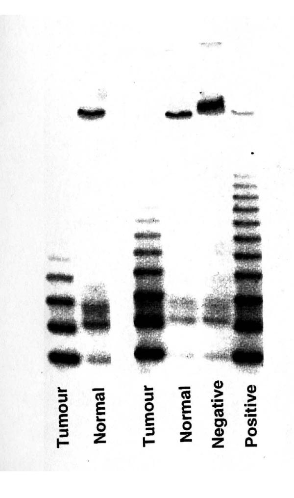
Telomerase activity was determined by the PCR-based TRAP assay as we described previously [18-20]. Figure 1 demonstrates typical telomerase positive and negative results.
Figure 1.
TRAP results in two breast tumours (TRAP +ve) and adjacent non-cancerous breast tissue (TRAP -ve). "Positive and negative" represent controls (squamous cell carcinoma of the head and neck and lysis buffer respectively). This figure shows the electrophoresis of PCR products on 10% polyacrylamide gel visualized by phospho-imager. Telomerase activity is indicated by the generation of a DNA ladder. The intensity of the bands correlates with telomerase activity.
Immunohistochemistry
Staining
4 μm sections were cut from standard paraffin wax blocks and mounted onto Superfost Plus slides. The sections were placed in 60°C oven overnight and dewaxed in xylene and placed in absolute alcohol for 2 minutes.
Endogenous peroxidase activity was blocked using methanol and hydrogen peroxide. The sections were washed in water. Heat mediated antigen retrieval was performed by pressure cooking Tris EDTA Citrate Buffer (pH = 7.8) for 15 minutes. The sections were then placed in the pressure-cooked Tris EDTA Citrate buffer and further pressure-cooked for 8 minutes. The sections were then placed in Tris buffer.
The rabbit affinity-purified polyclonal TGF beta RII antibody (L-21: sc-400, Santa Cruz Biotech) in a dilution of 1 in 300 was then applied and sections were incubated for 30 minutes, then washed in Tris buffer.
The second layer which consisted of peroxidase labelled polymer conjugated to goat antirabbit immunoglobulins (DAKO EnVision+ System Peroxidase, DAB, Code K4011) was then added and incubated for 30 minutes. The slides were then rinsed with Tris buffer. Liquid DAB+ substrate chromogen solution (3,3'-diaminobenzidine chromogen solution, DAKO EnVision+ System Peroxidase, DAB, Code K4011) was then applied for 5–10 minutes after which the slides were rinsed with water. The nuclei were counterstained using haematoxylin. The slides were then dehydrated, cleared and a coverslip was applied.
Scoring
The scoring system described by Gobbi et al [20] was used for scoring. TGF beta – RII staining is mostly cytoplasmic and was considered positive when the epithelial elements stained for TGF beta RII. The endothelial cells of blood vessels were used as internal positive control. The percentage of positive cells in breast cancers was assessed as 1 = <25%, 2 = 25–75% and 3 = > 75%. The intensity of positive staining was grouped into weak = 1, moderate = 2 and strong = 3 by comparing with the internal control. The score for intensity was added to the score for percentage of positive cells. Thus the minimum score was 2 and the maximum score was 6.
Statistical analysis
We hypothesised that telomerase reactivation in human breast cancer was associated with reduced immunohisto-chemical expression of TGF beta RII. Fisher's exact test was used to examine the association between telomerase activity and TGF beta RII expression. A p-value < 0.05 was considered significant.
Results
24 infiltrating breast carcinomas were included in this study. The median patients' age was 60 years (range: 37–87 years). Telomerase activity was detected in 17 (71%) of 24 tumours.
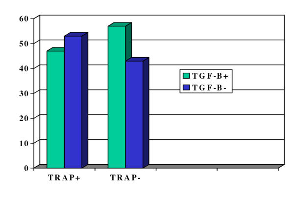
TGF beta RII was detected in all lesions. The percentage of stained cells ranged from 1–100%. According to the percentage and intensity of staining, the tumours were divided into two groups: group 1 (n = 12) with strong staining in >75% of cells (score = 6) and group 2 (n = 12) with weak to moderate staining in 1–75% of cells (score <6). The results are summarised in table 1 and figure 2. There was no significant association between telomerase activity and down-regulation of TGF beta RII expression (p = 1.0).
Table 1.
Telomerase activity and TGF beta RII expression (p = 1.0).
| Parameter | Telomerase Positive | Telomerase Negative | Total |
| TGF beta RII Strong Positive | 8 (47%) | 4 (57%) | 12 |
| TGF beta RII Moderate/Weak Positive | 9 (53%) | 3 (43%) | 12 |
| Total | 17 | 7 | 24 |
Figure 2.
Telomerase and TGF beta RII expression in human breast cancer. The 'y' axis shows the percentage of positive samples.
Discussion
This pilot study shows no significant association between telomerase reactivation and TGF beta RII expression in human breast cancer specimens. However, telomerase negative lesions were more likely to have a strong staining for TGF beta RII. Our initial hypothesis was that telomerase reactivation in human breast cancer was associated with reduced immunohisto-chemical expression of TGF beta is based on the fact that both telomerase activation [4-8] and genetic mutations related to TGF beta, its receptors or intracellular signalling molecules [18,20] are associated with carcinogenesis. The results of this study are not consistent with the in vitro results reported by Yang et al showing that autocrine TGF beta suppresses telomerase activity and hTERT expression in human cancer cell lines [19]. There are, however, two limitations in the present study. Firstly, the sample size is small (n = 24). Secondly, TGF beta RII and telomerase expressions were measured using two different methodologies. TGF beta RII expression was determined by IHC using a polyclonal antibody, whereas telomerase expression was measured using a sensitive PCR-based functional assay (TRAP). Furthermore, IHC results depend upon the specificity of the antibody used and this can be variable.
Moreover, immunohistochemical expression of a protein does not always correlate with its functional activity. Other possible explanations of this lack of association may be the fact that we determined the expression of TGFbeta RII rather than TGF beta itself. Because more than one receptor, in addition to Smad, are involved in the mechanism of action of TGF beta, therefore receptor II expression may not be a true reflection of TGF beta activity. These contradicting results could also be explained on the basis that carcinogenesis results from mutations in any component of the TGF beta functional pathway [9,17]. This suggests that the expression of TGF beta will be totally different from the expression of its receptors in terms of its relation to carcinogenesis. The study of Yang et al [19] was conducted on cell lines under controlled physiological conditions whereas this study was performed on human breast cancer specimens where the cancer cells are influenced by numerous growth factors, hormones and cytokines acting via endocrine and paracrine pathways. We have recently demonstrated that hTERT mRNA expression correlates with telomerase activity in human breast cancer [21], therefore the above hypothesis can be examined by determining the mRNA expression of hTERT, TGF beta, TGF beta RI and RII using quantitative reverse transcriptase PCR (QRT-PCR) methodology.
Contributor Information
Abd E Elkak, Email: aelkak2000@yahoo.co.uk.
Robert F Newbold, Email: robert.newbold@brunel.ac.uk.
Valene Thomas, Email: val.thomas@stgeorges.nhs.uk.
Kefah Mokbel, Email: kefahmokbel@hotmail.com.
References
- Morin GB. The human telomere terminal transferase enzyme is a ribonucleoprotein that synthesises TTAGGG repeats. Cell. 1989;59:521–9. doi: 10.1016/0092-8674(89)90035-4. [DOI] [PubMed] [Google Scholar]
- Counter CM, Avilion AA, LeFeuvre CE, et al. Telomere shortening associated with chromosome instability is arrested in immortal cells which express telomerase activity. EMBO J. 1992;11:1921–1929. doi: 10.1002/j.1460-2075.1992.tb05245.x. [DOI] [PMC free article] [PubMed] [Google Scholar]
- Blackburn EH. Structure and formation of telomeres. Nature. 1991;350:569–73. doi: 10.1038/350569a0. [DOI] [PubMed] [Google Scholar]
- Kim NW, Piatyszek MA, Prowse KR, et al. Specific association of human telomerase activity with immortal cells and cancer. Science. 1994;266:2011–15. doi: 10.1126/science.7605428. [DOI] [PubMed] [Google Scholar]
- Rhyu MS. Telomeres, Telomeres and immortality. J Natl Cancer Inst. 1995;87:884–94. doi: 10.1093/jnci/87.12.884. [DOI] [PubMed] [Google Scholar]
- Mokbel K, Ghilchik M, Williams G, et al. The association between telomerase activity and hormone receptor status and p53 expression in breast cancer. Int J Surg Investig. 2000;1:509–16. [PubMed] [Google Scholar]
- Mokbel K, Parris CN, Ghilchik M, et al. The association between telomerase activity, histopathological parameters and Ki-67 expression in invasive breast cancer. Am J Surg. 1999;178:69–72. doi: 10.1016/S0002-9610(99)00128-2. [DOI] [PubMed] [Google Scholar]
- Mokbel K, Parris CN, Ghilchik M, et al. Telomerase activity and lymphovascular invasion in breast cancer. Eur J Surg Oncol. 2000;26:30–3. doi: 10.1053/ejso.1999.0736. [DOI] [PubMed] [Google Scholar]
- Blobe GC, Schiemann WP, Lodish HF. Role of transforming growth factor beta in human disease. N Engl J Med. 2000;342:1350–8. doi: 10.1056/NEJM200005043421807. [DOI] [PubMed] [Google Scholar]
- Clark DA, Coker R. Transforming growth factor beta (TGF beta) Int J Biochem Cell Biol. 1998;30:293–8. doi: 10.1016/S1357-2725(97)00128-3. [DOI] [PubMed] [Google Scholar]
- Miyazono K. Positive and negative regulation of TGF beta signalling. J Cell Sci. 2000;113:1101–9. doi: 10.1242/jcs.113.7.1101. [DOI] [PubMed] [Google Scholar]
- Wang X-F, Lin HY, Ng-Eaton E, et al. Expression cloning and characterization of the TGF beta type III receptor. Cell. 1991;67:797–805. doi: 10.1016/0092-8674(91)90074-9. [DOI] [PubMed] [Google Scholar]
- Massague J. TGF beta signal transduction. Ann Rev Biochem. 1998;67:753–791. doi: 10.1146/annurev.biochem.67.1.753. [DOI] [PubMed] [Google Scholar]
- Lin HY, Wang X-F, Ng-Eaton E, et al. Expression cloning of the TGF beta type II receptor, a functional transmembrane serine/ threonine kinase. Cell. 1992;68:775–785. doi: 10.1016/0092-8674(92)90152-3. [DOI] [PubMed] [Google Scholar]
- Huse M, Chen Y-G, Massague J, et al. Crystal Structure of the cytoplasmic domain of type I TGF beta receptor in complex with FKBP 12. Cell. 1999;96:425–436. doi: 10.1016/s0092-8674(00)80555-3. [DOI] [PubMed] [Google Scholar]
- Wrana JL, Attisano L, Carcamo J, et al. TGF beta signals through a heteromeic protein kinase receptor complex. Cell. 1992;71:1003–1014. doi: 10.1016/0092-8674(92)90395-s. [DOI] [PubMed] [Google Scholar]
- Grady WM, Myeroff LL, Swinler SE, et al. Mutational inactivation of transforming growth factor beta receptor type II in microsatellite stable colon cancers. Cancer Res. 1999;59:320–4. [PubMed] [Google Scholar]
- Hojo M, Morimoto T, Maluccio M, et al. Cyclosporine induces cancer progression by a cell autonomous mechanism. Nature. 1999;397:530–4. doi: 10.1038/17401. [DOI] [PubMed] [Google Scholar]
- Yang H, Kyo S, Takatura M, et al. Autocrine transforming growth factor beta suppresses telomerase activity and transcription of human telomerase reverse transcriptase in human cancer cells. Cell Growth Differ. 2001;12:119–27. [PubMed] [Google Scholar]
- Gobbi H, Arteaga CL, Jensen RA, et al. Loss of expression of transforming growth factor beta type II receptor correlates with high tumour grade in human breast in-situ and invasive carcinomas. Histopathology. 2000;36:168–77. doi: 10.1046/j.1365-2559.2000.00841.x. [DOI] [PubMed] [Google Scholar]
- Kirkpatrick KL, Clarke G, Ghilchik M, Newbold RF, Mokbel K. Telomerase activity correlates with hTERT mRNA expression in human breast cancer. Eur J Surg Oncol. 2002. [DOI] [PubMed]


