Abstract
CD1d-restricted T cells contribute to tumor protection, but their precise roles remain unclear. Here we show that tumor cells engineered to secrete granulocyte–macrophage colony-stimulating factor induce the expansion of CD1d-restricted T cells through a mechanism that involves CD1d and macrophage inflammatory protein 2 expression by CD8α–, CD11c+ dendritic cells (DCs). The antitumor immunity stimulated by vaccination with irradiated, granulocyte-macrophage colony-stimulating factor-secreting tumor cells was abrogated in CD1d- and Jα281-deficient mice, revealing a critical role for CD1d-restricted T cells in this response. The loss of antitumor immunity was associated with impaired tumorinduced T helper 2 cytokine production, although IFN-γ secretion and cytotoxicity were preserved. DCs from immunized CD1d-deficient mice showed compromised maturation and function. Together, these results delineate a role for CD1d-restricted T cell–DC cross talk in the shaping of antitumor immunity.
CD161+ Vα14Jα281 invariant (iNKT) cells are involved in a wide variety of immune responses because of their expression of diverse cytokines, chemokines, and surface molecules (1). Vα14Jα281 T cells and their human CD161+ Vα24JαQ T cell counterparts are activated specifically by a glycolipid antigen (2–4) presented by the nonpolymorphic class Ib molecule CD1d (5, 6). Several studies indicate that Vα14Jα281 T cells participate in tumor protection. Experiments using either antibody-mediated depletion or Vα14Jα281-deficient mice have shown that these NKT cells play important roles in the antitumor effects stimulated by low doses of IL-12 (7–11). Moreover, Vα14Jα281 T cells are also required for protection against tumor development induced by chemical carcinogens (12, 13). The antitumor functions of CD1d-restricted T cells are augmented by α-galactosylceramide, an activating glycolipid antigen presented by CD1d, through a mechanism that involves dendritic cell (DC) production of IL-12 (14–16). The antitumor activities of Vα14Jα281 T cells include perforin-dependent NK-like cytotoxicity, IFN-γ production, and the stimulation of CD8+ T lymphocytes (2, 17).
The recognition that DCs play decisive roles in the priming of antigen-specific responses has led to the design of numerous protocols exploiting these cells for the induction of antitumor immunity (18). Either the ex vivo manipulation of DCs or the in vivo administration of DC-activating cytokines can enhance tumor rejection in model systems (19, 20). In this context, we have demonstrated that vaccination with irradiated tumor cells engineered to secrete granulocyte–macrophage colony-stimulating factor (GM-CSF) stimulates potent, specific, and long-lasting antitumor immunity in multiple murine tumor models (21). Although both GM-CSF and Flt3 ligand (FL) induced the marked expansion of DCs, vaccination with irradiated, GM-CSF-secreting tumor cells generated more potent antitumor immunity than vaccination with irradiated, FL-secreting tumor cells (22). The superior efficacy of the GM-CSF-based vaccine was associated with high-level CD1d expression on CD8α–, CD11c+ DCs.
While DCs are required for the efficient priming of antigen-specific lymphocyte responses, T cells in turn contribute to DC maturation (23, 24). In this regard, we and others recently showed that both human and murine CD1d-restricted NKT cells express multiple cytokines, chemokines, and surface proteins that are involved in the maturation of myeloid-type DCs (25, 26). This finding, together with the high level of CD1d expression on GM-CSF-stimulated DCs, motivated us to characterize DC function and the generation of antitumor immunity in CD1d-deficient mice. The experiments presented here reveal a role for CD1d-restricted T cell–DC cross talk in the shaping of antitumor immunity.
Methods
Mice. Eight- to 12-week-old female C57BL/6 mice were purchased from Taconic Farms. The CD1d null allele, generated in a 129 × C57BL/6 founder (27, 28), was backcrossed seven generations into the C57BL/6 strain. Homozygous CD1d-deficient mice were obtained from littermate pairings and used to generate heterozygote CD1d-deficient and WT controls. Jα281-deficient mice on a C57BL/6 background have been described (2, 7). All mouse experiments were approved and conducted under Institutional Animal Care and Use Committee guidelines.
Tumor Models. B16-F10 melanoma cells (syngeneic to C57BL/6 mice) were maintained in DMEM containing 10% (vol/vol) FCS and penicillin/streptomycin. GM-CSF-secreting B16 cells were generated as described (22). For the vaccination experiments, mice were immunized s.c. on the abdominal wall with 5 × 10⁵ irradiated (35 Gy), GM-CSF-secreting B16 cells and challenged 7 days later with 1 × 10⁶ live, WT B16 cells injected s.c. on the back. Animals were considered tumor free if they did not develop tumors during 60 days of observation. For fluorescence-activated cell sorting (FACS) experiments mice were challenged with live B16 melanomas engineered to secrete either GM-CSF or FL, and tumors were allowed to grow to 1.5–2 cm in diameter (10–14 days). Splenocytes were then harvested and stained with the appropriate antibodies for FACS analysis or cell sorting.
FACS Analysis. Fluorescent staining of splenocyte populations (depleted of erythrocytes with ammonium chloride) was performed by using FITC-, phycoerythrin-, or CyChrome-conjugated mAbs to T cell receptor (TCR), CD3, CD11c, CD11b, I-Ab, CD8α, CD1d, CD4, CD161, CD80, and CD86 obtained from PharMingen. Stained cells were analyzed on a FACScan cytometer (Becton Dickinson), and cell sorting was performed by using a MoFlo cytometer (Cytomation, Fort Collins, NJ). MHC class II staining was performed with the mAb Y3P.
Northern Blots. Total RNA was prepared from 99% pure sorted DC subsets by using Qiagen (Chatsworth, CA) RNeasy kits. Equal amounts of total RNA were electrophoresed, transferred to nitrocellulose membranes, and probed with 32P-labeled macrophage inflammatory protein 2 (MIP-2) cDNA. Equivalent loading of RNA was confirmed by ethidium bromide staining of the gel and reprobing with 32P-labeled GAPDH.
Analysis of Invariant Vα14Jα281 TCR Frequency. Total RNA was isolated from spleens of individual mice by using TRIzol (GIBCO/BRL) according to the manufacturer's recommendations. First-strand cDNA synthesis was performed by using oligo(dT) as a primer for reverse transcription of 2 μg of total RNA in a 50-μl reaction mixture using Moloney murine leukemia virus-reverse transcriptase (Life Technologies/GIBCO/BRL, Gaithersburg, MD). Quantitative analysis of Vα14Jα281 T cell frequency was done by using multiplex RT-PCR by comparing the intensity of the TCRα-chain CDR3 band with the invariant NKT cell-specific band, as described (29–31).
Cellular Assays. Mixed leukocyte reactions (MLRs) were performed as described (22). For peptide presentation experiments, mice were inoculated s.c. on the back with 1 × 10⁶ live, GM-CSF-secreting B16 cells, and their spleens were harvested when tumors reached 1.5–2 cm. CD11c+ DCs were isolated by using MACS CD11c Microbeads (Miltenyi Biotec, Auburn, CA) as per the manufacturer's specifications. For mixed lymphocyte experiments, the purified DCs (3 × 10³ per well) were cocultured in sextuplicate in 96-well Falcon 3077 U-bottom plates containing responder cells (5 × 10⁴ per well) from each strain of mouse, and proliferation was determined by 3H-thymidine incorporation at the end of 5 days. Antigen-specific IL-2 secretion was performed by using bead-purified DCs (3 × 10³ per well) cocultured in sextuplicate in 96-well Falcon 3077 U-bottom plates with 2 × 10⁴ cells of either the I-Ab:Eα52–68-specific hybridoma 1H3.1 or TCR transfectant H30 (32, 33) with or without increasing doses of Eα52–68 peptide (Dana–Farber Cancer Institute Molecular Biology Core Facility). IL-2 secretion was measured after 48 h by using the OptEIA mouse IL-2 ELISA kit from PharMingen as per the manufacturer's specifications. The 1H3.1 and H30 clones were kind gifts of Charles Janeway, Jr. (Yale University, New Haven, CT).
Cytokine Assays. Tumor-induced cytokine production was measured as described (22). Briefly, splenocytes were harvested 7 or 8 days after vaccination with irradiated, GM-CSF-secreting B16 cells, depleted of erythrocytes, and cultured (1 × 10⁶ cells) with irradiated (100 Gy) B16 cells (1 × 10⁵) in 2 ml of complete medium supplemented with 10 units/ml IL-2. Supernatants were harvested after 5 days and assayed for GM-CSF, IL-5, IL-10, IL-13, and INF-γ by ELISA using the appropriate mAbs [Endogen (Woburn, MA), PharMingen, and R & D Systems].
Cytotoxicity. Splenocytes were harvested 7–10 days after vaccination with irradiated, GM-CSF-secreting B16 cells, depleted of erythrocytes, and cultured (1 × 10⁶ cells) with irradiated (100 Gy) B16 cells (1 × 10⁵) in 2 ml of complete medium supplemented with 10 units/ml IL-2. A total of 2 × 10⁵ IFN-γ-treated B16 targets were labeled with 100 μCi of 51Cr for 2 h and plated at 103 cells per well in U-bottom 96-well plates. Effector cells were added in quadruplicate at varying effector-to-target ratios. 51Cr activity in supernatants taken 4 h later was measured in a gamma counter (Packard). Maximal and spontaneous lysis was determined by the addition of 4% Triton X-100 or media to the targets. The percentage of specific 51Cr release was calculated as 100 × (sample count–background count)/(maximal count–background count).
Statistics. A two-tailed Fisher's exact test was used for experiments examining tumor development after vaccination with cytokine-secreting tumor cells. All other comparisons were made with a Mann–Whitney test.
Results
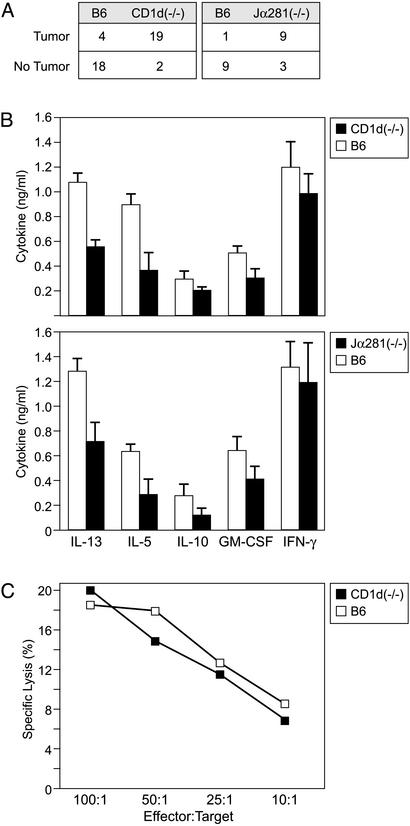
iNKT Cells Are Required for GM-CSF-Secreting Tumor Cell Vaccines. To examine the functional importance of CD1d in antitumor immunity and DC activation, we introduced a CD1d null allele onto the C57BL/6 background. Homozygous CD1d- or Jα281-deficient mice and their WT littermates were vaccinated with irradiated, GM-CSF-secreting B16 melanoma cells and challenged 1 week later with WT B16 cells. Whereas vaccinated WT mice efficiently rejected WT tumor challenge, as reported (21), vaccinated CD1d-(P < 0.01) and Jα281-deficient (P < 0.01) mice failed to generate protective immunity, with nearly all mutant animals developing tumors at the challenge site (Fig. 1A).
Fig. 1.
Vaccination with irradiated, GM-CSF-secreting B16 melanoma cells is abrogated in CD1d-deficient mice. (A) Female C57BL/6 WT, CD1d-, or Jα281-deficient mice were immunized with irradiated, GM-CSF-secreting B16 cells and challenged 1 week later with WT B16 cells. Vaccination with irradiated, WT B16 tumor cells failed to elicit protective immunity in any strain, and CD1d was not expressed on WT or transduced melanomas (data not shown). (B) Splenocytes isolated from vaccinated CD1d- or Jα281-deficient mice secreted significantly less (P < 0.05 Mann–Whitney test) IL-13, IL-5, IL-10, and GM-CSF, but equivalent amounts of IFN-γ, compared with splenocytes isolated from vaccinated WT mice. (C) CD1d-deficient mice show intact anti-B16 melanoma cytotoxicity. Splenocytes were harvested after vaccination and cultured in vitro for 5 days with irradiated B16 cells; 51Cr assays were performed against B16 targets.
To delineate the basis for the compromised vaccination in CD1d-deficient mice, the development of antitumor effectors was characterized. Because immunization with irradiated, GM-CSF-secreting tumor cells stimulated a broad T helper (Th) 1 and Th2 cytokine response (22, 34), splenocytes from vaccinated mice were harvested and their production of cytokines after coculture with irradiated, WT B16 cells was measured. Although CD1d- and Jα281-deficient mice and WT controls generated equivalent levels of IFN-γ, iNKT cell-deficient animals generated reduced levels of IL-5, IL-10, IL-13, and GM-CSF (Fig. 1B). IL-4 was not detected in any of the cultures (data not shown). The attenuated production of Th2 cytokines by iNKT cell-deficient mice was unexpected, as previous studies failed to reveal compromised Th2 responses (35–37). Consistent with the unaltered production of IFN-γ, however, vaccinated CD1d-deficient mice generated antitumor cytotoxic responses that were equivalent to WT littermates (Fig. 1C).
CD8α–, CD11c+ DCs Stimulated by GM-CSF Express CD1d and MIP-2. The impaired Th2 cytokine production in vaccinated iNKT cell-deficient mice was similar to the response we previously observed in WT mice vaccinated with irradiated, FL-secreting tumor cells (22). This earlier work also revealed that FL-stimulated DCs showed heterogeneous CD1d expression in contrast to the uniform, high-level CD1d expression on GM-CSF-stimulated DCs. Together, these findings suggested that GM-CSF and FL might differ in their abilities to activate CD1d-restricted T cells.
To explore this idea further, live B16 cells secreting either GM-CSF or FL were injected into WT mice, and their spleens were harvested after 2 weeks. As reported, the inoculation of either tumor line resulted in the marked expansion of CD11c+ DCs (22). However, GM-CSF-secreting tumor cells stimulated exclusively CD8α–, CD11c+ DCs, whereas FL-secreting tumor cells stimulated a mixture of CD8α–, CD11c+, CD8α+, and CD11c+ DCs (Fig. 2 A and B). Although the CD8α–, CD11c+ DCs induced by GM-CSF expressed CD1d, only the CD8α+, CD11c+ DCs induced by FL expressed CD1d, consistent with other experiments involving the injection of recombinant FL protein (38).
Fig. 2.
Myeloid-type DCs recruited by GM-CSF express CD1d and MIP-2. (A) Expression of CD1d on DCs expanded by GM-CSF or FL. The intensity of CD1d on CD8α+ and CD8α– DCs was determined by first gating on CD11c+ MHC class II+ DCs (Left) and then displaying CD1d versus CD8α staining (Right). A representative FACS from one of five experiments with similar findings is shown. (B) CD1d is expressed at significantly higher levels (*, P < 0.01, n = 5) on CD8α– DCs induced by GM-CSF treatment compared with CD8α– DCs induced by FL treatment. (C) The relative frequency of invariant Vα14Jα281 TCRα-chain transcripts in the spleens of mice challenged with GM-CSF- or FL-secreting tumor cells was determined by quantitative CDR3 spectratyping. One 2-fold dilution for equivalent amounts of total TCRβ-chain was assayed, and samples are shown next to serial dilutions of cDNA from the CD1d-restricted iNKT hybridoma DN32.d3 (8). (D) FACS analysis for CD161+ T cells in the spleens of mice with GM-CSF-secreting B16 cells are compared with mice with FL-secreting B16 cells (*, P < 0.01, n = 5). (E) Northern blot analysis of MIP-2 mRNA in DC subsets induced by FL or GM-CSF. Splenocytes were first sorted for CD11c+ and MHC class II bright, and then as CD8α+ or CD8α–. Total RNA isolated from sorted cells was probed with 32P-labeled MIP-2 or GAPDH cDNA.
Although both GM-CSF and FL elicited the expansion of DCs expressing CD1d, only GM-CSF stimulated an increase in the frequency of splenic Vα14Jα281 iNKT cells, as measured by both PCR and flow cytometry (Fig. 2 C and D). Because DCs produce specific chemokines during their maturation and migration (39), we investigated whether MIP-2, a chemokine recently reported to be important for the recruitment of CD1d-restricted iNKT cells (40), was differentially associated with DC subsets induced by GM-CSF or FL. RNA was thus isolated from sorted DC populations and MIP-2 expression assessed by Northern blot analysis. MIP-2 transcripts were found only in the CD8α–, CD11c+ DCs induced by GM-CSF (Fig. 2E).
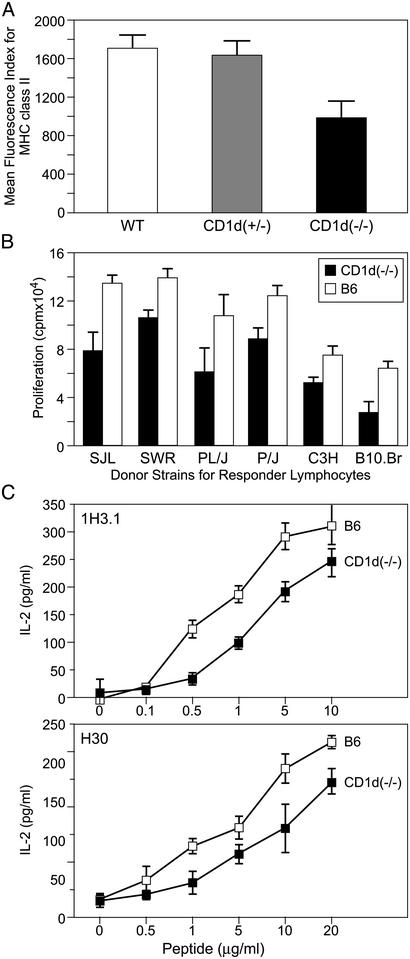
CD8α–, CD11c+ DC Maturation and Function Are Impaired in CD1d-Deficient Mice. Because CD1d-restricted iNKT cells express a panel of gene products important for the recruitment and activation of myeloid-type DCs (25, 26), the question of whether CD1d-restricted T cells contribute to DC activation in this tumor vaccination model was examined. A hallmark of DC maturation, a process required for the maximal stimulation of T cells, is an increase in cell surface MHC class II expression (41). FACS analysis of splenocytes harvested 14 days after injection of live, GM-CSF-secreting B16 cells showed comparable numbers of CD8α–, CD11c+ DCs in WT and CD1d-deficient mice (data not shown). However, MHC class II levels were significantly decreased on the DCs derived from CD1d-deficient mice when compared with WT or heterozygous controls (Fig. 3A).
Fig. 3.
DCs from CD1d-deficient mice are functionally impaired. (A) GM-CSF-secreting B16 melanomas recruit myeloid-type DCs that express significantly less cell surface MHC class II. WT, CD1d-heterozygous, and CD1d-deficient C57BL/6 mice were inoculated with live, GM-CSF-secreting B16 cells, and the expression of MHC class II on splenic myeloid-type DCs was determined 14 days later (P < 0.01 comparing CD1d–/–, n = 11 vs. CD1d+/–, n = 6, or CD1d+/+, n = 11). (B) MLRs induced by DCs are impaired in CD1d-deficient mice. Splenic DCs were isolated from mice injected with live, GM-CSF-secreting tumor cells and used to stimulate in vitro MLRs with splenocytes from SJL, SWR, PL/J, P/J, C3H, and B10.BR donors. Shown is a representative example from one of four experiments with identical patterns for MLR responses. (C) Presentation of peptide antigen to either the I-Ab:Ea52–68-specific hybridoma 1H3.1 or TCR transfectant H30 (32, 33) by DCs isolated from CD1d-deficient mice is impaired when compared with DCs isolated from WT mice. The data presented are representative of three experiments.
To determine whether the diminished MHC class II expression could be associated with impaired antigen-presenting cell function, DCs were harvested from CD1d-deficient mice and WT controls after injection of GM-CSF-secreting B16 tumor cells, and their ability to promote T cell responses in vitro was evaluated. Consistent with the observed MHC class II low surface phenotype, CD8α–, CD11c+ DCs isolated from CD1d-deficient mice were less efficient stimulators in MLRs than DCs from WT animals (Fig. 3B). Although MLRs are thought to depend on DC initiation of alloreactivity (41, 42), it is important to note that DCs from CD1d-deficient mice cannot stimulate iNKT cells in the responding population. Thus, a loss of iNKT cell-derived cytokines may also contribute to the diminished proliferative responses in these assays.
To assess the ability of CD8α–, CD11c+ DCs to function in an antigen-specific fashion, their ability to present the Eα52–68 peptide to the I-Ab-restricted hybridoma 1H3.1 and TCR transfectant H30 was examined (32, 33). Consistent with the results of the MLR, CD8α–, CD11c+ DCs isolated from CD1d-deficient mice were less efficient than WT controls in activating the Eα52–68-restricted hybridoma and transfectants (Fig. 3C).
Discussion
These investigations were undertaken in an effort to learn more about the roles of CD1d-restricted T cells in tumor immunity. Previous work illustrated that invariant NKT cells may inhibit chemical carcinogenesis and contribute to the therapeutic actions of IL-12 and α-galactosylceramide (7–9, 12–16). However, other studies suggested that CD1d-restricted T cells may attenuate tumor protection (43). These contrasting findings might reflect differential regulation of iNKT cell activities. Because DCs modulate iNKT cell functions (25, 26, 44–46), we examined in CD1d- and Jα281-deficient mice the generation of tumor immunity in response to GM-CSF-secreting melanoma cells. This immunization strategy stimulates the recruitment and maturation of myeloid-type DCs (22), rendering the system informative for exploring potential DC–iNKT cell interactions in vivo.
Our experiments demonstrate that CD1d-restricted T cells are required for tumor protection and optimal Th2 cytokine production as a consequence of GM-CSF-based cancer vaccines. Whereas invariant iNKT cells secrete IFN-γ and manifest NK-like cytotoxicity after IL-12 or α-galactosylceramide administration (14–16, 47), CD1d- and Jα281-deficient mice immunized with GM-CSF-secreting tumors developed comparable levels of IFN-γ and cytotoxicity as WT controls. These findings are consistent with previous reports showing that iNKT cells are a major source of IL-4 early after immune challenge (48). The association of impaired Th2 responses with compromised tumor rejection further suggests that tumor-associated cytotoxicity and IFN-γ secretion (49) may not be sufficient for maximal tumor immunity. Although preliminary studies indicate that host-derived GM-CSF or IL-5 individually are not required for efficient tumor protection (N.M. and G.D., unpublished data), we speculate that the coordinated loss of GM-CSF, IL-5, IL-10, and IL-13 contributes to the diminished tumor immunity. Other investigations also implicate important roles for Th2 cytokines in GM-CSF-based vaccines (50), and the adoptive transfer of Th2 cells can mediate tumor destruction (51). Nonetheless, the effects of Th2 cytokines may depend on specific characteristics of the tumor model, as iNKT cell-derived IL-13 inhibited immunity against a viral-associated neoplasm (43).
Because the impaired Th2 responses of vaccinated iNKT cell-deficient mice were similar to those previously observed in WT mice vaccinated with irradiated, FL-secreting cells (22), we hypothesized that GM-CSF and FL might differ in their abilities to activate iNKT cells. Indeed, only GM-CSF augmented the numbers of CD1d-restricted T cells in vivo, consistent with the failure of FL-induced splenic DCs to stimulate invariant iNKT cells in vitro (45). Moreover, the CD8α–, CD11c+ DCs generated with GM-CSF expressed high levels of CD1d and MIP-2, a chemokine involved in iNKT cell recruitment (40).
Our experiments further illustrate that CD1d-restricted T cells in turn contribute to DC maturation and function. GM-CSF stimulated DCs harvested from the spleens of CD1d- and Jα281-deficient mice showed reduced MHC class II expression and were less potent in stimulating MLRs and peptide-specific T cell responses compared with WT controls. These findings extend previous reports showing that invariant iNKT cells express multiple cytokines, chemokines, and surface proteins that modulate DC function (25, 26). The iNKT cell–DC cross talk revealed in these studies may also be operative in other immune responses, including the stimulation by α-galactosylceramide of CD4+ and CD8+ T lymphocytes and B cells in vivo (17, 52–55). Lastly, the tumor vaccine model reported here should prove useful for dissecting the roles of specific molecules expressed by CD1d-restricted T cells in DC activation (56).
Acknowledgments
We thank Drs. Mark Atkinson, Tetsu Kawano, Diane Mathis, Joan Stein-Streilein, and Yvonne van der Wal for critical reading of the manuscript and Dr. Patricia Bernardo (Department of Biostatistics, Dana–Farber Cancer Institute) for assistance with statistical analysis. This work was supported by National Institutes of Health Grants RO1 AI45051 (to S.B.W.), R35 CA47554 (to J.L.S.), and CA74886 (to G.D.), the Swiss National Science Foundation (to N.M. and S.G.), the Swiss Cancer League (to S.G.), Ter Meulen Fund and Royal Netherlands Academy of Arts and Sciences (to E.E.S.N.), and the Cancer Research Institute/Partridge Foundation and a Leukemia and Lymphoma Specialized Center of Research in Myeloid Leukemia Award (to G.D.). G.D. is a Clinical Scholar of the Leukemia and Lymphoma Society.
Abbreviations: DC, dendritic cell; GM-CSF, granulocyte–macrophage colony-stimulating factor; FL, Flt3 ligand; FACS, fluorescence-activated cell sorting; TCR, T cell receptor; MIP-2, macrophage inflammatory protein 2; MLR, mixed leukocyte reaction; Th, T helper.
References
- 1.Bendelac, A., Rivera, M. N., Park, H.-S. & Roark, J. H. (1997) Annu. Rev. Immunol. 15, 535–562. [DOI] [PubMed] [Google Scholar]
- 2.Kawano, T., Cui, J., Koezuka, Y., Toura, I., Kaneko, Y., Motoki, K., Ueno, H., Nakagawa, R., Sato, H., Kondo, E., et al. (1997) Science 278, 1626–1629. [DOI] [PubMed] [Google Scholar]
- 3.Zeng, Z.-H., Castano, A. R., Segelke, B. W., Stura, E. A., Peterson, P. A. & Wilson, I. A. (1997) Science 277, 339–345. [DOI] [PubMed] [Google Scholar]
- 4.Naidenko, O. V., Maher, J. K., Ernst, W. A., Sakai, T., Modllin, R. L. & Kronenberg, M. (1999) J. Exp. Med. 190, 1069–1079. [DOI] [PMC free article] [PubMed] [Google Scholar]
- 5.Balk, S. P., Bleicher, P. A. & Terhorst, C. (1989) Proc. Natl. Acad. Sci. USA 86, 252–256. [DOI] [PMC free article] [PubMed] [Google Scholar]
- 6.Balk, S. P., Bleicher, P. A. & Terhorst, C. (1991) J. Biol. Chem. 146, 768–774. [PubMed] [Google Scholar]
- 7.Cui, J., Shin, T., Kawano, T., Sato, H., Kondo, E., Toura, I., Kaneko, Y., Koseki, H., Kanno, M. & Taniguchi, M. (1997) Science 278, 1623–1626. [DOI] [PubMed] [Google Scholar]
- 8.Anzai, R., Seki, S., Ogasawara, K., Hashimoto, W., Sugiura, K., Sato, M., Kumagai, K. & Takeda, K. (1996) Immunology 88, 82–89. [DOI] [PMC free article] [PubMed] [Google Scholar]
- 9.Kawamura, T., Takeda, K., Mendiratta, S. K., Kawamura, H., Van Kaer, L., Yagita, H., Abo, T. & Okumura, K. (1998) J. Immunol. 160, 16–19. [PubMed] [Google Scholar]
- 10.Smyth, M. J., Taniguchi, M. & Street, S. E. (2000) J. Immunol. 165, 2665–2670. [DOI] [PubMed] [Google Scholar]
- 11.Takeda, K., Hayakawa, Y., Atsuta, M., Hong, S., Van Kaer, L., Kobayashi, K., Ito, M., Yagita, H. & Okumura, K. (2000) Int. Immunol. 12, 909–914. [DOI] [PubMed] [Google Scholar]
- 12.Smyth, M. J., Thia, K. Y. T., Street, S. E. A., Cretney, E., Trapani, J. A., Taniguchi, M., Tetsu, K., Pelikan, S. B., Crowe, N. Y. & Godfrey, D. I. (2000) J. Exp. Med. 191, 661–668. [DOI] [PMC free article] [PubMed] [Google Scholar]
- 13.Crowe, N. Y., Smyth, M. J. & Godfrey, D. I. (2002) J. Exp. Med. 196, 119–127. [DOI] [PMC free article] [PubMed] [Google Scholar]
- 14.Kitamura, H., Iwakabe, K., Yahata, T., Nishimura, S., Ohta, A., Ohmi, Y., Sato, M., Takeda, K., Okumura, K., Van Kaer, L., et al. (1999) J. Exp. Med. 189, 1121–1128. [DOI] [PMC free article] [PubMed] [Google Scholar]
- 15.Tomura, M., Yu, W.-G., Ahn, H.-J., Yamashita, M., Yang, Y.-F., Ono, S., Hamaoka, T., Kawano, T., Taniguchi, M., Koezuka, Y. & Fujiwara, H. (1999) J. Immunol. 163, 93–101. [PubMed] [Google Scholar]
- 16.Toura, I., Kawano, T., Akutsu, Y., Nakayama, T., Ochiai, T. & Taniguchi, M. (1999) J. Immunol. 163, 2387–2391. [PubMed] [Google Scholar]
- 17.Nishimura, T., Kitamura, H., Iwakabe, K., Yahata, T., Ohta, A., Sato, M., Takeda, K., Okumura, K., Van Kaer, L., Kawano, T., et al. (2000) Int. Immunol. 12, 987–994. [DOI] [PubMed] [Google Scholar]
- 18.Young, J. W. & Inaba, K. (1996) J. Exp. Med. 183, 7–11. [DOI] [PMC free article] [PubMed] [Google Scholar]
- 19.Mayordomo, J. I., Zorina, T., Storkus, W. J., Zitvogel, L., Celluzzi, C., Falo, L. D., Melief, C. J., Ildstad, S. T., Kast, W. M., Deleo, A. B. & Lotze, M. T. (1995) Nat. Med. 1, 1297–1302. [DOI] [PubMed] [Google Scholar]
- 20.Lynch, D. H., Andreasen, A., Maraskovsky, E., Whitmore, J., Miller, R. E. & Schuh, J. C. (1997) Nat. Med. 3, 625–631. [DOI] [PubMed] [Google Scholar]
- 21.Dranoff, G., Jaffee, E., Lazenby, A., Golumbek, P., Levitsky, H., Brose, K., Jackson, V., Hamada, H., Pardoll, D. & Mulligan, R. C. (1993) Proc. Natl. Acad. Sci. USA 90, 3539–3543. [DOI] [PMC free article] [PubMed] [Google Scholar]
- 22.Mach, N., Gillessen, S., Wilson, S. B., Sheehan, C., Mihm, M. & Dranoff, G. (2000) Cancer Res. 60, 3239–3246. [PubMed] [Google Scholar]
- 23.Rissoan, M. C., Soumelis, V., Kadowaki, N., Grouard, G., Briere, F., de Waal Malefyt, R. & Liu, Y. J. (1999) Science 283, 1183–1186. [DOI] [PubMed] [Google Scholar]
- 24.Shreedhar, V., Moodycliffe, A. M., Ullrich, S. E., Bucana, C., Kripke, M. L. & Flores-Romo, L. (1999) Immunity 11, 625–636. [DOI] [PubMed] [Google Scholar]
- 25.Wilson, S. B., Kent, S. C., Horton, H. F., Hill, A. A., Bollyky, P. L., Hafler, D. A., Strominger, J. L. & Byrne, M. C. (2000) Proc. Natl. Acad. Sci. USA 97, 7411–7416. [DOI] [PMC free article] [PubMed] [Google Scholar]
- 26.Yang, O. O., Racke, F. K., Nguyen, P. T., Gausling, R., Severino, M. E., Horton, H. F., Byrne, M. C., Strominger, J. L. & Wilson, S. B. (2000) J. Immunol. 165, 3756–3762. [DOI] [PubMed] [Google Scholar]
- 27.Sonoda, K. H., Exley, M., Snapper, S., Balk, S. P. & Stein-Streilein, J. (1999) J. Exp. Med. 190, 1215–1226. [DOI] [PMC free article] [PubMed] [Google Scholar]
- 28.Exley, M. A., Bigley, N. J., Cheng, O., Tahir, S. M., Smiley, S. T., Carter, Q. L., Stills, H. F., Grusby, M. J., Koezuka, Y., Taniguchi, M. & Balk, S. P. (2001) J. Leukocyte Biol. 69, 713–718. [PubMed] [Google Scholar]
- 29.Gorski, J., Yassai, M., Zhu, X., Kissela, B., Kissella, B., Keever, C. & Flomenberg, N. (1994) J. Immunol. 152, 5109–5119. [PubMed] [Google Scholar]
- 30.Naumov, Y. N., Naumova, E. N. & Gorski, J. (1996) Hum. Immunol. 48, 52–62. [DOI] [PubMed] [Google Scholar]
- 31.Apostolou, I., Takahama, Y., Belmant, C., Kawano, T., Huerre, M., Marchal, G., Cui, J., Taniguchi, M., Nakauchi, H., Fournie, J. J., et al. (1999) Proc. Natl. Acad. Sci. USA 96, 5141–5146. [DOI] [PMC free article] [PubMed] [Google Scholar]
- 32.Chervonsky, A. V., Medzhitov, R. M., Denzin, L. K., Barlow, A. K., Rudensky, A. Y. & Janeway, C. A., Jr. (1998) Proc. Natl. Acad. Sci. USA 95, 10094–10099. [DOI] [PMC free article] [PubMed] [Google Scholar]
- 33.Viret, C., Wong, F. S. & Janeway, C. A., Jr. (1999) Immunity 10, 559–568. [DOI] [PubMed] [Google Scholar]
- 34.Soiffer, R., Lynch, T., Mihm, M., Jung, K., Rhuda, C., Schmollinger, J. C., Hodi, F. S., Liebster, L., Lam, P., Mentzer, S., et al. (1998) Proc. Natl. Acad. Sci. USA 95, 13141–13146. [DOI] [PMC free article] [PubMed] [Google Scholar]
- 35.Smiley, S. T., Kaplan, M. H. & Grusby, M. J. (1997) Science 275, 977–979. [DOI] [PubMed] [Google Scholar]
- 36.Chen, Y.-H., Chiu, N. M., Mandal, M., Wang, N. & Wang, C.-R. (1997) Immunity 6, 459–467. [DOI] [PubMed] [Google Scholar]
- 37.Mendiratta, S. K., Martin, W. D., Hong, S., Boesteanu, A., Joyce, S. & Van Kaer, L. (1997) Immunity 6, 469–477. [DOI] [PubMed] [Google Scholar]
- 38.Pulendran, B., Lingappa, J., Kennedy, M. K., Smith, J., Teepe, M., Rudensky, A., Maliszewski, C. R. & Maraskovsky, E. (1997) J. Immunol. 159, 2222–2231. [PubMed] [Google Scholar]
- 39.Sallusto, F. & Lanzavecchia, A. (1999) J. Exp. Med. 189, 611–614. [DOI] [PMC free article] [PubMed] [Google Scholar]
- 40.Faunce, D. E., Sonoda, K. H. & Stein-Streilein, J. (2001) J. Immunol. 166, 313–321. [DOI] [PubMed] [Google Scholar]
- 41.Banchereau, J. & Steinman, R. M. (1998) Nature 392, 245–252. [DOI] [PubMed] [Google Scholar]
- 42.Steinman, R. M. & Inaba, K. (1985) Crit. Rev. Immunol. 5, 331–348. [PubMed] [Google Scholar]
- 43.Terabe, M., Matsui, S., Noben-Trauth, N., Chen, H., Watson, C., Donaldson, D., Carbone, D., Paul, W. & Berzofsky, J. (2000) Nat. Immunol. 1, 515–520. [DOI] [PubMed] [Google Scholar]
- 44.Kadowaki, N., Antonenko, S., Ho, S., Rissoan, M. C., Soumelis, V., Porcelli, S. A., Lanier, L. L. & Liu, Y. J. (2001) J. Exp. Med. 193, 1221–1226. [DOI] [PMC free article] [PubMed] [Google Scholar]
- 45.Ikarashi, Y., Mikami, R., Bendelac, A., Terme, M., Chaput, N., Terada, M., Turz, T., Angevin, E., Lemonnier, F. A., Wakasugi, H. & Zitvogel, L. (2001) J. Exp. Med. 194, 1179–1186. [DOI] [PMC free article] [PubMed] [Google Scholar]
- 46.Naumov, Y. N., Bahjat, K. S., Gausling, R., Abraham, R., Exley, M. A., Koezuka, Y., Balk, S. B., Strominger, J. L., Clare-Salzer, M. & Wilson, S. B. (2001) Proc. Natl. Acad. Sci. USA 98, 13838–13843. [DOI] [PMC free article] [PubMed] [Google Scholar]
- 47.Kawano, T., Nakayama, T., Kamada, N., Kaneko, Y., Harada, M., Ogura, N., Akutsu, Y., Motohashi, S., Iizasa, T., Endo, H., et al. (1999) Cancer Res. 59, 5102–5105. [PubMed] [Google Scholar]
- 48.Yoshimoto, T. & Paul, W. E. (1994) J. Exp. Med. 179, 1285–1295. [DOI] [PMC free article] [PubMed] [Google Scholar]
- 49.Mosmann, T. R. & Coffman, R. L. (1989) Annu. Rev. Immunol. 7, 145–173. [DOI] [PubMed] [Google Scholar]
- 50.Hung, K., Hayashi, R., Lafond-Walker, A., Lowenstein, C., Pardoll, D. & Levitsky, H. (1998) J. Exp. Med. 188, 2357–2368. [DOI] [PMC free article] [PubMed] [Google Scholar]
- 51.Rodolfo, M., Zilocchi, C., Accornero, P., Cappetti, B., Arioli, I. & Colombo, M. P. (1999) J. Immunol. 163, 1923–1928. [PubMed] [Google Scholar]
- 52.Singh, N., Hong, S., Scherer, D. C., Serizawa, I., Burdin, N., Kronenberg, M., Koezuka, Y. & Van Kaer, L. (1999) J. Immunol. 163, 2373–2377. [PubMed] [Google Scholar]
- 53.Burdin, N., Brossay, L. & Kronenberg, M. (1999) Eur. J. Immunol. 29, 2014–2025. [DOI] [PubMed] [Google Scholar]
- 54.Carnaud, C., Lee, D., Donnars, O., Park, S. H., Beavis, A., Koezuka, Y. & Bendelac, A. (1999) J. Immunol. 163, 4647–4650. [PubMed] [Google Scholar]
- 55.Kitamura, H., Ohta, A., Sekimoto, M., Sato, M., Iwakabe, K., Nakui, M., Yahata, T., Meng, H., Koda, T., Nishimura, S., et al. (2000) Cell. Immunol. 199, 37–42. [DOI] [PubMed] [Google Scholar]
- 56.Banchereau, J., Briere, F., Caux, C., Davoust, J., Lebecque, S., Liu, Y. J., Pulendran, B. & Palucka, K. (2000) Annu. Rev. Immunol. 18, 767–811. [DOI] [PubMed] [Google Scholar]


