Abstract
Both the protective antigen (PA) and the poly(γ-d-glutamic acid) capsule (γdPGA) are essential for the virulence of Bacillus anthracis. A critical level of vaccine-induced IgG anti-PA confers immunity to anthrax, but there is no information about the protective action of IgG anti-γdPGA. Because the number of spores presented by bioterrorists might be greater than encountered in nature, we sought to induce capsular antibodies to expand the immunity conferred by available anthrax vaccines. The nonimmunogenic γdPGA or corresponding synthetic peptides were bound to BSA, recombinant B. anthracis PA (rPA), or recombinant Pseudomonas aeruginosa exotoxin A (rEPA). To identify the optimal construct, conjugates of B. anthracis γdPGA, Bacillus pumilus γdLPGA, and peptides of varying lengths (5-, 10-, or 20-mers), of the d or l configuration with active groups at the N or C termini, were bound at 5–32 mol per protein. The conjugates were characterized by physico-chemical and immunological assays, including GLC-MS and matrix-assisted laser desorption ionization time-of-flight spectrometry, and immunogenicity in 5- to 6-week-old mice. IgG anti-γdPGA and antiprotein were measured by ELISA. The highest levels of IgG anti-γdPGA were elicited by decamers of γdPGA at 10 –20 mol per protein bound to the N- or C-terminal end. High IgG anti-γdPGA levels were elicited by two injections of 2.5 μg of γdPGA per mouse, whereas three injections were needed to achieve high levels of protein antibodies. rPA was the most effective carrier. Anti-γdPGA induced opsonophagocytic killing of B. anthracis tox–, cap+. γdPGA conjugates may enhance the protection conferred by PA alone. γdPGA-rPA conjugates induced both anti-PA and anti-γdPGA.
Anthrax probably caused the “festering boils” of the people and cattle of Egypt described in the sixth plague of the Old Testament. After the discovery of Bacillus anthracis by Robert Koch in 1880 (1), Pasteur (2) developed a vaccine for sheep composed of chemically treated attenuated strains. Routine use of a noncapsulated strain has virtually eliminated anthrax among domesticated animals (3). In the only controlled study of an anthrax vaccine in humans, culture-supernatant from a cap–nonproteolytic strain that produced protective antigen (PA), conferred 92% efficacy among woolsorters (4). The Centers for Disease Control monitored the anthrax vaccine adsorbed (AVA) in industrial settings between 1962 and 1974: none of 34 cases occurred in fully vaccinated individuals. A similar vaccine is used in the U.K. (5). This and other evidence indicate that serum IgG anti-PA confers immunity to cutaneous and inhalational anthrax in humans (6, 7).
The structure and expression of the essential virulence factors of B. anthracis are controlled by two plasmids. pX01 encodes anthrax toxin (AT) composed of the PA (binding subunit of AT), and two enzymes known as lethal factor and edema factor (8, 9). Administration of AT to primates mimics the symptoms of anthrax (9). pX02 encodes the poly(γ-d-glutamic acid) (γdPGA) capsule of B. anthracis (10, 11). Other bacilli produce poly(γ-glutamic acid) (γPGA) but only B. anthracis synthesizes it entirely in the d conformation (12). γdPGA is a surface structure (13), inhibits in vitro phagocytosis and, when injected, is a poor immunogen even as a bacterial component (14–18); the protective effect of anti-γdPGA has not been reported. The capsule shields the vegetative form of B. anthracis from agglutination by monoclonal antibodies to its cell wall polysaccharide (19). Systemic infection with B. anthracis induces γdPGA antibodies (20). Antibodies to d-amino acid polymers may be induced in animals by injection of γdPGA methylated BSA complexes along with Freund's adjuvant, i.v. injections of a formalin-treated capsulated B. anthracis, or by peptidyl proteins (16, 21). We report the synthesis and evaluation of conjugates that induce γdPGA antibodies under conditions suitable for clinical use.
Experimental Procedures
Bacterial Strains. Bacillus pumilus strain Sh18 and B. anthracis strain A34, a pX01–, pX02+ variant derived from the Ames strain by repeated passage at 43°C, have been described (10, 22).
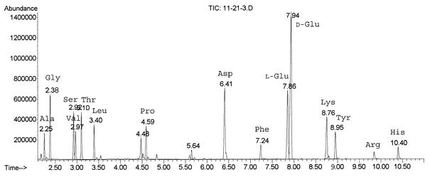
Analytic. Amino acid analyses were done by GLC-MS after hydrolysis with 6 M HCl, 150°C, 1 h, derivatization to heptafluorobutyryl R-(–)isobutyl esters, and assayed with a Hewlett–Packard apparatus (model HP 6890) with a HP-5 0.32 × 30 mm glass capillary column, temperature programming at 8°C per min, from 125°C to 250°C in the electron ionization (106 eV) mode (24). Under these conditions, we could separate d-glutamic acid from the l-enantiomer. The amount of each was calculated based on the ratio of d-glutamic acid relative to l-glutamic acid residues in the protein (Fig. 1). The number of peptide chains in l-peptide conjugates was calculated by the increase of total l-glutamic acid relative to aspartic acid. Protein concentration was measured by the method of Lowry (25), free ε amino groups were measured by Fields' assay (26), thiolation was measured by release of 2-pyridylthio groups (A343) (27), and hydrazide was measured as reported (28). SDS/PAGE used 14% gels according to the manufacturer's instructions. Double immunodiffusion was performed in 1.0% agarose gel in PBS.
Fig. 1.
GLC-MS analysis of rPA-Cys-Gly3-γdPGA10-C conjugate. Under the described conditions, l-Glu can be separated from d-Glu to calculate the number of γdPGA chains incorporated into the conjugate.
Matrix-Assisted Laser Desorption Ionization–Time-of-Flight (MALDI-TOF). Mass spectra were obtained with a PerSeptive BioSystems Voyager Elite DE-STR MALDI-TOF instrument (Applied Biosystems) operated in the linear mode, 25-kV accelerating voltage, and a 300-nsec ion extraction delay time. Samples for analysis were prepared by a “sandwich” of matrix and analyte. First, 1 μl of matrix (saturated solution of sinnapinic acid made in 1:1 CH3CN and 0.1% trifluroacetic acid) was dried on the sample stage. Second, 1 μl of sample and an additional 1 μl of matrix were applied. After the “sandwich” was dried, the sample was placed in the mass spectrometer.
Antigens. BSA (Sigma) was dialyzed against pyrogen-free water, sterile-filtered, and freeze-dried. Recombinant PA (rPA) from B. anthracis and recombinant exoprotein A (rEPA) from Pseudomonas aeruginosa were prepared and characterized (29, 30).
γPGA was extracted from the culture supernatant of B. anthracis or B. pumilus by cetavlon precipitation, acidification to pH 1.5, precipitation with ethanol, and passage through a 2.5 × 100-cm Sepharose CL-4B column in 0.2 M NaCl (23). Their compositions were confirmed by 1H-NMR and 13C-NMR, and their enantiomeric conformations were compared by GLC-MS spectroscopy.
Three types of γPGA peptides (AnaSpec, San Jose, CA) were synthesized by the method of Merrifield with 5, 10, or 20 residues. Their purity and authenticity were verified by GLC-MS, liquid chromatography MS, and MALDI-TOF. The peptides were bound to the protein at the C or the N termini (-C indicates that the C terminus is free, and N-indicates that the amino terminus is free).
Type I, NBrAc-Gly3-γdPGAn-COOH(Br-Gly3-γdPGAn-C); NBrAc-Gly3-γlPGAn-COOH(Br-Gly3-γlPGAn-C).
Type II, NAc-l-Cys-Gly3-γ-d-PGAn-COOH(Cys-Gly3-γdPGAn-C); NAc-l-Cys-Gly3-γ-l-PGAn-COOH(Cys-Gly3-γlPGAn-C).
Type III, NAc-γdPGAn-Gly3-l-Cys-CONH2(N-γdPGAn-Gly3-Cys); NAc-γlPGAn-Gly3-l-Cys-CONH2(N-γlPGAn-Gly3-Cys).
Conjugations to rPA are described. BSA and rEPA were used in a similar manner. All reactions were conducted in a pH stat under argon.
Conjugation of rPA with B. anthracis γdPGA and with B. pumilus γDLPGA. rPA was derivatized with adipic acid dihydrazide with modifications (28). The pH was maintained at 7.0, and 0.1 M 1-ethyl-3-(3-dimethylaminopropyl)carbodiimide·HCl (EDAC) was used. The product, rPA-AH, contained 2.0–4.7% hydrazide. γPGA was bound to rPA-AH or rEPA-AH with 0.01 M EDAC, the reaction mixture was passed through a 1 × 90-cm Sephacryl S-1000 column in 0.2 M NaCl, and fractions reacting with anti-PA and anti-γdPGA by an identity line were pooled.
Conjugation of Type-I Peptide with rPA via Thioether Bond. Step 1. Step 1 consisted of derivatization of rPA with N-hydroxysuccinimide ester of 3-(2-pyridyl dithio)-propionic acid (SPDP). To rPA (30 mg) in 1.5 ml of buffer A′ (PBS/3% glycerol/0.005 M EDTA, pH 7.6), SPDP (10 mg) in 50 μl of dimethyl sulfoxide was added in 10-μl aliquots and reacted for 1 h at pH 7.6. The product, 2-pyridyldithio-propionyl-rPA (PDP-rPA), was passed through a 1 × 48-cm Sephadex G-50 column in buffer A (PBS/0.05% glycerol/0.005 M EDTA, pH 7.6), and protein-containing fractions were pooled and assayed for thiolation, antigenicity, and molecular mass (27).
Step 2. Step 2 consisted of conjugation of PDP-protein with type-I peptide. PDP-protein (24 mg) in 2 ml of buffer A was treated with 50 mM dithiotreitol for 30 min at room temperature and passed through a 1 × 48-cm Sephadex G-50 column in buffer A. Fractions containing the 3-thiopropionyl-ε-Lys-NH2-rPA (rPA-SH) were collected and concentrated to 1.5 ml, and glycerol was added to a final concentration of 3%. Br-Gly3-γ-dPGAn-C, 10 mg in 1 ml of buffer A, was adjusted to pH 7.6 with 1 M NaOH, and rPA-SH was added, incubated for 1 h at room temperature (31), transferred to a vial, capped, and tumbled overnight at room temperature. Bromoacetamide, 0.5 mg in 50 μl of buffer A, was added to block unreacted thiols. After 30 min, the reaction mixture was passed through a 1 × 90-cm Sephacryl S-200 column in buffer B (0.01 M phosphate/0.2 M NaCl/0.05% glycerol, pH 7.2). Fractions containing protein-PGA were polled and assayed for peptide and protein concentration, antigenicity, and molecular mass.
Products. BSA contains 60, rPA contains 58, and rEPA contains 15 mol of Lys per mol of protein, respectively. Under the above conditions, 28 of 60 ε-Lys-NH2 of BSA, 50–55 of 58 of rPA and 15 of 15 of rEPA were derivatized with N-hydroxysuccinimide ester of 3-(2-pyridyl dithio)-propionic acid with retention of their antigenicity. Conjugation of BSA-SH, rPA-SH and rEPA-SH with type-I peptides yielded: BSA-SH/Gly3-γdPGAn-C; BSA-SH/Gly3-γlPGAn-C; rEPA-SH/Gly3-γdPGAn-C; rPA-SH/Gly3-γdPGAn-C.
Conjugation with Type-II and -III Peptides. Step 1. Derivatization of protein with succinimidyl 3-(bromoacetamido) propionate (SBAP). rPA (30 mg) in 1.5 ml of buffer A′ was adjusted to pH 7.2. SBAP, 11 mg in 50 μl dimethyl sulfoxide, was added in 10 μl aliquots (31). After 60 min, the reaction mixture was passed through a 1 × 90-cm Sepharose CL-6B column in buffer B. Fractions containing bromoacetamidopropionyl-ε-Lys-NH-rPA (Br-rPA) were collected and assayed for protein, free -NH2, antigenicity, and molecular mass.
Step 2. Step 2 involved conjugation of Br-protein with type-II and -III peptides. Type-II or -III peptides (5–15 mg in 1 ml buffer A) were adjusted to pH 7.6 with 1 M NaOH and Br-protein (25 mg) in 1.5 ml buffer A′ was added. After 1 h, the reaction mixture transferred to a vial, capped, and tumbled overnight at room temperature. 2-Mercaptoethanol (1 μl) was added to quench the remaining bromoacetyl groups in Br-protein. After 30 min, the reaction mixture was passed through a 1 × 90-cm Sepharose CL-6B column in buffer B. Fractions containing protein-PGA were polled and assayed for peptide and protein concentration, antigenicity, and molecular mass.
Products. Under these conditions, 50–55 of 58 and 15 of 15 residues of ε-Lys-NH2 of rPA and rEPA, respectively, were modified with succinimidyl 3-(bromoacetamido) propionate. Conjugation of Br-rPA and Br-rEPA with type-II peptides yielded four conjugates:
rPA/S-Cys-Gly3-γdPGAn-C.
rPA/S-Cys-Gly3-γlPGAn-C.
rEPA/S-Cys-Gly3-γdPGAn-C.
rEPA/S-Cys-Gly3-γlPGAn-C.
Conjugation of Br-rPA and Br-rEPA and with type-III peptides yielded four conjugates:
N-γdPGAn-Gly3-Cys-S/rPA.
N-γlPGAn-Gly3-Cys-S/rPA.
N-γdPGAn-Gly3-Cys-S/rEPA.
N-γlPGAn-Gly3-Cys-S/rEPA.
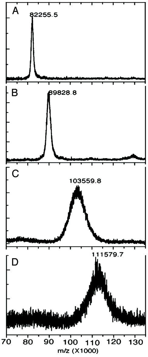
All eight conjugates precipitated with an identity reaction with their protein and γPGA antisera by immunodiffusion. Representative analysis by MALDI-TOF is shown in Fig. 2.
Fig. 2.
MALDI-TOF spectra. (A) rPA. (B) Br-rPA. (C and D) rPA-Cys-Gly3-γdPGA10-C conjugate containing an average of 11 γdPGA chains (C) or 16 γdPGA chains per rPA (D).
Immunization. Five- to six-week-old female National Institutes of Health Swiss–Webster mice were immunized s.c. three times at 2-week intervals with 2.5 μg of PGA as a conjugate in 0.1 ml of PBS, and groups of 10 mice were exsanguinated 7 days after the second or third injections (28). Controls received PBS.
Antibodies. Serum IgG antibodies were measured by ELISA (32). Nunc Maxisorb plates were coated with γdPGA, 20 μg/ml PBS, or 4 μg of rPA per ml of PBS (determined by checkerboard titration). Plates were blocked with 0.5% BSA (or with 0.5% HSA for assay of BSA conjugates) in PBS for 2 h at room temperature. A MRX Dynatech reader was used. Antibody levels were calculated relative to standard sera: for γdPGA, a hyperimmune murine serum prepared by multiple i.p. injections of formalin-treated B. anthracis strain A34 and assigned a value of 100 ELISA units; for PA, a mAb containing 4.7 mg of Ab per ml (33). Results were computed with an ELISA data processing program provided by the Biostatistics and Information Management Branch, Centers for Disease Control (34). IgG levels are expressed as geometric mean (GM).
Opsonophagocytosis. Spores of B. anthracis strain A34 were maintained at 5 × 10⁸ spores per ml in 1% phenol. The human cell line HL-60 (CCL240, American Type Culture Collection) was expanded and differentiated by dimethyl formamide into 44% myelocytes and metamyelocytes and 53% band and polymorphonuclear leukocytes (PMN). PMN were at an effector/target cell ratio of 400:1. PMN were centrifuged and resuspended in opsonophagocytosis buffer (Hanks' buffer with Ca2+, Mg2+, and 0.1% gelatin; Life Technologies, Grand Island, NY) to 2 × 10⁷. Spores were cultivated at 5 × 10⁷ per ml for 3 h, 20% CO2, and diluted to 5 × 10⁴ per ml. Sera were diluted 2-fold with 0.05 ml in opsonophagocytosis buffer in 24-well tissue culture plates (Falcon) and 0.02 ml containing ≈103 bacteria added to each well. The plates were incubated at 37°C, 5% CO2 for 15 min. A 0.01-ml aliquot of colostrum-deprived baby calf serum (used as a complement) and 0.02 ml of HL-60 suspension containing 4 × 10⁵ cells was added to each well and incubated at 37°C for 45 min, 5% CO2, with mixing at 220 rpm in a Minitron incubator shaker (Infors AG, Bottmingen, Switzerland). A 0.01-ml aliquot from each well was added to tryptic soy agar (Difco) at 50°C, and colony-forming units were determined the next morning. Opsonophagocytosis was defined by ≥50% killing compared with the growth in control wells (35).
Statistics. ELISA values are expressed as the GM. An unpaired t test was used to compare GMs in different groups of mice.
Results
Characterization of γPGA Conjugates. The PGA/protein ratio was assessed by MALDI-TOF spectrometry that provided molecular mass of the conjugates and by GLC-MS that provided the amount of bound PGA (Fig. 1 and 2). The two methods corroborated each other.
Serum IgG Anti-γdPGA. Native γdPGA from the capsule of B. anthracis elicited trace levels of antibodies after the third injection. All of the conjugates, in contrast, elicited IgG anti-γdPGA after two injections (Table 1). Conjugates of B. anthracis γdPGA and of B. pumilus γd(60%)l(40%)PGA elicited IgG anti-γdPGA of intermediate levels after two injections with a booster after the third. Precipitates were formed during the synthesis of both conjugates resulting in low yields.
Table 1. Composition and serum GM IgG anti-γdPGA and anti-carrier protein (ELISA) elicited by conjugates in mice of BSA, rEPA, and rPA.
|
Anti-γDPGA*
|
Anti-protein†
|
|||||
|---|---|---|---|---|---|---|
| Conjugate | Mol γDPGA per mol protein | Protein/γDPGA (wt/wt) | Second injection | Third injection | Second injection | Third injection |
| γDPGA-B. anthracis | NA | NA | 0.3 | 4.4 | NA | NA |
| rEPA-AH/γDPGA-B. anthracis | NA | 1:0.29 | 695 | 2,312 | ND | ND |
| rPA-AH γDPGA-B. anthracis | NA | 1:4.42 | 1,325 | 3,108 | ND | ND |
| BSA-SH/Gly3-γDPGA10-C‡ | 7 | 1:0.14 | 134 | 1,984 | ND | ND |
| BSA-SH/Gly3-γDPGA10-C | 18 | 1:0.35 | 1,882 | 1,821 | ND | ND |
| BSA-SH/Gly3-γDPGA10-C | 25 | 1:0.49 | 2,063 | 2,780 | ND | ND |
| BSA-SH/Gly3-γLPGA10-C | 7 | 1:0.14 | 261 | 618 | ND | ND |
| rEPA/Cys-Gly3-γDPGA10-C | 7 | 1:0.14 | 479 | 4,470 | ND | ND |
| rEPA-SH/Gly3-γDPGA5-C | 17 | 1:0.17 | 502 | 1,168 | ND | ND |
| rEPA-SH/Gly3-γDPGA10-C | 9 | 1:0.18 | 931 | 3,193 | ND | ND |
| rEPA-SH/Gly3-γDPGA20-C | 5 | 1:0.19 | 749 | 2,710 | ND | ND |
| rPA/Cys-Gly3-γDPGA5-C | 32 | 1:0.26 | 2,454 | 4,560 | 0.06 | 8.5 |
| rPA/Cys-Gly3-γDPGA10-C | 16 | 1:0.26 | 9,091 | 11,268 | 1.30 | 59.3 |
| rPA/Cys-Gly3-γDPGA20-C | 14 | 1:0.44 | 742 | 3,142 | 0.01 | 4.5 |
| rPA/Cys-Gly3-γDPGA5-N | 22 | 1:0.18 | 3,149 | 3,460 | 3.70 | 95.0 |
| rPA/Cys-Gly3-γDPGA10-N | 21 | 1:0.33 | 5,489 | 7,516 | 0.10 | 2.2 |
| rPA/Cys-Gly3-γDPGA20-N | 8 | 1:0.25 | 2,630 | 5,461 | 0.05 | 4.9 |
| rPA-SH/Gly3-γDPGA5-C | 15 | 1:0.12 | 1,813 | 3,607 | 0.27 | 19.7 |
| rPA-SH/Gly3-γDPGA10-C | 11 | 1:0.18 | 10,460 | 9,907 | 0.50 | 102.0 |
| rPA-SH/Gly3-γDPGA10-C | 14 | 1:0.22 | 4,378 | 7,206 | 0.34 | 66.3 |
| rPA-SH/Gly3-γDPGA20-C | 4 | 1:0.13 | 2,655 | 4,069 | 0.90 | 32.2 |
| rPA-SH/Gly3-γDPGA20-C | 8 | 1:0.25 | 9,672 | 7,320 | 0.22 | 189.0 |
| rPA/Cys-Gly3-γLPGA20-N | 22 | 1:0.70 | 24 | 79 | 0.14 | 3.0 |
| rPA/Cys-Gly3-γLPGA20-C | 24 | 1:0.76 | 155 | 437 | 0.31 | 7.8 |
NA, not applicable; ND, not done.
γDPGA from B. anthracis, strain A34, 2.5 μg as a conjugate used for injection, antibodies by ELISA expressed as EU.
Antibodies by ELISA expressed as μg Ab/ml.
C or N refers to the free amino acid on the γPGA bound to the protein.
IgG anti-γdPGA levels induced by the different conjugates overlapped. The highest levels were achieved with peptide decamers, 16 mol per protein for rPA/Cys-Gly3-γdPGA10-C, 11 and 14 mol per protein for rPA-SH/Gly3-γdPGA10-C. rPA was a more effective carrier than rEPA or BSA. With the exception of rPA-SH/Gly3-γdPGA10-C, with 11 chains per protein, all conjugates elicited a rise (mostly nonsignificant) after the third injection. Conjugates prepared with l peptides bound at either the C or N terminus induced low levels of IgG anti-γdPGA.
Table 2 shows the dose-response of two γdPGA conjugates with rPA or rEPA as the carrier; both peptides had 20 residues and similar numbers of chains per protein. Again, rPA was a more effective carrier than rEPA. The lowest dose (2.5 μg) of rPA-SH/Gly3-γdPGA-C elicited the highest level of IgG anti-γdPGA (9,152 ELISA units), the levels declined ≈1/2 at the 20-μg dose. rEPA-SH/Gly3-γdPGA-C, in contrast, elicited similar levels at all dosages.
Table 2. Dose/immunogenicity relation of conjugates prepared with 20-mers of γdPGA bound to rPA or recombinant rEPA.
| Conjugate | Mol γDPGA per mol protein | Protein/γDPGA (wt/wt) | Dose per mouse, μg γDPGA | Anti-γDPGA third injection |
|---|---|---|---|---|
| rPA-SH/Gly3-γDPGA20-C | 8 | 1:0.25 | 2.5 | 9,152 |
| 5 | 7,070 | |||
| 10 | 3,487 | |||
| 20 | 4,901 | |||
| rEPA-SH/Gly3-γDPGA20-C | 6 | 1:0.23 | 2.5 | 1,956 |
| 5 | 2,393 | |||
| 10 | 2,639 | |||
| 20 | 2,834 |
Five- to six-week-old National Institutes of Health Swiss—Webster mice (n = 10) injected s.c. with 0.1 ml of the conjugates every 2 weeks apart and exsanguinated 7 days after the third injection. IgG anti-γDPGA was measured by ELISA, and the results are expressed as the GM (9,152 vs. 3,487, P = 0.003; 9,152 vs. 4,901, P = 0.04; 9,152 vs. 1,956, P < 0.0001; 7,070 vs. 2,393, P < 0.0001).
Serum IgG Anti-Carrier Protein. With a few exceptions, both the length and number of γdPGA chains per protein were related to the level of IgG antiprotein. Conjugates prepared with γdPGA containing 20 residues elicited low levels of protein antibodies, although in one case the induced GM level was the highest. In general, conjugates prepared with 5 or 10 residues and with ≤15 chains per protein elicited the highest levels of IgG protein antibodies.
Opsonophagocytic Activity of Mouse Antisera. Sera from normal mice or those immunized with rEPA or rPA did not have opsonophagocytic activity (not shown). Table 3 shows a rough correlation between the level of IgG anti-γdPGA and opsonophagocytosis in mice immunized with BSA-SH/Gly3-γdPGA10-C or BSA-SH/Gly3-γdPGA10-C (r = 0.7, P = 0.03). Addition of γdPGA from B. anthracis to the immune sera showed a dose-related reduction of the opsonophagocytic titer of ≈60% (not shown).
Table 3. Opsonophagocytic activity and IgG anti-γdPGA (ELISA) elicited by BSA-SH/Gly3-γdPGA10-C.
| Sera | IgG anti-γdPGA | Reciprocal opsonophagocytic titer |
|---|---|---|
| 1196G | 407 | Not detected |
| 1195C | 1,147 | 640 |
| 1197B | 3,975 | 2,560 |
| 1190H | 3,330 | 2,560 |
| 1194D | 3,278 | 2,560 |
| 1193B | 3,178 | 2,560 |
| 1194G | 3,277 | 2,560 |
| 1191J | 5,191 | 5,120 |
Correlation coefficient between ELISA and reciprocal opsonophagocytic titer is 0.7, P = 0.03.
Discussion
Worldwide control of anthrax has been achieved in most countries by education, immunization of domesticated animals with attenuated (noncapsulated) strains of B. anthracis, and immunization of at-risk individuals with PA (AVA) and with antibiotics. Deliberate contamination of the mail with B. anthracis spores prompted improvement of AVA because (i) consistency of production is difficult because there is no measurement of PA or of other components in the vaccine that cause local and systemic reactions; these problems should be solved by replacing AVA with a purified PA: (ii) administration of AVA by s.c. injections at 0, 2, and 4 weeks and 6, 12, and 18 months with yearly boosters was designed for rapid induction of immunity in at-risk individuals (36). Replacement of this schedule with that used for primary immunization with DTP will require only three injections 2 months apart for the primary series, and a booster at 1 year will yield higher levels of anti-PA (37). (iii) The safety, immunogenicity, and efficacy of AVA in children have not been characterized.
The level of antibodies to PA required to confer immunity to B. anthracis seems high and may be difficult to maintain. Addition of anti-γdPGA-induced opsonophagocytic killing could augment protection mediated by anti-PA against the high levels of spores that might occur during a bioterrorist attack.
γdPGA and polysaccharides share some properties: (i) both are multivalent; (ii) both resist host enzyme catalysis; (iii) a hexamer of either occupies the antibody-combining site (38); (iv) covalent binding to a protein confers enhanced immunogenicity and T cell dependence (39–42); and (v) conjugates of synthetic saccharides as well as of γdPGA elicited higher levels of antibodies than did the “natural” polymer (39).
The structure determines the optimal immunogenicity for conjugates composed of linear polymers (hapten) bound at one point to a protein (39–42): (i) the hapten lengths should be sufficient to occupy the antibody combining site; and (ii) a critical density of the hapten on the carrier protein is necessary to form both aggregates with the surface Ig receptor and to permit interaction of the protein carrier fragments with T cells. As with synthetic oligosaccharides of Shigella dysenteriae type 1 (39), the results presented here and additional data not shown suggest that a decamer of γdPGA, bound to the protein at a density of 10–15 per mol of protein, would provide maximal immunogenicity.
Similar to the surface polysaccharides of capsulated pathogens, the level of vaccine-induced IgG antibodies and opsonophagocytosis were correlated. The finding that anti-γdPGA mediates in vitro opsonophagocytosis and possibly protection against challenge with B. anthracis warrants clinical evaluation of γdPGA conjugates vaccines.
Acknowledgments
We thank Dana Hsu, Jeanne B. Kaufman, Christopher Mocca, Chunyan Gou, and Robin Roberson for their technical assistance, Susan Welkos for providing anti-γPGA serum, and Robert Austrian for reviewing the manuscript.
Abbreviations: PA, protective antigen; MALDI-TOF, matrix-assisted laser desorption ionization–time-of-flight; γdPGA, poly(γ-d-glutamyl) capsule from Bacillus anthracis; rEPA, recombinant Pseudomonas aeruginosa exoprotein A; rPA, recombinant Bacillus anthracis PA; AVA, anthrax vaccine adsorbed; rPA-SH, 3-thiopropionyl-ε-Lys-NH2-rPA; GM, geometric mean.
References
- 1.Koch, R. (1877) Beitr. Biol. Pflanzen 2, 277–310. [Google Scholar]
- 2.Pasteur, M. L. & Chamberland, M. M. (1881) C. R. Acad. Sci. 92, 429–435. [Google Scholar]
- 3.Sterne, M. (1939) Onderstepoort J. Vet. Sci. Anim. Ind. 13, 307–312. [Google Scholar]
- 4.Brachman, P. S., Gold, H., Plotkin, S. A., Fekety, R., Werrin, M. & Ingraham, N. R. (1962) Am. J. Public Health 52, 632–645. [DOI] [PMC free article] [PubMed] [Google Scholar]
- 5.Anonymous (1965) Br. Med. J. 5464, 717–718.5825408 [Google Scholar]
- 6.Gladstone, G. P. (1946) Br. J. Exp. Pathol. 27, 394–318. [PMC free article] [PubMed] [Google Scholar]
- 7.Auerbach, S. & Wright, G. G. (1955) J. Immunol. 75, 129–133. [PubMed] [Google Scholar]
- 8.Mikesell, P., Ivins, B. E., Ristoph, J. D. & Dreier, T. M. (1983) Infect. Immun. 39, 371–376. [DOI] [PMC free article] [PubMed] [Google Scholar]
- 9.Klein, F., Hodges, D. R, Mahlandt, B. G, Jones, W. I., Haines, B. W. & Lincoln, R. E. (1962) Science 138, 1331–1333. [DOI] [PubMed] [Google Scholar]
- 10.Green, B. D., Battisti, L., Koehler, T. M., Thorne, C. B. & Ivins, B. E. (1985) Infect. Immun. 49, 291–297. [DOI] [PMC free article] [PubMed] [Google Scholar]
- 11.Kovács, J. & Bruckner, V. (1952) J. Chem. Soc., 4255–4259.
- 12.Etinger-Tulcynska, R. (1933) Z. Hyg. Infektionskr. 114, 769–775. [Google Scholar]
- 13.Zwartouw, H. T. & Smith, H. (1956) Biochem. J. 63, 437–442. [DOI] [PMC free article] [PubMed] [Google Scholar]
- 14.Makino, S., Sasakawa, C., Uchida, I., Terakado, N. & Yoshikawa, M. (1988) Mol. Microbiol. 2, 371–376. [DOI] [PubMed] [Google Scholar]
- 15.Eisner, V. M. (1959) Schweiz. Z. Path. Bakt. 22, 129–144. [PubMed] [Google Scholar]
- 16.Ezzell, J. W., Abshire, T. G., Little, S. F., Lidererding, B. C. & Brown, C. (1990) J. Clin. Microbiol. 28, 223–231. [DOI] [PMC free article] [PubMed] [Google Scholar]
- 17.Ostroff, G., Axelrod, D. R. & Bovarnick, M. (1958) Proc. Soc. Exp. Biol. Med. 99, 345–347. [DOI] [PubMed] [Google Scholar]
- 18.Dintzis, H. M., Symer, D. E., Dintzis, R. Z., Zawadzke, L. E. & Berg, J. M. (1993) Proteins 16, 306–308. [DOI] [PubMed] [Google Scholar]
- 19.Sirisanthana, T., Nelson, K. E., Ezzell, J. W. & Abshire, T. G. (1988) Am. J. Trop. Med. Hyg. 39, 575–581. [DOI] [PubMed] [Google Scholar]
- 20.Sage, H. J., Deutsch, G. F., Fasman, G. D. & Levine, L. (1964) Immunochemistry 1, 133–144. [DOI] [PubMed] [Google Scholar]
- 21.Goodman, J. W., Nitecki, D. E. & Stoltenberg, I. M. (1968) Biochemistry 7, 706–710. [PubMed] [Google Scholar]
- 22.Myerowitz, R. L., Gordon, R. E. & Robbins, J. B. (1973) Infect. Immun. 8, 896–900. [DOI] [PMC free article] [PubMed] [Google Scholar]
- 23.Troy, F. A. (1973) J. Biol. Chem. 248, 305–315. [PubMed] [Google Scholar]
- 24.MacKenzie, S. L. (1987) J. Assoc. Off. Anal. Chem. 70, 151–160. [PubMed] [Google Scholar]
- 25.Lowry, O. H. Rosenbrough, N., Farr, A. L. & Randall, R. J. (1951) J. Biol. Chem. 193, 266–273. [PubMed] [Google Scholar]
- 26.Fields, R. (1971) Biochem. J. 124, 581–590. [DOI] [PMC free article] [PubMed] [Google Scholar]
- 27.Carlsson, J., Drevin, H. & Axen, R. (1978) Biochem. J. 173, 723–737. [DOI] [PMC free article] [PubMed] [Google Scholar]
- 28.Schneerson, R., Barrera, O., Sutton, A. & Robbins, J. B. (1980) J. Exp. Med. 152, 361–376. [DOI] [PMC free article] [PubMed] [Google Scholar]
- 29.Ramirez, D. M., Leppla, S. H., Schneerson, R. & Shiloach, J. (2002) J. Ind. Microbiol. Biotechnol. 28, 232–238. [DOI] [PubMed] [Google Scholar]
- 30.Johansson, H. J., Jagersten, C. & Shiloach, J. (1996) J. Biotechnol. 48, 9–14. [DOI] [PubMed] [Google Scholar]
- 31.Inman, J. K., Highnest, P. F., Kolodny, N. & Robey, F. A. (1991) Bioconj. Chem. 2, 458–463. [DOI] [PubMed] [Google Scholar]
- 32.Taylor, D. N., Trofa, A. C., Sadoff, J., Chu, C., Bryla, D., Shiloach, J., Cohen, D., Ashkenazi, S., Lerman, Y., Egan, W., et al. (1993) Infect. Immun. 61, 3678–3687. [DOI] [PMC free article] [PubMed] [Google Scholar]
- 33.Little, S. F., Leppla, S. H. & Cora, E. (1988) Infect. Immun. 56, 1807–1813. [DOI] [PMC free article] [PubMed] [Google Scholar]
- 34.Plikaytis, B. D., Holder, P. F. & Carlone, G. M. (1996) User's Manual 12 (Centers for Disease Control, Atlanta), Version 1.00.
- 35.Romero-Steiner, S., Liutti, D., Pais, L. B., Dykes, J., Anderson, P., Whitin, J. C., Keyserling, H. L. & Carlone, G. M. (1997) Clin. Diagn. Lab. Immunol. 4, 415–433. [DOI] [PMC free article] [PubMed] [Google Scholar]
- 36.Puziss, M. & Wright, G. G. (1962) J. Bacteriol. 85, 230–236. [DOI] [PMC free article] [PubMed] [Google Scholar]
- 37.Pittman, P. R., Mangiafico, J. A., Rossi, C. A., Cannon, T. L., Gibbs, P. H., Parker, G. W. & Friedlander, A. M. (2001) Vaccine 19, 213–216. [DOI] [PubMed] [Google Scholar]
- 38.Kabat, E. A. (1983) Prog. Immunol. 5, 67–85. [Google Scholar]
- 39.Pozsgay, V., Chu, C., Pannell, L., Wolfe, J., Robbins, J. B. & Schneerson, R. (1999) Proc. Natl. Acad. Sci. USA 96, 5194–5197. [DOI] [PMC free article] [PubMed] [Google Scholar]
- 40.Lee, W. Y. & Sehon, A. H. (1976) J. Immunol. 116, 1711–1718. [PubMed] [Google Scholar]
- 41.Dintzis, H. M., Dintzis, R. Z. & Vogelstein, B. (1976) Proc. Natl. Acad. Sci. USA 73, 3671–3675. [DOI] [PMC free article] [PubMed] [Google Scholar]
- 42.Anderson, P. W., Pichichero, M. E. A., Stein, E. C., Porcelli, S., Betts, R. F., Connuck, D. M., Korones, D., Insel, R. A., Zahradnik, J. M. & Eby, R. (1989) J. Immunol. 142, 2464–2468. [PubMed] [Google Scholar]


