Abstract
After a period of eccentric exercise of elbow flexor muscles of one arm in young, adult human subjects, muscles became fatigued and damaged. Damage indicators were a fall in force, change in resting elbow angle and delayed onset of soreness. After the exercise, subjects were asked to match the forearm angle of one arm, whose position was set by the experimenter, with their other arm. Subjects matched the position of the unsupported reference arm, when this was unexercised, with a significantly more flexed position in their exercised indicator arm. Errors were in the opposite direction when the reference arm was exercised. The size of the errors correlated with the drop in force. Less consistent errors were observed when the reference arm was supported. A similar pattern of errors was seen after concentric exercise, which does not produce muscle damage. The data suggested that subjects were using as a position cue the perceived effort required to maintain a given forearm angle against the force of gravity. The fall in force from fatigue after exercise meant more effort was required to maintain a given position. That led to matching errors between the exercised and unexercised arms. It was concluded that while a role for muscle spindles in kinaesthesia cannot be excluded, detailed information about static limb position can be derived from the effort required to support the limb against the force of gravity.
We have been studying the effects of eccentric exercise on muscle properties. Eccentric exercise is interesting because it is the only form of mild exercise which routinely produces muscle damage if the individual is not trained to be protected against it (Hough, 1902). As a result it leaves them stiff and sore the next day (Proske & Morgan, 2001). It has also been reported to produce a disturbance to proprioception; Saxton et al. (1995) found that eccentric exercise led subjects to make significant position matching errors in a forearm matching task. Several years ago we repeated those experiments under somewhat modified conditions and came up with a different result (Brockett et al. 1997). The primary motivation for the experiments reported here was to try to reconcile the various observations. The findings led us to adopt a rather different point of view.
In a forearm position matching task it was found that after a period of eccentric exercise which reduced muscle force by 40–50%, subjects made significant position matching errors. The errors were in the direction reported by Saxton et al. The size of the errors correlated with the drop in force. We concluded that the fall in force led to an increase in the effort required to maintain position of the limb against the force of gravity. This increase in effort led to the matching errors.
A preliminary report of this work was recently presented at a local scientific meeting (Walsh et al. 2004).
Methods
The study comprised three experiments, each of which included six subjects. For the 18 individuals, four females and 14 males, the range of ages was 22–64 years. All subjects gave their informed, written consent. The experiments were approved by the Monash University Committee for Human Experimentation and the experiments were consistent with the Declaration of Helsinki.
The subjects were required to attend a number of sessions. A control series of measurements was carried out, which helped to familiarize them with the equipment and gave them some experience in the procedures. For the eccentric exercise, measurements were made immediately before and after the exercise, then at 2, 24, 48, 72 and 96 h post-exercise. For the concentric exercise, measurements were made before and after the exercise and at 2 and 24 h post-exercise. Whether the right or left arm was exercised was varied at random between subjects.
Position matching task
Measurements were carried out as described by Brockett et al. (1997). Each subject's forearms were strapped to two light-weight paddles, hinged at one end, with the hinges aligned with the subject's elbow joint. The angle of the upper arm was kept at approximately 45 deg to the horizontal. Potentiometers attached to the paddle hinges provided an analog voltage signal proportional to joint angle. Voltage signals were recorded by computer.
In the first experiment, the experimenter moved the reference forearm from the horizontal position, in the direction of flexion, to a predetermined angle and the subject, who was blindfolded, was asked to hold the arm in that position. He or she was then asked to match the angle with the other arm, that is, to align the two forearms by voluntarily moving the indicator arm.
In the second experiment, the experimenter placed the reference arm at a set angle where position of the arm was maintained by means of a support. The subject therefore did not need to generate any effort to maintain the position of the reference arm. To ensure that a subject's reference arm remained relaxed during the matches, surface EMG was monitored for biceps. This was done with Ag–AgCl electrodes, attached to the skin with tape, with a solid gel contact point. EMG signals were amplified using an ADInstruments bioAmp and the amplified signal was fed through a speaker so that the blindfolded subject could hear it. Once a subject's reference arm had been placed in position he or she was instructed to relax, and keep the EMG signal at a minimum.
A third experiment used concentric rather than eccentric exercise and here position matching was done with the reference arm unsupported.
Arm position was defined by the angle subtended by the forearm to the horizontal, which was taken as zero. So an angle of 30 deg represented an elbow angle of 105 deg, given that the upper arm was at an angle of 45 deg. In the series involving eccentric exercise, matching angles of 30, 60 and 90 deg were used. For the concentric exercise 15, 30 and 45 deg were used. The choice of angles for each trial was varied at random, as was the choice of reference and indicator arms. Each trial was repeated five times for each of the three test angles for both right and left arms, making for a total of 30 trials.
Measurements of muscle properties after exercise
Maximum voluntary contraction
For measurement of maximum voluntary contraction (MVC), a paddle was moved into the vertical position, that is, an elbow angle of 90 deg, which approximates to the optimum angle for torque in elbow flexors (Weerakkody et al. 2003b). The paddle was held in that position by a horizontal metal bar which at its other end was attached to an isometric tension transducer. Subjects were required to flex their elbow maximally for three contractions, each separated by a 1 min rest period. The mean peak value of torque was used as the MVC measurement. In the experiments on eccentric exercise, MVC was measured before each position matching session. For the concentric exercise, additional measurements of MVC were made for the session immediately after the exercise. Here MVC was measured both before and after the position-matching task, to obtain a better indication of the rate of recovery from fatigue.
Resting elbow angle
Damage from eccentric exercise in elbow flexors leads the relaxed arm to adopt a more flexed position than normal (Jones et al. 1987). To determine whether some damage had been produced after the exercise in these experiments, measurements were made of resting elbow angle. For this, the standing subjects were asked to let their arms hang naturally by their sides. A goniometer was used to measure the excluded angle between the humerus and ulna at the lateral epicondyle.
Muscle tenderness
Measurements at 24 h after eccentric exercise showed that the muscle had developed areas of tenderness in response to local pressure (Weerakkody et al. 2003a). To quantify this, muscle tenderness measurements were made with a compression gauge which had a 1.5 cm diameter plunger and a force range of 1–60 N. Resolution over that range was ± 0.5 N. Experience had shown that the soreness was distributed unevenly across the muscle (Weerakkody et al. 2003a). Therefore at the start of each measurement session different regions were tested manually until, in the subject's view, the most sensitive spot had been identified. Measurements of pain threshold were made at this site and the value was recorded. For this the plunger was pushed slowly into the muscle and the force at which some pain was detected was recorded. If force values were > 30 N it was assumed that there was no soreness.
The exercise
Eccentric exercise
The exercise regime was similar to that described by Weerakkody et al. (2003b). Subjects were seated in the chair of a dynamometer (Biodex, Shirley, NY, USA) with the one hand grasping the arm attachment. The shoulder was strapped to the chair so that it did not move. Immediately before the exercise, MVC was measured with the dynamometer in isometric mode. During the exercise subjects were required to resist elbow extension carried out by the dynamometer, which was set to generate isokinetic movents at 60 deg s−1 with a yield at 30% MVC. Subjects were instructed to exert just enough force to slow the movement into extension but without stopping it. When the arm reached full extension the movement stopped, the subjects relaxed their arm, and the arm attachment was returned to its starting position by the operator. Subjects carried out five sets of 10 eccentric contractions with 20 s rests between each set. This comprised one exercise bout. Typically subjects completed four to five bouts depending on their fitness. As soon as subjects began to have difficulty in resisting the extension movements, the exercise was stopped.
Concentric exercise
In this task subjects were required to lift a weight which had been adjusted to represent a load of 30% of the isometric MVC for elbow flexors. Subjects were asked to do a series of lifts from full extension to full flexion. They were instructed to lift the weight by only flexing at the elbow, keeping their shoulder rigid. When the weight had been lifted to the point where the elbow was fully flexed, the experimenter lowered the weight again. Subjects carried out sets of 10 lifts, with a 15 s rest period between sets. They continued until they began to have difficulty lifting the weight. Subjects differed widely in their endurance, achieving between 50 and 200 lifts. They were then seated in front of the forearm matching gear and their arms were taped to the paddles. Typically by this time a small amount of force recovery had already taken place.
Statistical analysis
The data were analysed using the software package IgorPro v. 4.07 (WaveMetrics, Inc., Lake Oswego, OR, USA) running on an Apple iMac computer. All statistical analysis used the program Data Desk 5.0.1 (Data Description, Inc., Ithaca, NY, USA). Analysis consisted of an ANOVA with no interactions, followed by a Bonferroni post hoc test, if the ANOVA was significant. When subject was used as a factor, it was random and the other factors, time post-exercise and reference elbow angle were fixed factors. Position error versus MVC correlations used a general linear model. MVC was a continuous, random variable and reference angle was a discrete, fixed variable. Threshold for significance was taken as a P value of 0.05. Wherever mean values are cited they are given ± s.e.m. (standard error of the mean).
Results
Three different experiments were carried out. The first two involved eccentric exercise, the third concentric exercise. In the first experiment, during the forearm position-matching task, subjects were required to actively maintain the reference elbow angle, set by the experimenter, by contracting elbow flexors sufficiently to support the weight of the arm. They would then move their indicator arm to a position they considered matched that of the reference. In a second experiment, once the reference angle had been selected by the experimenter, the arm was placed in that position and kept there by means of a support. It meant that subjects did not have to exert any effort to maintain the reference position, so no effort cue would be available during matching. In the third experiment muscle fatigue was produced by concentric exercise rather than eccentric exercise and the position-matching test used an unsupported reference arm. Here the idea was tested that just fatigue was sufficient to produce position-matching errors, since it is known that concentric exercise does not produce any muscle damage (Newham et al. 1983a,b).
Indicators of damage and fatigue
Eccentric exercise
Before considering the effects of eccentric exercise on position sense it was necessary to confirm that the exercise had produced some muscle damage, since our initial hypothesis had been that any errors in position sense were attributable to the damaging effects of the exercise. Three indicators of damage were measured, the drop in MVC, elbow angle and muscle soreness.
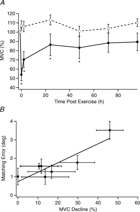
All subjects who underwent eccentric exercise showed a drop in MVC. The largest fall in MVC occurred immediately after the exercise, with a mean fall of 46 ± 7% (n = 6 subjects). Force then gradually recovered, until at 72 h it was no longer significantly different from control values (Fig. 1). An ANOVA with MVC of the exercised arm as dependent variable and subject and time post-exercise as factors, found that time was significant. A post hoc test on time post-exercise showed that MVC had fallen significantly below control values immediately after the exercise, at 2, 24 and 48 h.
Figure 1. Changes in MVC and the Error –MVC relation for position errors after eccentric exercise.
A, percent change in MVC of elbow flexor muscles over 4 days after eccentric exercise. The pre-exercise value was assigned 100%. Continuous line, exercised arm; dashed line, unexercised arm. Values are means (± s.e.m.) for 6 subjects. An asterisk indicates where values were significantly different from the control. B, relationship between the mean decline in MVC, expressed as a percentage of control values, and mean position matching errors (unsupported matches) between reference and indicator arms, given in degrees of forearm inclination above the horizontal. Matching errors were averaged across 5 trials, 3 angles and 6 subjects. The correlation was significant. The slope of the relation was 0.05 deg per percentage MVC decline.
There is evidence that after eccentric exercise there is a rise in whole-muscle passive tension (Whitehead et al. 2001, 2003). The rise in tension in elbow flexors leads the relaxed arm to adopt a more flexed position than normal (Jones et al. 1987). The shift in mean elbow angle observed here was 8 deg immediately after the exercise and it peaked at 12 deg at 24 h. An ANOVA with relaxed elbow angle of the exercised arm as dependent variable and subject and time post-exercise as factors showed both to be significant. A Bonferroni post hoc test showed significant differences from control values immediately after the exercise and at 2, 24, 48 and 72 h. The values at 96 h were not significant. The unexercised arm showed shifts in mean elbow angle of less than 1 deg. The corresponding ANOVA showed that time was significant but the post hoc test did not show a significantly more flexed elbow at any particular time.
All subjects who underwent eccentric exercise showed evidence of tenderness next day, in response to muscle palpation, stretch or contraction. Pain threshold to local muscle pressure fell at 24 h by an average of 15 ± 4 N. Analysis (ANOVA plus post hoc test) showed tenderness at 24, 48 and 72 h to be significantly different from control values.
Concentric exercise
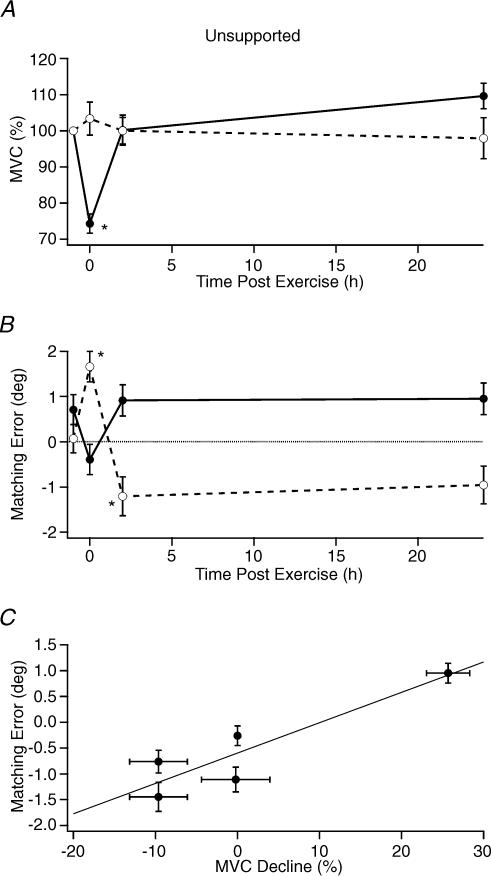
The drop in force after the concentric exercise was 26 ± 6% (n = 6). Unlike the eccentric exercise, force had recovered to control levels by 2 h post-exercise (Fig. 5). An ANOVA found time post-exercise to be a significant factor and post hoc tests revealed that MVC immediately after the exercise was significantly different from control values.
Figure 5. Position matching errors after concentric exercise.
A, changes in MVC for 6 subjects after a period of concentric exercise. The value measured before the exercise was assigned 100%. Measurements were carried out before the exercise, immediately afterwards and at 2 h and 24 h. Continuous line and filled symbols, MVC values in exercised arm. Dashed line and open symbols, unexercised arm. Asterisks indicate values significantly different from controls. B, mean values for 5 trials, 3 matching angles and 6 subjects of position matching errors, in degrees, between the two forearms after concentric exercise. Reference arm unsupported. Open circles, unexercised arm as reference, filled circles exercised arm as reference. C, plot of relationship between elbow matching errors and fall in MVC. The MVC decline was expressed as the percentage below the control value. Here matching errors were expressed for the exercised arm relative to the unexercised arm, rather than between indicator relative to the reference. It allowed pooling of errors for both arms and calculation of a mean. The four values are from before the exercise, immediately afterwards, at 2 h and at 24 h. The correlation was significant. The slope had a value of 0.053 deg per percentage MVC. In the figure, all values are means (± s.e.m.) across 2 arms, 3 angles, 5 trials and 6 subjects. Asterisks indicate points significantly different from control values.
For the six subjects who underwent concentric exercise, some reported a slight soreness at the end of the exercise, but this was gone within a few minutes. Concentric exercise was not accompanied by any change in relaxed elbow angle.
Experiment 1 – Position matching after eccentric exercise with the reference arm unsupported
Here six subjects participated. In this initial series it was found that the ability of subjects to match forearm angles under control conditions varied considerably from subject to subject with an error range of 2–8 deg. In the later experiments subjects went through an initial screening process and after a familiarization procedure using an active matching trial, only those able to achieve an accuracy of ± 2 deg were selected for further measurements.
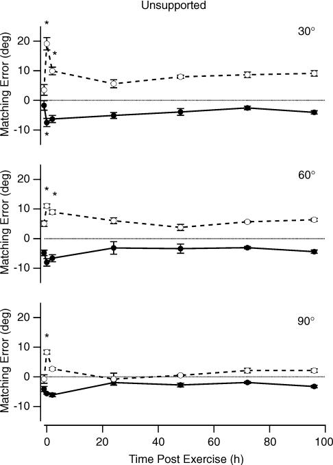
Immediately after the eccentric exercise, subjects made systematic position matching errors. When the exercised arm was the indicator, it tended to adopt a more flexed position than that maintained by the unexercised reference. The reverse trend was seen when the exercised arm was the reference. The indicator arm tended to adopt a more extended position in what the subject perceived to be an accurate match (Fig. 2). A second consistent trend was the finding that at small forearm angles, that is, with the forearm more nearly horizontal, errors tended to be larger than when the arm was closer to the vertical (Fig. 2).
Figure 2. Position matching errors after eccentric exercise.
Forearm position matching errors for one subject with their reference arm unsupported. Forearm angles were expressed in degrees above the horizontal, which was assigned 0 deg. Three test angles were used, 30, 60 and 90 deg. The differences in positions of reference and indicator arms were given in degrees. When matching errors were in the direction of flexion, they were given a positive value, when they were in the direction of extension they were negative. Dashed line and open symbols, reference arm unexercised; continuous line and filled symbols, reference arm exercised. Zero error is indicated by the dotted line. Errors were measured before a period of eccentric exercise, immediately afterwards (0 h) and at 24, 48, 72 and 96 h. All values are means (± s.e.m.) across 5 trials and 3 angles. Asterisks indicate values significantly different from control.
An ANOVA, with the dependent variable being the error, expressed as the difference between exercised and unexercised arms, and factors being reference angle, time post-exercise and subject, found all to be significant. For time post-exercise, the post hoc test showed the immediately post-exercise values to be significantly different from controls, but no other times were significant with all P values greater than 0.9. For reference angle, using the whole data set, errors at 30 and 60 deg were not significantly different from each other, but errors at 90 deg were significantly smaller. When the errors at various times after the exercise were averaged across subjects, they were found to be correlated with the deficit in force, relative to its pre-exercise value (Fig. 1B). The slope of the relation was 0.05 deg per percentage MVC.
Experiment 2 – Position matching after eccentric exercise with the reference arm supported
A second group of six subjects participated in this experiment. Here position of the reference arm was maintained by means of a support and the subject was instructed to keep that arm fully relaxed during the matching. It meant that there would be no effort cues coming from the reference arm.
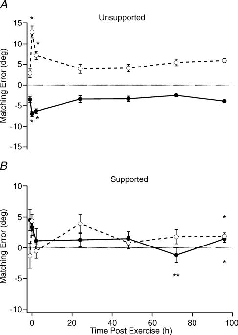
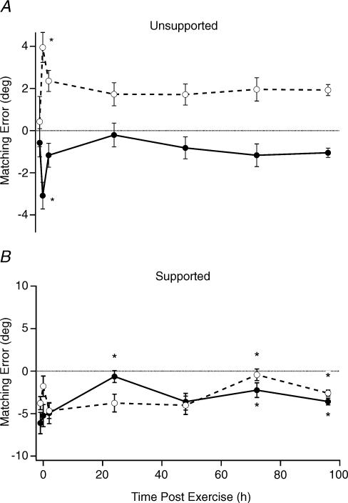
Subjects were less sure about the position of their arms when the reference remained relaxed. This became apparent even before the exercise. Control mean matching errors for trials with an unsupported reference arm were 0.6 ± 0.7 deg when one arm was the reference and 0.4 ± 1.2 deg when the other arm was the reference. When one arm was supported, errors were 6.1 ± 1.3 deg with one arm as reference and 3.8 ± 0.8 deg with the other as reference. Immediately following the exercise, there were some small errors, particularly for the condition where the exercised arm was the indicator, but these were not significant. With this protocol, positioning of the indicator arm was still done voluntarily by the subject. So for the indicator arm an effort cue was still available. However any alteration of the sense of effort as result of fatigue and damage from the exercise would not have been apparent when the exercised arm acted as the reference.
Data for one subject showed that when errors for the three angular positions were pooled, they were not significant at the peak of fatigue (Fig. 3). Indeed, this was the case for the pooled data from the six subjects (Fig. 4), which showed variations in matching errors during the days after force had recovered that were larger than the errors when the force drop was greatest, immediately after the exercise. The points significantly different from control values, as indicated by post hoc test, are shown in Fig. 4. A comparison of errors pre-exercise with errors immediately afterwards gave P = 0.58 when the indicator arm was exercised and P = 0.99 when the reference arm was exercised.
Figure 3. Matching errors for unsupported and supported reference arms.
Pooled data for position matching errors for the 3 matching angles for one subject. A, the subject maintained the reference angle voluntarily by contracting elbow flexor muscles (Experiment 1). B, the reference position was maintained by a support and the subjects were asked to fully relax their reference arm (Experiment 2). In each panel, dashed lines and open circles are values when the unexercised arm was the reference; continuous lines and filled circles are when the exercised arm was the reference. The dotted line indicates zero error. All values are means (± s.e.m.) across 5 trials and 3 angles. Asterisks indicate points significantly different from control.
Figure 4. Pooled data for matching errors.
Pooled matching errors for 6 subjects, each of which was measured 5 times at each of 3 matching angles before and at various times after a period of eccentric exercise. A, reference arm was unsupported. B, reference arm supported. All values are means (± s.e.m.) across 6 subjects, 5 trials and 3 angles. Continuous line and filled symbols, reference arm exercised; dashed line and open symbols, reference arm unexercised. Dotted line indicates zero error. Asterisks alongside values in A and above or below values in B indicate values significantly different from controls.
Experiment 3 – Concentric exercise
Because it was more difficult to get large falls in force with concentric exercise, when compared with eccentric exercise, it was decided to do the matching with forearm angles closer to the horizontal (15, 30 and 45 deg). The rationale was that if an effort signal was involved in position sense this would be larger for forearm angles where the gravity vector was close to its peak. This was also the angle range where there were large changes in torque due to the muscle's length–tension relation. It was argued that any disturbance of effort by fatigue might therefore be more readily detected at these smaller angles.
As expected, fatigue from concentric exercise was less and of much shorter duration than after eccentric exercise. Immediately after the exercise MVC had dropped by 26 ± 6% (n = 6) and it had returned to control levels 1 h later. During the post-exercise period subjects exhibited position matching errors, although these were smaller than after eccentric exercise. Errors immediately after the exercise, when the unexercised arm was the reference, were significant (ANOVA plus post hoc test). When the exercised arm was the reference, the ANOVA still showed time post-exercise to be a significant predictor of matching errors but the post hoc test revealed no individual point as significant. The errors were in the direction predicted by the effort:position matching hypothesis (Fig. 5). When the exercised arm was the reference, the indicator adopted a more extended position with a mean error of –0.4 ± 3 deg immediately after the exercise (6 subjects, 5 trials, 3 angles). When the unexercised arm was the reference, the indicator adopted a more flexed position with a mean error of +1.6 ± 0.3 deg.
Combining matching errors for the exercised and unexercised arms, and averaging across subjects for both reference and indicator conditions showed a significant dependence on MVC (ANOVA). The slope of the relation was 0.053 deg per percentage MVC (Fig. 5). Because different matching angles were used for the concentric and eccentric experiments, a direct comparison between them was not possible.
Discussion
The working hypothesis that provided the motivation for these experiments was that eccentric exercise in elbow flexor muscles would produce muscle damage and any subsequent errors in position sense would be attributable to the damage. Position sense has traditionally been assigned to muscle spindles (McCloskey, 1978; Matthews, 1988; Gandevia, 1996). We speculated that if ordinary muscle fibres were damaged by eccentric exercise, it was conceivable that the intrafusal fibres of muscle spindles, too, could become damaged. This would lead to a disturbance of spindle function and consequently errors in position sense. Indeed, we had considered this a possibility previously (Brockett et al. 1997). In the event, the data presented here do not support such an explanation.
Perhaps the most important finding of this study was that position errors were significant when the drop in force was at its maximum, regardless of whether it had been generated by eccentric or concentric exercise. Furthermore, the sizes of the matching errors correlated with the fall in force (Figs 1 and 5), suggesting that the drop in MVC was responsible for matching errors. Given that both kinds of exercise produced errors, it implied that the muscle damage associated with eccentric exercise was not a contributing factor, other than by its effect on muscle force. It is known that while concentric exercise leads to muscle fatigue there is no evidence of damage (Newham et al. 1983a,b).
We have recently carried out a series of animal experiments in which we eccentrically exercised the medial gastrocnemius muscle of the anaesthetized cat using fusimotor-strength stimulation. The exercise was accompanied by evidence of muscle damage. Yet testing the responses of muscle spindles to stretch and fusimotor stimulation before and after the exercise revealed no evidence of a change in responsiveness (Gregory et al. 2004). It was concluded that eccentric exercise did not damage muscle spindles.
When we generate a given level of force in elbow flexors of one arm and try to match it with the other arm, we appear not to match the force but the effort required to generate it (see, for example, Carson et al. 2002; Weerakkody et al. 2003b). The present-day view is that the sensation of effort is generated centrally as a result of interactions between sensory and motor cortex (McCloskey et al. 1983; Gandevia, 2001). The sense of effort has been referred to as deriving from a corollary discharge or efference copy of the motor command (Matthews, 1988). Here we propose that the effort required to maintain position of the arm against the force of gravity provides us with information about its location in space.
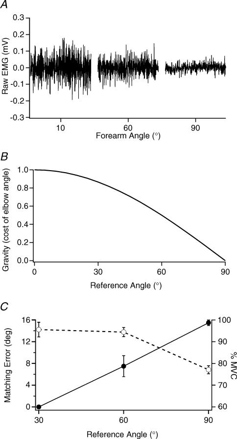
For the unsupported matching task, a consistent finding was that when the unexercised arm was the reference and matching was done with the fatigued indicator, this adopted a more flexed position with respect to the reference (Figs 1 and 3). Mean peak error for the six subjects was 4.0 ± 0.7 deg (Fig. 4). So, if the subject was using the perceived effort in the two arms to achieve a match, they placed the fatigued arm more nearly vertically. Our interpretation is that the exercise had disturbed the relationship between force and effort so that more effort was now required to maintain position of the arm at a given forearm angle. Adoption of a more vertical position requires less effort, for two reasons. First, the vector component of the force of gravity on the arm is less (Fig. 6). Second, a more vertical position is closer to the elbow flexors' optimum length for active tension (Fig. 6). Thus in matching efforts subjects placed the fatigued arm in a position where the force generated by its elbow flexors was sufficient to bear its weight. The relationship between the flexing torque required to support the arm and forearm angle has the shape of a half-sinusoid (cosine), with forces peaking when the arm was horizontal, where the gravitational vector was largest (Fig. 6). The change in torque with angle becomes steeper for larger forearm angles. We interpret the smaller errors seen with larger matching angles (Fig. 2) as due to the more precise information subjects received about the position of their forearm over this range.
Figure 6. Biceps EMG, the force of gravity and matching errors for different forearm angles.
A, surface EMG from biceps of an unsupported, unexercised arm recorded for 3 different forearm angles, relative to the horizontal (10, 60 and 90 deg). B, estimated changes in the force of gravity with elbow angle over the range 0 to 90 deg. The changes were calculated based on the assumption that the vector acting on the arm was proportional to the cosine of the angle of the forearm to the horizontal. C, mean position matching errors for 30, 60 and 90 deg (dashed line) are shown together with the changes in MVC at these angles (continuous line). The position matching errors are means from 5 trials and 6 subjects and were measured immediately after a period of eccentric exercise. The unexercised arm was the reference and it was unsupported. The length dependence of MVC for elbow flexors used data from eight subjects. Peak MVC was at 90 deg. The 30 deg value was obtained by extrapolation (Weerakkody et al. 2003b; Fig. 6).
When the exercised arm was the reference, in the unsupported task (Figs 2 and 3), the unexercised indicator adopted a more extended position. Our explanation is that the reference effort signal was larger than normal, because, as a result of fatigue, the tension generated by a given effort was less. Because of the larger reference effort, the unexercised matching arm adopted a position where more effort would be required to maintain its position. A more extended arm is subject to a larger vector component of the force of gravity and elbow flexors are further down the ascending limb of their length–tension relation. The errors with the fatigued arm acting as the reference were generally smaller, with a mean of 3.1 ± 0.6 deg, perhaps because in the direction of extension the length–tension relation for elbow flexors becomes steeper and so the effort required to generate a given level of force is proportionately greater.
To put our ideas about the sense of effort to the test we devised an experiment in which there was no reference effort cue. This was done by supporting the reference arm. The data from this experiment showed that with the reference arm supported, subjects no longer made consistent errors between the two sides. The errors observed immediately after the exercise, when force levels had significantly fallen, were less than the random errors observed during the following days (Figs 3 and 4).
There were some small, if non-significant, errors immediately after the exercise when the exercised arm was the indicator. These persisting errors were perhaps the result of the subjects using their memory of the relationship between effort and position, in the absence of a reference cue. This led to errors because elbow flexors were fatigued.
Following the concentric exercises, errors measured with the reference arm unsupported, were in the anticipated direction and they were larger when the unexercised arm was the reference, compared with errors when the reference arm had been exercised. There was also a 10% increase in MVC in the exercised arm at 24 h, perhaps because of a training effect.
Our findings are consistent with the results of Saxton et al. (1995). The opposite result observed by Brockett et al. (1997), we attribute to the fact that effects of eccentric exercise in one arm were compared with concentric exercise in the other. No comparisons were made with unexercised arms. In addition, the exercise was rather mild, leading to a drop in MVC which was greater for the concentric exercise (14%) than the eccentric exercise (10%). The greater fatigue after concentric exercise would account for the direction of the errors.
We are therefore suggesting that the effort required to maintain the arm against the force of gravity provides a positional cue. This idea is not new (Weber, 1922; Soechting, 1982; Watson et al. 1984; Worringham & Stelmach, 1985). Its consideration has recently been brought into focus by reports of the difficulties encountered by astronauts in carrying out motor tasks in the absence of visual control in a low-gravity environment (Young et al. 1993; McCall et al. 2001). In other experiments, by changing the force of gravity, positional illusions are experienced (Lackner & Graybiel, 1981). Removing the influence of gravity by supporting the arm (Paillard & Brouchon, 1968) or by using a counterweight (Gooey et al. 2000) leads to less consistent matching. The idea of effort-related positional errors has also been invoked in circumstances where gravity was not a factor. Paralysis of the extraocular muscles led to a displacement of the visual field and a perceived displacement of targets. Focusing on targets further out in the peripheral field required more effort and led to larger perceived displacements (Stevens, 1978).
A role for muscle spindles in kinaesthesia remains undisputed. The experiments of Goodwin et al. (1972) have demonstrated the powerful illusions of position and movement of a limb produced by muscle vibration. The receptors responsible are most likely muscle spindles (Roll et al. 1989). But because of the fusimotor innervation of spindles, there are difficulties in assigning to them a role in static position sense (McCloskey et al. 1983). Our current working hypothesis is that when the arm is moved into position, the movement information comes largely from muscle spindles, with additional contributions from skin and joint receptors. Once the arm is in position, the effort required to keep it there provides detailed positional information. How the force–effort relationship is calibrated, remains unclear. Perhaps it involves tendon organs. There are, of course, other circumstances in which tendon organs are likely to play a more major role in signalling force, for example, during the tonic vibration reflex (McCloskey et al. 1974).
Consistent with the idea that position information and movement information have different origins, Sittig et al. (1985) found subjects' perception of velocity and position both affected by muscle vibration. By appropriate interrogation, subjects could readily switch between the real and illusory positions, as though they were accessing two different sources of information. More recent experiments on repetitive hand reaching movements in the absence of vision have shown hand position drifting while distance and direction remain accurate. It was concluded that position and movement information are controlled by distinct neural mechanisms (Brown et al. 2003).
It could be argued that the pattern of positional errors observed in this study is the result of fatigue-related changes in the responses of muscle spindles. Whenever we carry out a voluntary contraction there is coactivation of skeletomotor and fusimotor neurones (Vallbo, 1974). When the fatigued arm was the reference, because of the greater activation required to maintain its position, spindle firing rates would be expected to be higher. This would be matched by the indicator arm adopting a more extended position, where the muscle and its spindles were subjected to a greater degree of stretch. Similar arguments could be used to explain errors in the opposite direction when the unfatigued arm was the reference.
However it is unlikely that the fusimotor-evoked increases in spindle firing provide a positional signal. We know that carrying out a voluntary isometric contraction, where spindles are also coactivated, produces no positional illusions. The present-day interpretation is that there is a central subtraction process of any fusimotor-evoked spindle activity from the length-related discharge (McCloskey et al. 1983). If that is the case, there should not be a fatigue-related change in spindle positional signal.
In summary, we have provided evidence from experiments on eccentric and concentric exercise that the effort required to hold our limbs in space provides us with static positional information. Little is known about the central mechanisms associated with generation of the sense of effort. One of the challenges for the future will be to determine how the effort signal is generated and how it combines with afferent information from the periphery to give us our kinaesthetic sense.
Acknowledgments
The work was carried out with support from the National Health and Medical Research Council of Australia. The authors would like to thank S. C. Gandevia for his comments on the manuscript.
References
- Brockett C, Warren N, Gregory JE, Morgan DL, Proske U. A comparison of the effects of concentric versus eccentric exercise on force and position sense at the human elbow joint. Brain Res. 1997;771:251–258. doi: 10.1016/s0006-8993(97)00808-1. [DOI] [PubMed] [Google Scholar]
- Brown LE, Rosenbaum DA, Sainburg RL. Limb position drift: implications for control of posture and movement. J Neurophysiol. 2003;90:3105–3118. doi: 10.1152/jn.00013.2003. [DOI] [PMC free article] [PubMed] [Google Scholar]
- Carson RG, Riek S, Shahbazpour N. Central and peripheral mediation of human force sensation following eccentric or concentric contractions. J Physiol. 2002;539:913–925. doi: 10.1113/jphysiol.2001.013385. [DOI] [PMC free article] [PubMed] [Google Scholar]
- Gandevia SC. Kinesthesia: roles for afferent signals and motor commands. In: Rowell LB, Shepherd J T, editors. Handbook of Physiology. New York: Oxford University Press; 1996. pp. 128–172. section 12, Exercise: Regulation and Integration of Multiple Systems, [Google Scholar]
- Gandevia SC. Spinal and supraspinal factors in human muscle fatigue. Physiol Rev. 2001;81:1725–1789. doi: 10.1152/physrev.2001.81.4.1725. [DOI] [PubMed] [Google Scholar]
- Goodwin GM, McCloskey DI, Matthews PB. The contribution of muscle afferents to kinaesthesia shown by vibration induced illusions of movement and by the effects of paralysing joint afferents. Brain. 1972;95:705–748. doi: 10.1093/brain/95.4.705. [DOI] [PubMed] [Google Scholar]
- Gooey K, Bradfield O, Talbot J, Morgan DL, Proske U. Effects of body orientation, load and vibration on sensing position and movement at the human elbow joint. Exp Brain Res. 2000;133:340–348. doi: 10.1007/s002210000380. [DOI] [PubMed] [Google Scholar]
- Gregory JE, Morgan DL, Proske U. Responses of muscle spindles following a series of eccentric contractions. Exp Brain Res. 2004 doi: 10.1007/s00221-004-1838-9. (in press) [DOI] [PubMed] [Google Scholar]
- Hough T. Ergographic studies in muscular soreness. Am J Physiol. 1902;7:76–92. [Google Scholar]
- Jones DA, Newham DJ, Clarkson PM. Skeletal muscle stiffness and pain following eccentric exercise of the elbow flexors. Pain. 1987;30:233–242. doi: 10.1016/0304-3959(87)91079-7. [DOI] [PubMed] [Google Scholar]
- Lackner JR, Graybiel A. Illusions of postural, visual, and aircraft motion elicited by deep knee in the increased gravitoinertial force phase of parabolic flight. Evidence for dynamic sensory-motor calibration to earth gravity force levels. Exp Brain Res. 1981;44:312–316. doi: 10.1007/BF00236568. [DOI] [PubMed] [Google Scholar]
- Matthews PBC. Proprioceptors and their contributions to somatosensory mapping: complex messages require complex processing. Can J Physiol Pharmacol. 1988;66:430–438. doi: 10.1139/y88-073. [DOI] [PubMed] [Google Scholar]
- McCall GE, Boorman GI, Goulet C, Roy RR, Edgerton VR. Bioastronautics Investigators' Workshop. Houston: USRA; 2001. Changes in neural control of movement in humans as a function of the gravitational environment; p. 101. [Google Scholar]
- McCloskey DI. Kinesthetic sensibility. Physiol Rev. 1978;58:763–820. doi: 10.1152/physrev.1978.58.4.763. [DOI] [PubMed] [Google Scholar]
- McCloskey DI, Ebeling P, Goodwin GM. Estimation of weights and tensions and apparent involvement of a ‘sense of effort’. Exp Neurol. 1974;42:220–232. doi: 10.1016/0014-4886(74)90019-3. [DOI] [PubMed] [Google Scholar]
- McCloskey DI, Gandevia S, Potter EK, Colebatch JG. Muscle sense and effort: motor commands and judgments about muscular contractions. Adv Neurol. 1983;39:151–167. [PubMed] [Google Scholar]
- Newham DJ, McPhail G, Mills KR, Edwards RH. Ultrastructural changes after concentric and eccentric contractions of human muscle. J Neurol Sci. 1983a;61:109–122. doi: 10.1016/0022-510x(83)90058-8. [DOI] [PubMed] [Google Scholar]
- Newham DJ, Mills KR, Quigley BM, Edwards RH. Pain and fatigue after concentric and eccentric muscle contractions. Clin Sci (Lond) 1983b;64:55–62. doi: 10.1042/cs0640055. [DOI] [PubMed] [Google Scholar]
- Paillard M, Brouchon M. Active and passive movements in the calibration of position sense. In: Freedman SJ, editor. The Neuropsychology of Spatially Oriented Behaviour. Homewood, IL, USA: Dorsey Press; 1968. pp. 37–55. [Google Scholar]
- Proske U, Morgan DL. Muscle damage from eccentric exercise: mechanism, mechanical signs, adaptation and clinical applications. J Physiol. 2001;537:333–345. doi: 10.1111/j.1469-7793.2001.00333.x. [DOI] [PMC free article] [PubMed] [Google Scholar]
- Roll JP, Vedel JP, Ribot E. Alteration of proprioceptive messages induced by tendon vibration in man: a microneurographic study. Exp Brain Res. 1989;76:213–222. doi: 10.1007/BF00253639. [DOI] [PubMed] [Google Scholar]
- Saxton JM, Clarkson PM, James R, Miles M, Westerfer M, Clark S, Donnelly AE. Neuromuscular dysfunction following eccentric exercise. Med Sci Sports Exerc. 1995;27:1185–1193. [PubMed] [Google Scholar]
- Sittig AC, Denier van der Gon JJ, Gielen CC. Separate control of arm position and velocity demonstrated by vibration of muscle tendon in man. Exp Brain Res. 1985;60:445–453. doi: 10.1007/BF00236930. [DOI] [PubMed] [Google Scholar]
- Soechting JF. Does position sense at the elbow reflect a sense of elbow joint angle or one of limb orientation? Brain Res. 1982;248:392–395. doi: 10.1016/0006-8993(82)90601-1. [DOI] [PubMed] [Google Scholar]
- Stevens J. The corollary discharge: is it a sense of position or a sense of space? Behav Brain Sci. 1978;1:163–165. [Google Scholar]
- Vallbo A. Human muscle spindle discharge during isometric voluntary contractions. Amplitude relations between spindle frequency and torque. Acta Physiol Scand. 1974;90:319–336. doi: 10.1111/j.1748-1716.1974.tb05594.x. [DOI] [PubMed] [Google Scholar]
- Walsh LD, Hesse CW, Morgan DL, Proske U. The role of effort in position sense. Proc Aust Neurosci Soc, Satellite Symposium on Motor Control. 2004:10. [Google Scholar]
- Watson JD, Colebatch JG, McCloskey DI. Effects of externally imposed elastic loads on the ability to estimate position and force. Behav Brain Res. 1984;13:267–271. doi: 10.1016/0166-4328(84)90169-4. [DOI] [PubMed] [Google Scholar]
- Weber CD. The properties of space and time in kinaesthetic fields of force. J Exp Psychol. 1922;38:597–606. [Google Scholar]
- Weerakkody NS, Percival P, Hickey MW, Morgan DL, Gregory JE, Canny BJ, Proske U. Effects of local pressure and vibration on muscle pain from eccentric exercise and hypertonic saline. Pain. 2003a;105:425–435. doi: 10.1016/S0304-3959(03)00257-4. [DOI] [PubMed] [Google Scholar]
- Weerakkody NS, Percival P, Morgan DL, Gregory JE, Proske U. Matching different levels of isometric torque in elbow flexor muscles after eccentric exercise. Exp Brain Res. 2003b;149:141–150. doi: 10.1007/s00221-002-1341-0. [DOI] [PubMed] [Google Scholar]
- Whitehead NP, Morgan DL, Gregory JE, Proske U. Rises in whole muscle passive tension of mammalian muscle after eccentric contractions at different muscle lengths. J Appl Physiol. 2003;95:1224–1234. doi: 10.1152/japplphysiol.00163.2003. [DOI] [PubMed] [Google Scholar]
- Whitehead NP, Weerakkody NS, Gregory JE, Morgan DL, Proske U. Changes in passive tension of muscle in humans and animals after eccentric exercise. J Physiol. 2001;533:593–604. doi: 10.1111/j.1469-7793.2001.0593a.x. [DOI] [PMC free article] [PubMed] [Google Scholar]
- Worringham CJ, Stelmach GE. The contribution of gravitational torques to limb position sense. Exp Brain Res. 1985;61:38–42. doi: 10.1007/BF00235618. [DOI] [PubMed] [Google Scholar]
- Young LR, Oman CM, Merfeld D, Watt D, Roy S, DeLuca C, Balkwill D, Christie J, Groleau N, Jackson DK. Spatial orientation and posture during and following weightlessness: human experiments on Spacelab Life Sciences 1. J Vestib Res. 1993;3:231–239. [PubMed] [Google Scholar]


