Abstract
Lack of the barley (Hordeum vulgare) seven-transmembrane domain MLO protein confers resistance against the fungal pathogen Blumeria graminis f. sp. hordei (Bgh). To broaden the basis for MLO structure/function studies, we sequenced additional mlo resistance alleles, two of which confer only partial resistance. Wild-type MLO dampens the cell wall-restricted hydrogen peroxide burst at points of attempted fungal penetration of the epidermal cell wall, and in subtending mesophyll cells, it suppresses a second oxidative burst and cell death. Although the Bgh-induced cell death in mlo plants is spatially and temporally separated from resistance, we show that the two processes are linked. Uninoculated mutant mlo plants exhibit spontaneous mesophyll cell death that appears to be part of accelerated leaf senescence. Mlo transcript abundance increases in response to Bgh, rice (Oryza sativa) blast, wounding, paraquat treatment, a wheat powdery mildew-derived carbohydrate elicitor, and during leaf senescence. This suggests a broad involvement of Mlo in cell death protection and in responses to biotic and abiotic stresses.
Homozygous mutant (mlo) alleles of the Mlo gene confer broad spectrum disease resistance to the biotrophic powdery mildew fungus, Blumeria graminis f. sp. hordei (Bgh; Jørgensen, 1992). The resistance is manifested in the failure of the fungus to penetrate the epidermal cell wall, and at these sites, cell wall remodeling and oxidative cross-linking processes fortify the cell wall (Thordal-Christensen et al., 1997; von Röpenack et al., 1998; Hückelhoven et al., 1999). Although cell wall reinforcement is likely to contribute to the resistant phenotype, other yet unknown molecular events may lead to abortion of fungal attack. Although the wild-type Mlo gene in effect acts as a negative regulator of a defense response, wild-type Ror1 and Ror2 are two genes that are required for full expression of mlo resistance to Bgh, but not race-specific resistance (Freialdenhoven et al., 1996; Peterhänsel et al., 1997). It is curious that powdery mildew-resistant mlo plants exhibit enhanced susceptibility to the fungal pathogens Magnaporthe grisea and Bipolaris sorokiniana (Jarosch et al., 1999; Kumar et al., 2001).
Mutant mlo plants exhibit a spontaneous mesophyll cell death phenotype that is compromised by ror1 and ror2 mutations (Wolter et al., 1993; Peterhänsel et al., 1997). In this respect, mlo mutations resemble other mutations known to enhance host cell death processes along with disease resistance (Shirasu and Schulze-Lefert, 2000). Many of these mutants constitutively express pathogen-related (PR) genes, but in barley (Hordeum vulgare) mlo, maize (Zea mays) lls1, and Arabidopsis lsd1 and edr1 plants, pathogen inoculation is required to trigger an enhanced defense response (Jabs et al., 1996; Frye and Innes, 1998; Simmons et al., 1998). These four genes have been isolated (Büschges et al., 1997; Dietrich et al., 1997; Gray et al., 1997; Frye et al., 2001), and encode dissimilar proteins.
Barley MLO is the prototype of a family of seven-transmembrane (7-TM) proteins that is found in higher plants and bryophytes, but not in prokaryotes, yeast, or animals (Büschges et al., 1997; Devoto et al., 1999). In Arabidopsis, the family comprises 15 members that share a common membrane-spanning topology (A. Devoto, H.A. Hartmann, P. Schulze-Lefert, and R. Panstruga, unpublished data). Sequence diversity between MLO family members within a species, their 7-TM domain topology, and their location in the plasma membrane are reminiscent of fungal and animal G-protein-coupled receptors (Bockaert and Pin, 1999; Devoto et al., 1999). However, recent data indicate that MLO-mediated defense suppression in barley functions independently of heterotrimeric G-proteins and that calmodulin interacts with MLO to dampen defense reactions against the powdery mildew fungus (Kim et al., 2002). In addition, a small GTP-binding protein of the barley RAC family (RACB) may play a role in MLO-mediated powdery mildew compatibility (Schultheiss et al., 2002). The exact biochemical mechanisms by which MLO proteins function have yet to be determined, and with the exception of barley MLO, none of the MLO proteins has been ascribed any biological function.
To widen the basis for structure/function analysis of the barley MLO protein, we describe previously unsequenced mlo resistance alleles, of which two confer partial Bgh resistance. We use the partial mlo resistance alleles and mutations in Ror genes to connect a previously unreported Bgh-triggered hydrogen peroxide (H2O2) burst at and cell death of mesophyll cells with the resistance response. The occurrence of spontaneous mesophyll cell death after leaves reach their final size, and the kinetics of leaf pigment removal in mlo plants suggest a role for wild-type Mlo in delaying leaf senescence. Mlo expression was found to be up-regulated in response to pathogen inoculation and under a range of other stress conditions. These data imply that the wild-type Mlo gene plays a broad role in cell death protection, defense, and stress response processes.
RESULTS
mlo-16 and mlo-30 Are Defective in Intron Splicing
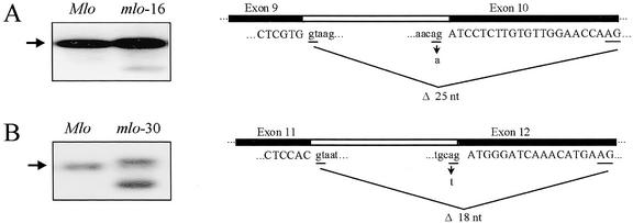
We added to the collection of characterized mlo resistance mutations (Büschges et al., 1997) by determining the DNA sequence of six additional mlo resistance alleles (designated mlo-12, -16, -27, -28, -29, and -30), each derived from chemical mutagenesis of susceptible Mlo wild-type plants (Habekuss and Hentrich, 1988; see “Materials and Methods”). All of the mutant alleles were found to contain single nucleotide substitutions in Mlo. Four of the mutations result in single amino acid changes, and they identify additional critical residues for MLO function in the second and third intracellular loop of the 7-TM protein (Table I). The two remaining mutations, present in mlo-16 and -30, are each located in intron sequences, and affect conserved nucleotides at intron-exon boundaries known to be critical for transcript splicing (Table I; Goodall and Filipowicz, 1991).
Table I.
Novel mlo alleles
| Allele | Mutant IDsa | Mutational Event at Mlo | Effect on MLO Protein | Protein Domain Affected | Bgh Resistance | Mutagen |
|---|---|---|---|---|---|---|
| mlo-12 | Do 4122 | C1396 → A | F240 → L | Second intracellular loop | Partial | Nitrosomethylurea |
| mlo-16 | Do 2376 | G1917 → Ab | Not applicable | Not applicable | Full | Ethymethane sulfonate (EMS) |
| mlo-27 | Do 2021c | G2004 → A | G318 → E | Third intracellular loop | Full | EMS |
| mlo-28 | Do 4228 | C1224 → T | T222 → I | Second intracellular loop | Partial | NaN3 |
| mlo-29 | This study | C2051 → T | P334 → L | Third intracellular loop | Full | NaN3 |
| mlo-30 | Do 2234, 2235 | A2242 → Td | Δ 6 amino acids | C terminus (intracellular) | Full | EMS |
Nucleotide nos. based on the genomic Mlo DNA sequence starting from the translational start site (ATG).
According to Habekuss and Hentrich (1988).
Nucleotide substitution in the conserved 3′ splice site of intron 9.
Also Do 2002, 2003, 2004, 2005, 2014, 2015, 2016, and 2019.
Nucleotide substitution in the conserved 3′ splice site of intron 11.
Reverse transcriptase (RT)-PCR from mlo-16 revealed one Mlo transcript with a 25-nucleotide deletion of exon 10 resulting from the use of a cryptic 3′-splice acceptor site in exon 10, and a transcript containing the entire unspliced intron 9 (Fig. 1A). Both of these mRNAs contain in-frame premature stop codons. RT-PCR from mlo-30 revealed one transcript containing an 18-nucleotide deletion of exon 12 sequences resulting from the use of a cryptic 3′-splice acceptor site in downstream exonic sequences, and another containing the entire unspliced intron 11 (Fig. 1B). These transcripts encode a protein with a six-amino acid in-frame deletion in the C-terminal cytoplasmic tail, and a severely truncated protein, respectively. RNA-blot analysis using an Mlo probe revealed additional transcripts in mlo-16 and -30, which were substantially smaller than the wild-type Mlo transcript, and which were not detected by RT-PCR (Fig. 1, A and B). In addition, correctly spliced transcripts were not detected for mlo-16 or for mlo-30 by RT-PCR. Thus, resistance of mlo-16 and -30 lines is due to severely reduced amounts of correctly spliced transcripts (encoding wild-type MLO) and/or compromised activity of abnormal MLO proteins resulting from incorrect splicing events.
Figure 1.
Characterization of two splicing-defective mlo mutant alleles. RNA-blot analysis (mRNA panels) and schematic representation of transcript splice products in mlo-16 (A) and mlo-30 genotypes (B). Northern-blot analysis was performed as described in “Materials and Methods” using wild-type Mlo plants as a control. Arrows indicate the Mlo wild-type transcript. Exonic sequences in the schematic representations are in uppercase and intron sequences in lowercase. Underlined dinucleotides in lowercase denotes 5′-splice donor and acceptor sites, and the underlined dinucleotides in uppercase represent the cryptic 3′-splice acceptor sites utilized in the mlo-16 and mlo-30 alleles. The arrows highlight the point mutations in mlo-16 (G→A) and mlo-30 (A→T) genotypes. Major splicing events in mutant cDNAs and corresponding effects on Mlo coding sequences are indicated by black lines.
Resistance Conferred by mlo-12 and mlo-28 Is Partial
Barley lines homozygous for the six newly sequenced mlo resistance alleles and 11 mlo resistance alleles sequenced by Büschges et al. (1997) were inoculated with fungal spores, and infection phenotypes were macroscopically and microscopically compared. Fifteen of the alleles (mlo-1, -3, -4, -5, -7, -8, -9, -10, -13, -16, -17, -26, 27, -29, and -30) allowed no fungal growth, whereas two of the newly sequenced alleles (mlo-12 and -28) allowed some sparse fungal hyphae growth. The size of the colonies on these partially resistant lines was smaller compared with wild-type Mlo plants at 7 d after fungal inoculation (Fig. 2). The complete and partial mlo-resistant lines, but not wild-type Mlo lines, showed host tissue necrosis at sites of attempted fungal invasion at 7 d after inoculation (Fig. 2). Frequencies of epidermal cell wall penetration by the fungus on the mlo-12 and -28 lines were determined to be between 18% and 35% (complete mlo resistance alleles = 0.8%; wild-type Mlo = 56%).
Figure 2.
Bgh-triggered cell death in a partially resistant mlo mutant. Macroscopic phenotypes of wild-type Mlo, partially and fully resistant mlo mutants 7 d after fungal challenge. In the Mlo genotype (compatible interaction), no sign of cell death is visible beneath the sporulating colony. In the mlo-28 mutant (partially resistant), necrotic mesophyll cells are visible beneath a small fungal colony, and in the mlo-5 null mutant (fully resistant), a cluster of necrotic mesophyll cells can be seen beneath the failed penetration attempt. Bar = 50 μm.
Bgh-Triggered H2O2 Production and Mesophyll Cell Death Is Coupled with mlo Resistance
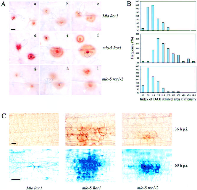
A cell wall-localized H2O2 burst occurs directly beneath sites of attempted epidermal cell penetration by Bgh in mlo-resistant and Mlo-susceptible barley lines (Freialdenhoven et al., 1996; Hückelhoven et al., 1999). This epidermal oxidative burst is greater in resistant mlo mutants than in susceptible (wild-type Mlo) plants and is modified by Ror genes (Hückelhoven et al., 2000). We quantified H2O2 accumulation 24 h after inoculation at sites of attempted epidermal cell wall penetration in near-isogenic genotypes containing wild-type or defective alleles of Mlo or Ror1, using computer image analysis of 3,3′-diamino-benzidine (DAB) polymerization (Fig. 3, A and B, and “Materials and Methods”). Measurements were taken at 24 h after inoculation, by which time the outcome of the penetration attempt has normally been determined. The susceptible Mlo Ror1 genotype and the partially susceptible double mutant mlo-5 ror1-2 produced similar levels of H2O2, whereas the fully resistant mlo-5 Ror1 genotype showed greater H2O2 levels (Fig. 3B), demonstrating a correlation between the production of cell wall-restricted H2O2 and resistance.
Figure 3.
Inactivation of the MLO protein leads to enhanced H2O2 accumulation at epidermal cells and to cell death in the mesophyll. A, In situ detection of H2O2 by DAB precipitation at 24 h after inoculation. For each genotype, three sites are shown to illustrate the range of DAB staining (area and intensity) observed at sites of attempted fungal penetration (white arrow). Bar = 10 μm. B, Quantitative analysis of DAB-stained areas beneath fungal appressorial germ tubes (A, white arrows). C, DAB staining in mesophyll cells beneath sites of attempted penetration (top) at 36 h following inoculation; retention of trypan blue 60 h after fungal challenge (bottom). In the fully compatible interaction (Mlo Ror1), no DAB staining or trypan blue retention was observed. DAB staining and trypan blue retention were found to be significantly reduced in the double mutant mlo-5 ror1-2. Bar in top = 10 μm; bar in bottom = 50 μm.
A second previously unreported oxidative burst occurs at mesophyll cells of mlo lines that subtend attacked epidermal cells (Fig. 3C, top). This H2O2 burst occurred approximately 36 h after spore inoculation, at least 16 h after the fungal penetration normally takes place. Approximately 60 h after spore inoculation, membranes of these mesophyll cells became irreversibly damaged as indicated by trypan blue retention (Fig. 3C, bottom). The mesophyll cell death response becomes macroscopically visible as localized necrosis 5 to 6 d after pathogen challenge (Fig. 2). DAB staining and trypan blue retention in the mesophyll were impaired in mlo-5 ror1-2 double mutants and were undetectable in wild-type Mlo Ror1 plants (Fig. 3C), demonstrating that the mesophyll oxidative burst and the mesophyll cell death response are under control of Mlo and Ror1. The partially resistant mutants mlo-12 and -28 also exhibited the H2O2 and cell death response in the subtending mesophyll cells (data not shown), further demonstrating a link between resistance and the mesophyll H2O2 and cell death responses.
Premature Death of mlo Mutant Leaves Mimics Senescence
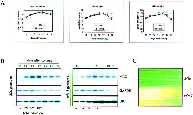
Axenically grown mlo mutants exhibit a spontaneous cell death phenotype, noticeable as premature leaf chlorosis (Wolter et al., 1993; Peterhänsel et al., 1997). We compared this premature death in mlo plants with senescence of the first leaf normally observed in wild-type (Mlo) genotypes by monitoring markers for the latter. Leaf chlorophyll a and b and total carotenoid content increased indistinguishably in Mlo and mlo-5 genotypes during the first 17 d after sowing, after which the pigments began decreasing at a faster rate in mlo-5 than in Mlo plants (Fig. 4A). Carotenoids declined only marginally faster in the mutant leaves, consistent with their known relative stability during senescence (Biswal, 1995).
Figure 4.
Accelerated leaf senescence in mlo mutants. A, Time course analysis of chlorophyll and carotenoid pigment content in Mlo wild-type and mlo-5 genotypes. First foliage leaves were collected at 2-d intervals from 9 to 21 d after sowing, and pigment analysis was carried out as described in “Materials and Methods.” Each time point in the graphs shows the mean (±sd) of six leaf samples. B, Time course analysis of MLO transcript abundance throughout first foliage leaf development in Mlo wild-type and mlo-5 genotypes. The same blot was probed sequentially with the MLO-, GAPDH-, and ubiquitin (UBI)-labeled cDNAs. The fold changes in MLO transcript abundance were calculated using the GAPDH signal as a control. C, Mid-portions of first foliage leaves primary leaves of 24-d-old seedlings of the Mlo (top) and mlo-5 (bottom) genotypes. Chlorosis (yellowing) and, at later time points, necrotic lesions are noticeable in the mlo-5 genotype.
We monitored Mlo, ubiquitin, and glyceraldehyde-3-P dehydrogenase (GAPDH) mRNA levels by analyzing RNA samples from the same time course experiment. Mlo transcript abundance showed a gradual increase during the first 15 d after sowing (10- to 12-fold) in Mlo and mlo-5 leaves, which preceded the onset of the chlorophyll decline (Fig. 4B). Along with Mlo transcripts, ubiquitin transcripts increased markedly, but only in the mlo-5 line, possibly reflecting elevated protein degradation in the mlo leaves (for review, see Belknap and Garbarino, 1996). GAPDH transcripts declined in the mlo-5 mutant around 19 d after sowing, presumably reflecting a reduction in anabolic activity. The molecular changes observed in mutant and wild-type leaves preceded the first appearance of chlorosis, between 21 and 22 d after sowing in the mlo-5 line (Fig. 4C).
Mlo Expression Is Inducible by Biotic and Abiotic Stress
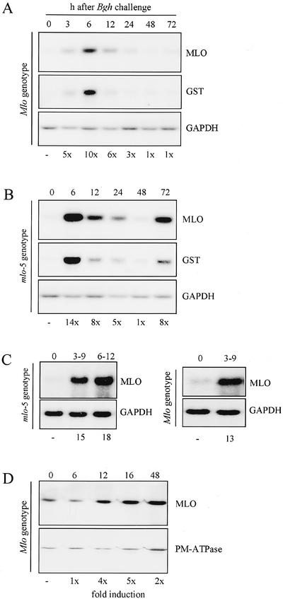
RNA-blot analysis showed that the Mlo mRNA in the leaf increased in abundance transiently after powdery mildew spore inoculation, indicating that Mlo expression is pathogen responsive (Fig. 5, A and B). This increase was observed in susceptible (Mlo) and resistant (mlo) genotypes, although the peak in abundance was more intense in the resistant genotype (14- versus 10-fold, respectively; Fig. 5, A and B). After returning to basal levels by 48 h, the Mlo transcripts increased again in abundance at 72 h, but only in the resistant genotype (Fig. 5, A and B). Mlo transcript changes followed an almost identical pattern to that of another pathogen-responsive gene encoding glutathione-S-transferase (GST; Fig. 5, A and B). A marked increase in Mlo transcript abundance was also detected in leaf epidermal peels of both genotypes, demonstrating pathogen-responsive gene expression in host cells that are in direct physical contact with the pathogen (Fig. 5C). Amounts of transcript accumulation were reflected by a 5-fold increase in MLO protein levels in enriched plasma membrane fractions, which peaked at 16 h after inoculation in the Mlo genotype (Fig. 5D; MLO-specific antibody described in Devoto et al., 1999). The time difference between transcript and protein accumulation peaks is consistent with the time required for polytopic membrane proteins to complete a series of maturation events, including endoplasmic reticulum insertion and trafficking via the Golgi to the plasma membrane (Hirschberg et al., 1998).
Figure 5.
Time course analysis of MLO transcript and MLO protein upon powdery mildew inoculation. A and B, Accumulation of MLO transcript following Bgh challenge in Mlo wild-type (A) and mlo-5 (B) leaves. The same blot was sequentially probed with the MLO, GST, and GAPDH cDNA probes. The fold changes in MLO transcript abundance were calculated using the GAPDH signal as a control. The ×1 level was defined as ratio MLO:GAPDH ratio as at time 0. Figures showing MLO transcript levels were calculated using three independent experiments. C, Accumulation of MLO transcript following Bgh challenge in epidermal peels (see “Materials and Methods”) of mlo-5 mutant (left) and Mlo wild-type (right) leaves. Abaxial leaf epidermal tissue was harvested during the indicated time intervals after spore inoculation, and the same blot was sequentially probed with the MLO and GAPDH cDNA probes. The fold changes in MLO transcript abundance were calculated using the GAPDH signal as a control. The ×1 level was defined as ratio MLO:GAPDH ratio as at time 0. D, Accumulation of MLO protein upon powdery mildew inoculation. Western blots of plasma membrane vesicle preparations from inoculated and uninoculated leaves were probed with the barley MLO-specific and the plasma membrane ATPase (PM-ATPase) antibodies. Positive signals were analyzed using a phosphor imager, and the fold MLO protein induction was calculated relative to the PM-ATPase signal. The ×1 level was defined as ratio MLO:PM-ATPase as at time 0. Figures showing MLO protein induction were calculated using two independent experiments.
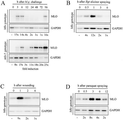
An increase in the amount of Mlo transcript was also observed after inoculation of barley with the rice (Oryza sativa) blast fungus M. grisea, indicating that Mlo expression is responsive to pathogens other than Bgh (Fig. 6A). However, unlike inoculation with Bgh, the second wave of transcript accumulation occurred in both genotypes instead of just the mlo-5 genotype. In the mlo-5 genotype, which is more susceptible to M. grisea (Jarosch et al., 1999), the second peak came approximately 72 h earlier than in the Mlo genotype, and continued increasing until at least 96 h after inoculation (Fig. 6A).
Figure 6.
Time course analysis of MLO transcript accumulation upon M. grisea challenge and abiotic stresses. A, Accumulation of MLO transcript upon challenge of barley Mlo wild-type (top) and mlo-5 (bottom) leaves with M. grisea. The same blot was sequentially probed with the MLO and GAPDH probes. Signals were analyzed using a phosphorimager, and fold Mlo transcript accumulation was calculated using the GAPDH signal as control. Figures showing MLO transcript accumulation were calculated using two independent experiments. B, Accumulation of MLO mRNA upon a carbohydrate elicitor derived from the wheat (Tritcum aestivum) powdery mildew fungus (Egt elicitor). The same blot was probed sequentially with the barley MLO and GAPDH cDNAs. C, Accumulation of MLO mRNA upon wounding. First foliage barley leaves were harvested from 8-d-old seedlings and were mechanically wounded. Representative data from two independent experiments are shown. D, Accumulation of MLO mRNA upon paraquat treatment. Paraquat was sprayed onto 8-d-old seedlings to generate an oxidative burst in chloroplasts. After spraying, plants were kept for 2 h in the dark and were then transferred to light to ensure homogenous distribution of the chemical in the seedlings. The same blot was probed sequentially with the barley MLO and GAPDH cDNAs.
We tested additional treatments: a soluble carbohydrate elicitor derived from the wheat powdery mildew B. graminis f. sp. tritici (Schweizer et al., 2000), leaf wounding, and the herbicide paraquat. Each of these treatments up-regulated Mlo expression, but the peak of transcript abundance occurred at least 3 h earlier compared with that observed with the fungal inoculations (Fig. 6, B–D).
DISCUSSION
MLO Plays a Broad Role in Modulating Defense Responses and Cell Death
Host cell death is often associated with plant pathogen resistance, but may not always be required for the resistance per se (for review, see Shirasu and Schulze-Lefert, 2000). In the case of mlo resistance against Bgh, the mesophyll cells directly underlying the attacked epidermal cell respond with an H2O2 burst and then die (Fig. 3C). The mesophyll cell death reaction is reduced in the partially susceptible mlo-5 ror1-2 genotype, is not observed in the wild-type Mlo genotype, and accompanies the mlo-12 and -28 partial resistance. The death of the mesophyll cells occurs after the fungal cell wall penetration attempt has halted (at about 60 versus 20 h) and the attacked epidermal cell remains alive, despite an earlier cell wall-localized H2O2 burst at the site of attempted penetration. Therefore, the mlo resistance is spatially and temporally separated from cell death, although the two processes are linked. Mlo is a suppressor of a defense reaction, such that loss of Mlo enhances the cell wall-restricted H2O2 burst (Fig. 3, A and B), the pathogen-induced expression of PR genes, and the size of fungal-induced papillae (Skou et al., 1984; Peterhänsel et al., 1997). Whether mlo plants exhibit mesophyll cell death because of the enhanced defense reaction (e.g. H2O2 production in the epidermis) or because the mlo mutation makes the mesophyll cells sensitive to cell death remains to be determined. It is not known whether the enhanced production of reactive oxygen species at sites of attempted fungal penetration in mlo mutants directly contributes to aborting fungal growth. In an alternate manner, the fungus may have a means of suppressing H2O2 production, which is less effective when the fungal growth is terminated early. The observation that the fully susceptible Mlo Ror1 genotype and the partially susceptible mlo-5 ror1-2 genotype produced similar levels of H2O2 in the epidermis (Fig. 3B) implies that factors other than reactive oxygen species must contribute to fungal abortion.
Perhaps unexpectedly, mutations in Mlo result in enhanced susceptibility to the rice blast fungus M. grisea (Jarosch et al., 1999), a hemibiotroph (Talbot and Foster, 2001), and the necrotrophic fungus B. sorokiniana (Kumar et al., 2001). It is an intriguing possibility that mlo mutations assist these pathogens in their necrotrophic lifestyles by triggering mesophyll cell death upon inoculation. In an alternate manner, wild-type Mlo may modulate different resistance mechanisms effective against biotrophic and necrotrophic fungi, in opposite directions.
In the absence of pathogens, first leaves of mlo plants become chlorotic and die several days earlier than those of Mlo plants (Fig. 4C). The premature leaf death resembled natural leaf senescence with regard to loss of pigments. Senescence of first (and second) foliage leaves in grasses (e.g. Hordeum vulgare and Avena sativa) has been well studied, and is a process that enables nutrients to be salvaged for incorporation into new growth. It begins soon after the leaves reach their final size (12–17 d after sowing), and involves a slow removal of chlorophyll and proteins (Miersch et al., 2000; Klerk and van Loon, 1997). Our data show that the onset of first foliage leaf senescence in mlo mutants remains unchanged while the speed is accelerated. Patches of mesophyll cell membrane damage appear in 18-d-old mlo-5 seedlings in a Ror1- and Ror2-dependent manner (Peterhänsel et al., 1997), coincident with the beginning of the leaf pigment decline reported in this study. It is possible that the accelerated senescence of mlo leaves is related to the mesophyll cell death clusters observed in leaves of an earlier age, as a cause or consequence of the latter. Irrespective of these interpretations, wild-type Mlo appears to play a broad role in delaying or preventing mesophyll cell death in pathogen-challenged and -nonchallenged leaves.
Control of Mlo Gene Expression
In wild-type Mlo plants, approximately 30% of fungal sporelings fail to penetrate the host epidermal cell wall as compared with 99.5% in mlo genotypes (Peterhänsel et al., 1997). Host reactions to Bgh, which are associated with mlo resistance such as vesicle congregation, papillae formation, a local cell wall-restricted H2O2 burst, and PR gene expression, are also observed in Mlo genotypes, albeit with less intensity (Peterhänsel et al., 1997). This suggests that wild-type Mlo is an incomplete suppressor of mlo resistance processes. Constitutive overexpression of wild-type Mlo in an Mlo background confers supersusceptibility to Bgh (Kim et al., 2002). Low constitutive levels of Mlo expression prior to Bgh-triggered induction of Mlo transcript accumulation may partly explain the incomplete resistance suppression by wild-type Mlo. At least part of the increase in Mlo expression appears to be taking place in the epidermis, which is the target tissue for Bgh (Fig. 5C). An increase in the plasma membrane MLO protein level becomes detectable by 12 h after inoculation (Fig. 5D), whereas penetration of the cell wall by Bgh occurs at about 15 h after inoculation (Peterhänsel et al., 1997). Therefore, the responsiveness of Mlo expression may be rapid enough to influence the level of Bgh compatibility in genotypes that encode an active MLO protein (Mlo genotype). Increased Mlo expression following pathogen attack may also be significant in protecting mesophyll cells from the H2O2 burst and mesophyll cell death responses otherwise seen in mlo genotypes from about 36 and 60 h after inoculation, respectively.
Before producing the appressorial germ tube, which contacts the leaf surface after about 10 h, Bgh spores produce a small primary germ tube that stops growing when it breaches the leaf surface after about 1 to 2 h (Carver et al., 1995). Therefore, the earliest observed increase in Mlo transcripts seen at 6 h after inoculation (Fig. 5, A and B) must occur before the attempted penetration, upon contact of the fungal spore or primary germ tube with the leaf.
H2O2, which is associated with and evokes defense and cell death processes in plants (Doke et al., 1996), is produced opposite to the Bgh primary and appressorial germ tubes (Thordal-Christensen et al., 1997). The more intense primary peak of Mlo induction in the mlo genotype corresponds with the more intense defense reaction in mlo plants, and the late induction peak observed in the mlo genotype only, at 72 h, corresponds with mesophyll H2O2 accumulation and cell death seen only in mlo plants. Elicitors and wounding are known to induce H2O2 production and other defense reactions (Doke et al., 1996; Lamb and Dixon, 1997), and the herbicide paraquat acts by triggering endogenous production of reactive oxygen species (Dodge, 1971). Induction of Mlo expression by these treatments is more rapid than that observed upon inoculation with Bgh or M. grisea, possibly reflecting the time needed for the pathogens to produce and transfer the microbial elicitor(s) to the host. Upon M. grisea inoculation, a second peak of Mlo induction occurs in mlo-5 and Mlo genotypes, although this begins about 72 h earlier in the mlo-5 genotype (Fig. 6A). In this case, Mlo induction is more conspicuously associated with the cell death that accompanies the interaction between the seminecrotroph M. grisea and the susceptible mlo genotype than the enhanced M. grisea resistance of the Mlo genotype (Jarosch et al., 1999). Mlo is also induced in first leaves soon after the leaves reach their final size (10-d-old seedlings), and the maximum of transcript abundance coincides with the onset of detectable molecular changes that are associated with senescence (Fig. 4). Thus, although the exact cues that lead to induction of Mlo expression are not yet known, these appear to be linked with cell death and/or defense processes.
It is curious that the induction of Mlo closely mirrors the expression of the cellular protectant GST (Fig. 5, A and B), suggesting that these two genes may be subject to similar control factors. Although the timing of this Bgh-induced transcript accumulation pattern appears to be different from those previously observed with defense-related genes, including PR-1, peroxidase, chitinase, and Phe ammonia lyase (Clark et al., 1994; Peterhänsel et al., 1997), they share a more intense accumulation in mlo compared with Mlo genotypes. The inducibility of Mlo expression under a range of conditions, plus the phenotypic consequences of mlo mutations, indicate a broad role for Mlo, which goes beyond modulation of Bgh resistance and associated cell death. This may have implications for elucidating the role(s) for the MLO protein family as a whole.
MATERIALS AND METHODS
Plant Material and Plant Treatments
Near-isogenic barley (Hordeum vulgare) lines cv Ingrid (susceptible wild type; Mlo Ror1 genotype), back cross Ingrid mlo-5 (fully resistant null mutant; mlo-5 Ror1 genotype; Büschges et al., 1997), and back cross Ingrid mlo-5 carrying the ror1-2 mutant allele (mlo-5 ror1-2 genotype; Freialdenhoven et al., 1996) were used for the experiments. Inoculation with Bgh spores was carried out as described by Freialdenhoven et al. (1996) using the powdery mildew isolate K1 (Hinze et al., 1991) on first leaves of 8-d-old seedlings grown under conditions described by Devoto et al. (1999). All experiments to investigate induction of Mlo transcript and MLO protein were performed at least three times. Epidermal peels of first leaves were obtained by making a transverse cut at the abaxial side and by removal of the epidermal layer using forceps. Inoculations with Magnaporthe grisea race 007 were carried out as described by Jarosch et al. (1999). Wheat (Triticum aestivum) powdery mildew elicitor kindly donated by Patrick Schweizer (IPK Gatersleben, Department of Cytogenetics, Germany; Schweizer et al., 2000) was sprayed on to 8-d-old barley seedlings at a concentration of 10 μg mL−1 Glc equivalents in 10 mm sodium phosphate buffer, pH 7.5, and 0.1% (w/v) Tween 20. Paraquat (methyl viologen; Sigma, St. Louis) was sprayed on to primary leaves of 8-d-old barley plants at a concentration of 50 μm in 10 mm sodium phosphate buffer, pH 7.5, and 0.1% (w/v) Tween 20, the plants were incubated in the dark for 2 h, and they were then transferred to light conditions (150 μmol photon m−2 s−1). Wounding was carried out by making transverse cuts with a scalpel blade on primary leaves of 8-d-old seedlings, and the leaves were then floated on water.
Molecular Analysis of mlo Alleles
The additional mlo mutant alleles sequenced in this study were originally isolated by Habekuss and Hentrich (1988) except mlo-29, which was isolated by James Orme in this study following sodium azide mutagenesis of cv Sultan 5 Mlo. Genomic DNA sequences of mlo alleles were obtained by direct sequencing of PCR products. Using three sets of Mlo-specific primers, three overlapping fragments (each approximately 1.0 kb in size) were amplified using Expand HiFi Taq Polymerase (Roche Molecular Biochemicals, Summerville, NJ). PCR products were gel purified and subjected to direct sequencing.
DAB and Trypan Blue Histochemical Analysis
DAB uptake was carried out as described by Thordal-Christensen et al. (1997), fixing leaf samples at 24, 36, and 48 h after inoculation. DAB quantification was performed using the Scion Image analysis software (http://www.scioncorp.com/) to analyze 75 random penetration sites from six independent leaves per genotype. The program evaluates the color intensity of the stained area, quantifies its size, and calculates an index by multiplying the two values. Trypan blue retention was performed as previously described (Peterhänsel et al., 1997) using leaf segments collected 60 h postinoculation. Fungal penetration efficiencies were determined by examination of Coomassie Brilliant Blue R250-stained, lactophenol-treated, primary leaves of inoculated 8-d-old seedlings as described in Freialdenhoven et al. (1996). Occurrence of an intracellular haustorium in attacked epidermal cells was used as a measure for successful fungal penetration at single plant fungus interaction sites.
Northern-Blot, RT-PCR, and Western-Blot Analysis
Total RNA was isolated using the TriReagent (Sigma) protocol and poly(A)+-enriched RNA fraction prepared using Oligotex columns (Qiagen, Valencia, CA). A final precipitation of the poly(A)+ RNA was performed using glycogen (1/50 of original volume) and ethanol 100% (v/v; 2.5 volumes). Northern-blot analysis was carried out using standard procedures. Probes consisted of the full-length Mlo cDNA (Büschges et al., 1997; GenBank accession no. Z83834), the full-length cDNA encoding barley GAPDH (Chojecki, 1986; GenBank accession no. M36650), the WIR5 wheat GST cDNA (Dudler et al., 1991; GenBank accession no. X56012), and the barley mub1 ubiquitin cDNA (Gausing and Jensen, 1990; GenBank accession no. M60175).
First-strand cDNA was synthesized from 100 ng of poly(A)+-enriched RNA using the oligo dT15 primer (Roche Molecular Biochemicals) and SuperScript RNase H− Reverse Transcriptase (Invitrogen, Carlsbad, CA). PCR was carried out using Mlo cDNA-specific primers and Expand HiFi Taq Polymerase (Roche Molecular Biochemicals). Western-blot analysis was performed as described previously (Devoto et al., 1999).
Chlorophyll and Carotenoid Quantification
Total chlorophyll a, chlorophyll b, and carotenoid levels were determined following the extraction procedure described by Wolfenden et al. (1988) with minor modifications. Three leaf discs (total fresh weight 0.25 g) were isolated from a primary leaf at 2-d intervals (expressed as days after sowing). Six leaves were analyzed at each time point. Data were analyzed and plotted using the Excel-5 program (Microsoft Co., Redmond, WA).
Footnotes
This work was supported by the Gatsby Charitable Organization and the Max-Planck Society (grants to P.S.-L.), by the Biotechnology and Biological Science Research Council (research grant no. 83/P09868 to P.P. and C.C.), and by the European Union-funded European Gramineae Mapping Project consortium (to F.Z.).
Article, publication date, and citation information can be found at www.plantphysiol.org/cgi/doi/10.1104/pp.010954.
LITERATURE CITED
- Belknap WR, Garbarino JE. The role of ubiquitin in plant senescence and stress responses. Trends Plant Sci. 1996;1:331–335. [Google Scholar]
- Biswal B. Carotenoid catabolism during leaf senescence and its control by light. J Photochem Photobiol B Biol. 1995;30:3–13. [Google Scholar]
- Bockaert J, Pin JP. Molecular tinkering of G protein-coupled receptors: an evolutionary success. EMBO J. 1999;18:1723–1729. doi: 10.1093/emboj/18.7.1723. [DOI] [PMC free article] [PubMed] [Google Scholar]
- Büschges R, Hollricher K, Panstruga R, Simons G, Wolter M, Frijters A, van Daelen R, van der Lee T, Diergaarde P, Groenendijk J et al. The barley Mlo gene: a novel control element of plant pathogen resistance. Cell. 1997;88:695–705. doi: 10.1016/s0092-8674(00)81912-1. [DOI] [PubMed] [Google Scholar]
- Carver T, Ingerson-Morris S, Thomas B, Zeyen R. Early interactions during powdery mildew infection. Can J Bot Suppl 1. 1995;73:S632–S639. [Google Scholar]
- Chojecki J. Identification and characterization of a cDNA clone for cytosolic glyceraldehyde-3-phosphate dehydrogenase in barley. Carlsberg Res Commun. 1986;51:203–210. [Google Scholar]
- Clark TA, Zeyen RJ, Smith AG, Carver TLW, Vance CP. Phenylalanine ammonia lyase mRNA accumulation, enzyme activity and cytoplasmic responses in barley isolines, differing at Ml-a and Ml-o loci, attacked by Erysiphe graminis f.sp. hordei. Physiol Mol Plant Pathol. 1994;44:171–185. [Google Scholar]
- Devoto A, Piffanelli P, Nilsson I, Wallin E, Panstruga R, Heijne G, Schulze-Lefert P. Topology, subcellular localization, and sequence diversity of the Mlo family in plants. J Biol Chem. 1999;274:34993–35004. doi: 10.1074/jbc.274.49.34993. [DOI] [PubMed] [Google Scholar]
- Dietrich RA, Richberg MH, Schmidt R, Dean C, Dangl JL. A novel zinc finger protein is encoded by the Arabidopsis LSD1 gene and functions as a negative regulator of plant cell death. Cell. 1997;88:685–694. doi: 10.1016/s0092-8674(00)81911-x. [DOI] [PubMed] [Google Scholar]
- Dodge AD. Mode of action of bipyridilium herbicides, paraquat and diquat. Endeavor. 1971;30:130–135. doi: 10.1016/0160-9327(71)90039-1. [DOI] [PubMed] [Google Scholar]
- Doke N, Miura Y, Sanchez LM, Park HJ, Noritake T, Yoshioka H, Kawakita K. The oxidative burst protects plants against pathogen attack: mechanism and role as an emergency signal for plant bio-defense: a review. Gene. 1996;179:45–51. doi: 10.1016/s0378-1119(96)00423-4. [DOI] [PubMed] [Google Scholar]
- Dudler R, Hertig C, Rebmann G, Bull J, Mauch F. A pathogen-induced wheat gene encodes a protein homologous to glutathione-S-transferases. Mol Plant-Microbe Interact. 1991;4:14–18. doi: 10.1094/mpmi-4-014. [DOI] [PubMed] [Google Scholar]
- Freialdenhoven A, Peterhänsel C, Kurth J, Kreuzaler F, Schulze-Lefert P. Identification of genes required for the function of non-race-specific mlo resistance to powdery mildew in barley. Plant Cell. 1996;8:5–14. doi: 10.1105/tpc.8.1.5. [DOI] [PMC free article] [PubMed] [Google Scholar]
- Frye CA, Innes RW. An Arabidopsis mutant with enhanced resistance to powdery mildew. Plant Cell. 1998;10:947–956. doi: 10.1105/tpc.10.6.947. [DOI] [PMC free article] [PubMed] [Google Scholar]
- Frye CA, Tang DZ, Innes RW. Negative regulation of defense responses in plants by a conserved MAPKK kinase. Proc Natl Acad Sci USA. 2001;98:373–378. doi: 10.1073/pnas.98.1.373. [DOI] [PMC free article] [PubMed] [Google Scholar]
- Gausing K, Jensen CB. Two ubiquitin-long-tail fusion genes arranged as closely spaced direct repeats in barley. Gene. 1990;94:165–171. doi: 10.1016/0378-1119(90)90383-3. [DOI] [PubMed] [Google Scholar]
- Goodall GJ, Filipowicz W. Different effects of intron nucleotide composition and secondary structure on pre-mRNA splicing in monocot and dicot plants. EMBO J. 1991;10:2635–2644. doi: 10.1002/j.1460-2075.1991.tb07806.x. [DOI] [PMC free article] [PubMed] [Google Scholar]
- Gray J, Close PS, Briggs SP, Joha GS. A novel suppressor of cell death in plants by the Lls1 gene of maize. Cell. 1997;89:25–31. doi: 10.1016/s0092-8674(00)80179-8. [DOI] [PubMed] [Google Scholar]
- Habekuss A, Hentrich W. Charakterisierung funktionell verschiedener mlo Mutanten durch Primärinfektion, Pustelwachstum, Inkubationszeit und Befallsverlauf. Berlin: Tag Ber Akad Landwirtsch-Wiss DDR; 1988. 272: S-229–S-237. [Google Scholar]
- Hinze K, Thompson RD, Ritter E, Salamini F, Schulze-Lefert P. Restriction fragment length polymorphism-mediated targeting of the mlo resistance locus in barley (Hordeum vulgare) Proc Natl Acad Sci USA. 1991;88:3691–3695. doi: 10.1073/pnas.88.9.3691. [DOI] [PMC free article] [PubMed] [Google Scholar]
- Hirschberg K, Miller CM, Ellenberg J, Presley JF, Siggia ED, Phair RD, Lippincott-Schwartz J. Kinetic analysis of secretory protein traffic and characterization of Golgi to plasma membrane transport intermediates in living cells. J Cell Biol. 1998;143:1485–1503. doi: 10.1083/jcb.143.6.1485. [DOI] [PMC free article] [PubMed] [Google Scholar]
- Hückelhoven R, Fodor J, Preis C, Kogel KH. Hypersensitive cell death and papilla formation in barley attacked by the powdery mildew fungus are associated with hydrogen peroxide but not with salicylic acid accumulation. Plant Physiol. 1999;119:1251–1260. doi: 10.1104/pp.119.4.1251. [DOI] [PMC free article] [PubMed] [Google Scholar]
- Hückelhoven R, Trujillo M, Kogel KH. Mutations in Ror1 and Ror2 genes cause modification of hydrogen peroxide accumulation in mlo-barley under attack from the powdery mildew fungus. Mol Plant Pathol. 2000;1:287–292. doi: 10.1046/j.1364-3703.2000.00032.x. [DOI] [PubMed] [Google Scholar]
- Jabs T, Dietrich RA, Dangl JL. Initiation of runaway cell death in an Arabidopsis mutant by extracellular superoxide. Science. 1996;273:1853–1856. doi: 10.1126/science.273.5283.1853. [DOI] [PubMed] [Google Scholar]
- Jarosch B, Kogel KH, Schaffrath U. The ambivalence of the barley Mlo locus: Mutations conferring resistance against powdery mildew (Blumeria graminis f. sp. hordei) enhance susceptibility to the rice blast fungus Magnaporthe grisea. Mol Plant-Microbe Interact. 1999;12:508–514. [Google Scholar]
- Jørgensen JH. Discovery, characterization and exploitation of Mlo powdery mildew resistance in barley. Euphytica. 1992;63:141–152. [Google Scholar]
- Kim MC, Panstruga R, Elliott C, Müller J, Devoto A, Yoon HW, Park H, Cho MJ, Schulze-Lefert P. Calmodulin interacts with MLO to regulate defense against mildew in barley. Nature. 2002;416:447–450. doi: 10.1038/416447a. [DOI] [PubMed] [Google Scholar]
- Klerk H, van Loon LC. Characteristics of protein turnover in the developing first leaf of oats (Avena sativa L.) J Plant Physiol. 1997;151:176–187. [Google Scholar]
- Kumar J, Hückelhoven R, Beckhove U, Nagarajan S, Kogel KH. A compromised Mlo pathway affects the response of barley to the necrotrophic fungus Bipolaris sorokiniana (Teleomorph: Cochliobolus sativus) and its toxins. Phytopathology. 2001;91:127–133. doi: 10.1094/PHYTO.2001.91.2.127. [DOI] [PubMed] [Google Scholar]
- Lamb C, Dixon R. The oxidative burst in plant disease resistance. Annu Rev Plant Physiol Plant Mol Biol. 1997;48:251–275. doi: 10.1146/annurev.arplant.48.1.251. [DOI] [PubMed] [Google Scholar]
- Miersch I, Heise J, Zelmer I, Humbeck K. Differential degradation of the photosynthetic apparatus during leaf senescence in barley (Hordeum vulgare L.) Plant Biol. 2000;2:618–623. [Google Scholar]
- Peterhänsel C, Freialdenhoven A, Kurth J, Kolsch R, Schulze-Lefert P. Interaction analyses of genes required for resistance responses to powdery mildew in barley reveal distinct pathways leading to leaf cell death. Plant Cell. 1997;9:1397–1409. doi: 10.1105/tpc.9.8.1397. [DOI] [PMC free article] [PubMed] [Google Scholar]
- Schultheiss H, Dechert C, Kogel KH, Hückelhoven R. A small GTP-binding host protein is required for entry of powdery mildew fungus into epidermal cells of barley. Plant Physiol. 2002;128:1447–1454. doi: 10.1104/pp.010805. [DOI] [PMC free article] [PubMed] [Google Scholar]
- Schweizer P, Kmecl A, Carpita N, Dudler R. A soluble carbohydrate elicitor from Blumeria graminis f. sp. tritici is recognised by a broad range of cereals. Physiol Mol Plant Pathol. 2000;56:157–167. [Google Scholar]
- Shirasu K, Schulze-Lefert P. Regulators of cell death in disease resistance. Plant Mol Biol. 2000;44:371–385. doi: 10.1023/a:1026552827716. [DOI] [PubMed] [Google Scholar]
- Simmons C, Hantke S, Grant S, Johal GS, Briggs SP. The maize lethal leaf spot 1 mutant has elevated resistance to fungal infection at the leaf epidermis. Mol Plant-Microbe Interact. 1998;11:1110–1118. [Google Scholar]
- Skou J, Jørgensen J, Lilholt U. Comparative studies on callose formation in powdery mildew compatible and incompatible barley. Phytopath Z. 1984;109:147–168. [Google Scholar]
- Talbot NJ, Foster AJ. Genetics and genomics of the rice blast fungus Magnaporthe grisea: developing an experimental model for understanding fungal diseases of cereals. Adv Bot Res. 2001;34:263–287. [Google Scholar]
- Thordal-Christensen H, Zhang Z, Wei Y, Collinge DB. Subcellular localization of H2O2 in plants: H2O2 accumulation in papillae and hypersensitive response during the barley-powdery mildew interaction. Plant J. 1997;11:1187–1194. [Google Scholar]
- von Röpenack E, Parr A, Schulze-Lefert P. Structural analyses and dynamics of soluble and cell wall-bound phenolics in a broad spectrum resistance to the powdery mildew fungus in barley. J Biol Chem. 1998;273:9013–9022. doi: 10.1074/jbc.273.15.9013. [DOI] [PubMed] [Google Scholar]
- Wolfenden J, Robinson DC, Cape JN, Paterson IS, Francis BJ, Mehlhorn H, Wellburn AR. Use of carotenoid ratios, ethylene emissions and buffer capacities for the early diagnosis of forest decline. New Phytol. 1988;109:85–95. [Google Scholar]
- Wolter M, Hollricher K, Salamini F, Schulze-Lefert P. The mlo resistance alleles to powdery mildew infection in barley trigger a developmentally controlled defense mimic phenotype. Mol Gen Genet. 1993;239:122–128. doi: 10.1007/BF00281610. [DOI] [PubMed] [Google Scholar]


