Abstract
Cerebellar Purkinje cells integrate motor information conveyed by excitatory synaptic inputs from parallel and climbing fibres. Purkinje cells abundantly express B-type G-protein-coupled γ-aminobutyric acid receptors (GABABR) that are assumed to mediate major responses, including postsynaptic modulation of the synaptic inputs. However, the identity and function of effectors operated by GABABR are not fully elucidated. Here we characterized an inwardly rectifying current activated by baclofen (Ibacl), a GABABR agonist, in cultured mouse Purkinje cells using a ruptured-patch whole-cell technique. Ibacl is operated by GABABR via Gi/o-proteins, as it is not inducible in pertussis-toxin-pretreated cells. Ibacl is carried by K+ because its reversal potential shifts with the equilibrium potential of K+. Ibacl is blocked by 10−3m Ba2+ or Cs+, and 10−8m tertiapin-Q. Upon the onset and offset of a hyperpolarizing step, Ibacl is activated and deactivated, respectively, with double-exponential time courses (time constants, <1 ms and 30–80 ms). Based on similarities in the above properties, G-protein-coupled inwardly rectifying K+ (GIRK) channels are thought to be responsible for Ibacl. Perforated-patch recordings from cultured Purkinje cells demonstrate that Ibacl hyperpolarizes the resting potential and the peak level achieved by glutamate-evoked potentials initiated in the dendrites. Moreover, cell-attached recordings from Purkinje cells in cerebellar slices demonstrate that Ibacl impedes spontaneous firing. Therefore, Ibacl may reduce the postsynaptic and intrinsic excitability of Purkinje cells under physiological conditions. These findings give a new insight into the role of GABABR signalling in cerebellar information processing.
The cerebellar cortex plays central roles in motor coordination and learning (Thach et al. 1992; Llinas & Welsh, 1993; Kawato et al. 2003; Ohyama et al. 2003). Purkinje cells are the sole output neurones of the cerebellar cortex, and they integrate motor information conveyed by excitatory synaptic inputs from parallel and climbing fibres (Palay & Chan-Palay, 1974; Llinas et al. 2003). Purkinje cells express a very high density of B-type Gi/o-protein-coupled γ-aminobutyric acid receptor (GABABR) (Jones et al. 1998; Kaupmann et al. 1998; Kuner et al. 1999). GABABR in Purkinje cells may be activated by not only GABA synaptically released from the innervating interneurones (Llinas et al. 2003), but also by GABA spillover from synapses between neighbouring neurones (Hirono et al. 2001). Thus, GABABR is assumed to mediate major cellular functions in Purkinje cells. A previous study (Kawaguchi & Hirano, 2000) reported that GABABR activation leads to suppression of rebound potentiation of inhibitory synaptic inputs to Purkinje cells (Kano et al. 1992). Moreover, GABABR is thought to play a role in the postsynaptic modulation of the excitatory synaptic inputs because the peak subcellular density of GABABR is found at the excitatory synapses (Fritschy et al. 1999; Ige et al. 2000; Kulik et al. 2002). For example, GABABR activation may lead to the enhancement of the postsynaptic responses mediated by type-1 metabotropic glutamate receptors (Hirono et al. 2001). However, the identity and physiological actions of effectors operated by GABABR are not fully elucidated in Purkinje cells.
In this study, we explored a GABABR-operated ion current presumably produced by G-protein-coupled inwardly rectifying K+ (GIRK) channels (Bichet et al. 2003) in Purkinje cells. GIRK channels are gated by the βγ subunit complex released from activated Gi/o-proteins (Sadja et al. 2003), and they form a major class of GABABR-operated effectors in various central neurones (Jan & Jan, 1997). GIRK channels can produce a large inward K+ current at membrane potentials (Em) more negative than the equilibrium potential of K+ (EK, ∼−100 mV), while producing only a small outward K+ current at more positive membrane potentials (Bichet et al. 2003). Thus, K+ conductance through GIRK channels at physiological membrane potentials is much smaller than the maximum. Nevertheless, this conductance is often capable of hyperpolarizing the resting potential (Erest) and attenuating excitatory postsynaptic potentials (Jan & Jan, 1997; Mark & Herlitze, 2000; Seeger & Alzheimer, 2001). A functional GIRK channel is a tetramer consisting of various combinations of subunits termed GIRK1–4 (Kir3.1–3.4)(Wischmeyer et al. 1997; Bichet et al. 2003). Some in situ hybridization and immunohistochemical studies detect the mRNAs and proteins of GIRK subunits (Karschin et al. 1996; Iizuka et al. 1997; Lauritzen et al. 1997; Murer et al. 1997) except GIRK1 (Liao et al. 1996; Ponce et al. 1996; Miyashita & Kubo, 1997) in Purkinje cells. In certain heterologous expression systems, GIRK2–4 can form functional homo- and heteromeric channels (Wischmeyer et al. 1997). However, macroscopic currents produced by these channels are much smaller than that of GIRK1-containing channels (Wischmeyer et al. 1997). Therefore, it is important to functionally determine at the whole-cell level whether GABABR activation induces a significant inwardly rectifying K+ current in Purkinje cells.
We used a ruptured-patch whole-cell voltage-clamp technique to extract an inwardly rectifying current activated by baclofen (Ibacl), a GABABR-selective agonist (Bowery, 1993). We characterized the basic properties of Ibacl, including the Gi/o-protein-dependence, carrier ion species, pharmacological profile and kinetics. These results demonstrate that Purkinje cells are equipped with an inwardly rectifying K+ current most likely produced by GIRK channels. These results also offer a useful tool for studying G-protein signalling in Purkinje cells because the amplitude of GIRK currents directly reflects the activity of G-proteins (Sadja et al. 2003). Moreover, we assessed the possible physiological contribution of Ibacl to regulation of the postsynaptic and intrinsic excitability of Purkinje cells using a perforated-patch whole-cell technique in cultured cell preparations, and a cell-attached technique in cerebellar slice preparations. These findings give a new insight into the role of GABABR signalling in cerebellar information processing.
Methods
Cultured cell preparation
Cerebellar Purkinje cells from C57BL/6 mice were cultured as described elsewhere (Tabata et al. 2000). Briefly, perinatal embryos were removed by Caesarian section from pregnant mice, which were deeply anaesthetized, and killed with diethylether. The embryos were deeply anaesthetized by cooling in chilled phosphate-buffered saline, and then killed by decapitation. The cerebella from these embryos were dissociated with trypsin, and plated onto plastic dishes (diameter, 35 mm; Falcon 3001). The neurones were cultured in an ultra-low-serum, hormone/nutrient-supplemented Dulbecco's modified Eagle's medium for 10 days to 3 weeks. Purkinje cells were identified by their large somata (15–30 μm) and thick primary dendrites.
Cerebellar slice preparation
C57BL/6 mice (4–5 weeks old) were deeply anaesthetized with diethylether, and killed by cervical dislocation. Parasagittal cerebellar slices (250 μm thick) were prepared, using a vibrating slicer (VT-1000S, Leica, Wetzlar, Germany), as described elsewhere (Kano et al. 1995).
Electrophysiology
We measured whole-cell currents from the somata of cultured Purkinje cells using a ruptured-patch voltage-clamp technique (Marty & Neher, 1995) (25–26°C). The pipette solution consisted of (mm): 130 d-gluconate potassium salt, 10 NaCl, 10 Hepes, 0.5 ethyleneglycol-bis-(β-aminoethylether)N,N,N′,N′-tetraacetic acid, 4 Mg-ATP and 0.4 Na2-GTP; pH was adjusted to 7.3 with KOH or d-gluconic acid; the total concentrations of K+ and Mg2+ were adjusted to 150.6 and 5.2 mm with KCl and MgCl2, respectively. The recording chamber (culture dish) was perfused at a rate of 1–2 ml min−1 with a saline whose standard composition was (mm): 116 NaCl, 16 KCl, 1.1 NaH2PO4, 23.8 NaHCO3, 2 CaCl2, 0.3 MgCl2, 5.5 d-glucose and 5 Hepes; pH was adjusted to 7.3 with HCl. Voltage-gated Na+ channels and ionotropic receptors for glutamate and GABA were always blocked by supplementing the saline with (μm): 0.5 tetrodotoxin (TTX), 1000 kynurenic acid (dissolved with 1 eq. NaOH) and 10 (–)-bicuculline methochloride. Current signals were low-pass filtered at 0.5–1 kHz, and sampled at 2–20 kHz using a voltage-clamp amplifier (EPC 8 or 9/2, HEKA, Lambrecht, Germany) driven by PULSE software (version 8.31 or 8.53, HEKA). Command potentials were corrected for a liquid junction potential between the pipette solution and the saline, and given to the examined cell, employing electronic compensation (by 60%) of the series resistance (typically 5–20 MΩ). The capacitance cancellation circuitry was adjusted to erase the slowest component of the capacitive currents (monitored with 5 mV voltage steps; typically 20–70 pF; Tabata et al. 2000), which is thought to reflect the capacitance of the soma and proximal dendrites (Llano et al. 1991). Upon membrane rupture, we repeatedly monitored the amplitude of current responses at an interval of 30–60 s until it became stable. We then consecutively obtained basal and test records, applying the control and drug-containing saline to the cell, respectively. When a drug had a detectable effect on a current response, we checked the recovery of the response, washing out the drug with the control saline (30–60 s). Only if the effect of the drug was at least partially reversible, the case was adopted for the following analyses.
We measured the somatic Em of cultured Purkinje cells using a perforated-patch current-clamp technique (Marty & Neher, 1995) (25–26°C). The pipette solution consisted of (mm): 140 d-gluconate potassium salt, 10 NaOH, 10 Hepes and 8 MgCl2; pH was adjusted to 7.3 with HCl; 0.005 vol. dimethylsulfoxide solution of amphotericin B (0.2 mg μl−1) was added before recordings. The ionic composition of the chamber-perfusing saline was the same as that used for the voltage-clamp measurements, except that the total concentration of K+ was reduced to 3 mm. Toxins included in the saline are specified in the corresponding figure legends. Current stimulation and Em signal acquisition were performed using the fast current-clamp circuitry of the EPC-9/2 amplifier driven by the Pulse software. Signals were low-pass filtered at 5–10 kHz, and sampled at 10–20 kHz. We started recording after the series resistance decreased below 90 MΩ. The measured Em was corrected for a liquid junction potential between the pipette solution and the saline.
We monitored the spontaneous firing of Purkinje cells in cerebellar slices by measuring transient inward currents corresponding to action potentials under voltage clamp (we hereafter term this current a spike current) in a cell-attached mode (Hamill et al. 1981) (24–26°C). The pipette solution consisted of (mm): 125 NaCl, 2.5 KCl, 10 Hepes, 2 CaCl2 and 1 MgSO4; pH was adjusted to 7.3 with NaOH. The recording chamber (volume, 1 ml) was perfused at a rate of 0.8 ml min−1 with a saline whose standard composition was (mm): 125 NaCl, 2.5 KCl, 1.25 NaH2PO4, 26 NaHCO3, 2 CaCl2, 1 MgSO4, and 20 d-glucose (bubbled with 95% O2 and 5% CO2). Toxins included in the saline are specified in the corresponding figure legend. The command potential was set to 0 mV. Current signals were low-pass filtered at 2 kHz, and sampled at 20 kHz using a voltage-clamp amplifier (Axopatch-1D, Axon Instruments) driven by PULSE software (version 8.54, HEKA).
Drug preparation and application
Baclofen (0417, Tocris, Avonmouth, Bristol, UK), (–)-N6-(2-phenylisopropyl)adenosine (R-PIA; P-4532, Sigma-Aldrich), BaCl2, CsCl, l-glutamate sodium salt (G-1626, Sigma), (–)-bicuculline methochloride (0131, Tocris) and CGP55845A (gift from Novartis, Basel, Switzerland) were dissolved into water to concentrations 1000–10 000 higher than the final levels, and kept at ≤4°C until use. Tertiapin-Q (TQ; 1316, Tocris) was dissolved into water to concentrations 300–1000 times higher than the final level, and kept at ≤−20°C until use. (RS)-α-Amino-3-hydroxy-5-methyl-4-isoxazolepropionic acid (AMPA; 0169, Tocris) was dissolved into the saline to a concentration of 10 mm, and kept at −20°C until use. 6-Cyano-7-nitroquinoxaline-2,3-dione (CNQX; 0190, Tocris) was dissolved into dimethylsulfoxide to a concentration of 100 mm, and kept at 4°C until use. Pertussis toxin (PTX; 516560, Calbiochem, San Diego, CA, USA) was reconstituted into the culture medium to a concentration of 50 μg·ml−1, and kept at 4°C until use.
In the ruptured-patch recordings, saline containing the drugs or the control agent (0.001 vol. of water) was applied locally to the whole of the examined Purkinje cell through a theta-tube (BT150-10, Sutter, Novato, CA, USA) under the control of gravity. In some experiments, baclofen and TQ were bath-applied with the chamber-perfusing saline. BaCl2 and CsCl were added to both the chamber-perfusing and locally applied saline. For pretreatment with PTX, the drug (0.5 μg ml−1) was added to the culture dishes ≥20 h before recordings.
In the perforated-patch recordings, the glutamate- or AMPA-containing saline was applied locally to the dendrites of the examined Purkinje cell through a pipette (tip diameter, < 1 μm) attached to a pressure ejection system (∼0.35 kg cm−2, PicoSpritzer III, Parker, Fairfield, NJ, USA).
In the cell-attached recordings, CNQX, bicuculline, TQ and CGP55845A were bath-applied with the chamber-perfusing saline.
Data analysis
A baclofen- or R-PIA-induced current was extracted as a difference between the basal and test records (see above) of the whole-cell currents. The amplitude of the extracted component was expressed by the mean current level over a range of test potentials from −129.5 to −131.5 mV (referred to as the amplitude at −130 mV). The reversal potential (Erev) was estimated from the null-current point of a line fitted to the local region (5–10 mV wide) of a I–V plot using Igor software (versions 4.00–5.01, WaveMetrics, Lake Oswego, OR, USA). The equilibrium potentials of K+ and Na+ (EK and ENa, respectively) were estimated using Nernst equation, assuming the equality of ion activity coefficients between the intra- and extracellular sides. For analysing the kinetics of a voltage step-activated current response, a double-exponential rise function (I(t) =a1[1 − exp(−t/τ1)]+a2[1 − exp(−t/τ2)]+c, where I(t), a, τ and c are current level at time t, amplitude, time constant and basal current level, respectively), and a double-exponential decay function [I(t) =a1 exp(−t/τ1) +a2 exp(−t/τ2) +c)] were fitted to the activation phase (50–100 ms) and deactivation phase (15–100 ms), respectively, using the Igor software.
The Erest and peak amplitude of action potentials are expressed by the mean values of five records obtained during a 75 s period. The peak level and amplitude of glutamate- or AMPA-evoked potentials (GEPs and AEPs, respectively) are expressed by the mean values of six records obtained during a 100 s period.
Spike currents were analysed, using Mini Analysis Program (version 5.1.1, Synaptosoft, Decatur, GA, USA). Inter-spike current interval (ISCI) was measured as an interval between the maximal inward deflections of consecutive spike currents. The peak amplitude of a spike current is measured as a difference from the mean prespike level (length, 2 ms) to the maximal inward deflection. These measures are expressed by the mean values of all of the events in a 9 s record collected during a 3 min period.
Groups of numerical data are presented as means ± s.e.m. Differences between raw values were tested by Student's paired or unpaired t test (with analysis of variance (ANOVA) for more than two groups). Differences between percentage-scored values were tested by rank sum test.
Results
Extraction of a GABABR-operated inwardly rectifying current
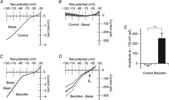
To explore a possible GABABR-operated inwardly rectifying K+ current in cerebellar Purkinje cells, we measured the whole-cell currents activated by a voltage ramp (Fig. 1). The Em was first held at −31 mV for 100 ms to facilitate the inactivation of depolarization-activated currents, and then ramped to −131 mV at a rate of −100 mV s−1. In the control saline, the total currents consisted of an apparently inwardly rectifying component at test potentials more negative than −45 mV, and an apparently outwardly rectifying component at more positive test potentials (Fig. 1A and C, Basal). A major part of the apparently inwardly rectifying component is attributable to the slow activation of the hyperpolarization-activated mixed-cation current (Ih) (Crepel & Penit-Soria, 1986; Li et al. 1993) because of its pharmacological profile (analysed in Fig. 4A and B). The remaining part may include constitutively active inwardly rectifying K+ current (Liesi et al. 2000) presumably produced by IRK channels (Falk et al. 1995; Miyashita & Kubo, 1997). The apparently outwardly rectifying component may include the ‘tail’ currents of the depolarization-activated currents. Continuous application of the control saline (2 min) did not change the overall I–V relation of the total currents (Fig. 1A, Control; Fig. 1B). By contrast, baclofen, a GABABR-selective agonist (3 μm) augmented the inwardly rectifying component of the total current (Fig. 1C). We extracted the Ibacl as a difference between the total currents recorded before and at the end of a 2 min application of baclofen (3 μm) (Fig. 1D). Ibacl displayed a Erev (−60.4 ± 3.6 mV, n = 11) close to the EK (−57.6 mV, Fig. 1D, arrow) and inward rectification (more clearly discernible in Fig. 3B). Induction of Ibacl with 3 μm baclofen was partially or completely reversible (Fig. 1C, ‘Wash’). When compared at a test potential of −130 mV, the amplitude of Ibacl (256.4 ± 54.2 pA, n = 12) was significantly larger than a change in the amplitude of the total currents following a 2 min application of the control saline (−14.3 ± 12.9 pA, n = 10; Fig. 1E). These results exclude that Ibacl is an artefact. In experiments described in the following sections, we did not use higher concentrations of baclofen because their effect was often irreversible, and thus not readily distinguished from artefacts. The results in Fig. 1 clearly demonstrate that GABABR activation induces an inwardly rectifying K+ current in cerebellar Purkinje cells.
Figure 1. GABABR-operated inwardly rectifying current in cerebellar Purkinje cells.
A and B, local application of the control saline does not induce an inwardly rectifying current in cultured Purkinje cells. A, sample voltage ramp-activated currents recorded from a cell consecutively before (Basal) and at the end of (Control) a 2 min application of the control saline. The equilibrium potential of K+(EK) =−57.6 mV. In this and the following figures, the test potential was ramped from −31 to −131 mV at a rate of −100 mV s−1, and the resultant current was converted to a I–V plot. B, mean difference of the basal and control currents; grey lines, ± s.e.m.; n = 10. C and D, baclofen, a GABABR-selective agonist, induces an inwardly rectifying current (Ibacl) in cultured Purkinje cells. C, sample voltage-ramp-activated currents recorded from a cell consecutively before (Basal), at the end of (Baclofen) and after (Wash) a 2 min application of 3 μm baclofen. EK =−57.6 mV. D, mean Ibacl (grey lines, ± s.e.m.) extracted by subtracting the basal currents from the baclofen currents. This extraction procedure is used throughout this study. n = 12. Note that the reversal potential (Erev) of Ibacl is close to the EK (arrow). E, mean amplitudes at −130 mV of the currents induced by the control saline (2 min, n = 10) and Ibacl (baclofen, 3 μm, 2 min, n = 12). Inward deflection is taken as positive. Error bars, ± s.e.m.; **P < 0.01, Student's unpaired t test.
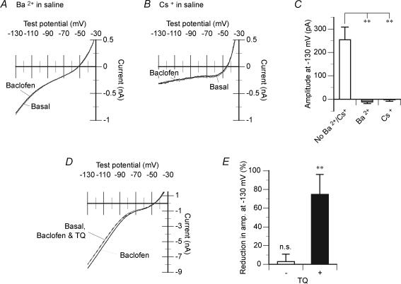
Figure 4. Pharmacology of Ibacl.
A–C, Ibacl is not inducible in the continuous presence of extracellular Ba2+ or Cs+. A and B, sample voltage-ramp-activated currents recorded from single cultured Purkinje cells consecutively before (Basal) and at the end of (Baclofen) a 2 min application of 3 μm baclofen in the saline containing 1 mm BaCl2 (A) or 3 mm CsCl (B). In addition to the effect on Ibacl, Cs+ also blocks the inwardly rectifying component of basal current; note that this component is relatively small in the CsCl-containing saline (B) compared with that in the CsCl-free saline (cf. Figs 1A and C, Basal; see Results for more detailed explanation). C, mean amplitudes of Ibacl at −130 mV (baclofen, 3 μm, 2 min) measured in the normal (n = 12, reproduced from Fig. 1E), 1 mm BaCl2-containing (n = 9) and 3 mm CsCl-containing (n = 16) saline. Error bars, ± s.e.m.**P < 0.01, ANOVA and Student's unpaired t test. D–E, the GIRK channel antagonist TQ reduces Ibacl. D, sample voltage-ramp-activated currents recorded from a cultured Purkinje cell consecutively before (grey line, Basal) and during application of the labelled drugs. In this experiment, we observed the induction of Ibacl at the end of a 2 min application of 3 μm baclofen alone (continuous black line) and then repeatedly monitored at an interval of 30 s the reduction of Ibacl, coapplying 3 μm baclofen and 30 nm TQ (dashed black line). The effect of TQ reached a maximum within 30–90 s of the coapplication onset. E, mean reduction in the amplitude of Ibacl at −130 mV caused by coapplication of baclofen and TQ (3 μm and 30 nm, respectively, n = 10, ‘+’). For each cell, the maximal effect was expressed as a percentage of the amplitude of Ibacl induced by baclofen alone (3 μm, 2 min). For control, mean reduction of Ibacl was measured during prolonged application of baclofen alone (3 μm, 30–120 s, n = 6, −). Error bars, ± s.e.m.; n.s., P > 0.05; **P < 0.01; Student's paired t test.
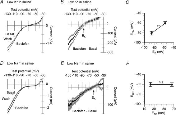
Figure 3. Carrier ion species of Ibacl.
A–C, the Erev of Ibacl shifts with the EK. A, sample voltage-ramp-activated currents recorded from a cultured Purkinje cell consecutively before (grey line, Basal), at the end of (continuous line, Baclofen) and after (dashed line, Wash) a 2 min application of 3 μm baclofen in the saline with reduced KCl (5.4 mm). EK =−85.5 mV. B, mean Ibacl (baclofen, 3 μm, 2 min) in the 5.4 mm KCl-containing saline. Grey lines, ± s.e.m., n = 10. Note that the Erev of Ibacl is close to EK (arrow). C, mean Erev of Ibacl (baclofen, 3 μm, 2 min) measured in the saline containing 5.4 mm KCl (n = 10) and 16 mm KCl (n = 11, data taken from Fig. 1D) plotted against the corresponding EK. Error bars, ± s.e.m.; **P < 0.01, Student's unpaired t test. D–F, the Erev of Ibacl does not shift with equilibrium potential of Na+ (ENa). D, sample voltage-ramp-activated currents recorded from a cultured Purkinje cell consecutively before (grey line, Basal), at the end of (continuous line, Baclofen) and after (dashed line, Wash) a 2 min application of 3 μm baclofen in the saline with reduced NaCl (25.9 mm; by equimolar replacement with N-methyl-d-glucamine). ENa =+22.5 mV. E, mean Ibacl (baclofen, 3 μm, 2 min) in the 25.9 mm NaCl-containing saline. Grey lines, ± s.e.m.; n = 7. Note that the Erev of Ibacl is close to the EK (−57.6 mV, arrow) but not ENa. F, mean Erev of Ibacl (baclofen, 3 μm, 2 min) measured in the saline containing 25.9 mm NaCl (n = 7) and 141.9 mm NaCl (n = 11, data taken from Fig. 1D) plotted against the corresponding ENa. Error bars, ± s.e.m.; n.s., P > 0.05, Student's unpaired t test.
Gi/o-protein-dependence of Ibacl
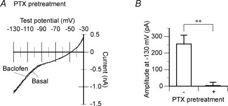
Cerebellar Purkinje cell may express GIRK channels (see Introduction) which can be operated by GABABR via Gi/o-proteins (Sadja et al. 2003). We examined the Gi/o-protein-dependence of Ibacl (Fig. 2). To this end, we pretreated cultured Purkinje cells for ≥20 h prior to the recordings with PTX, which uncouples Gi/o-proteins from G-protein-coupled receptors. In these cells, baclofen (3 μm) failed to induce Ibacl (Fig. 2A). A change in the amplitude of the total currents following a 2 min application of baclofen (3 μm) in the PTX-pretreated cells (3.6 ± 20.8 pA, n = 7) was significantly smaller than the amplitude of Ibacl in the untreated cells (254.6 ± 54.2 pA, n = 12; Fig. 2B). This result clearly demonstrates that GABABR operates Ibacl via Gi/o-proteins.
Figure 2. Gi/o-protein dependence of Ibacl.
Ibacl is not inducible in cultured Purkinje cells pretreated with pertussis toxin (PTX), a Gi/o-protein inhibitor (0.5 μg ml−1, ≥20 h). A, sample voltage-ramp-activated currents recorded from a cell consecutively before (Basal) and at the end of (Baclofen) a 2 min application of 3 μm baclofen. EK =−57.6 mV. B, mean amplitude at −130 mV of Ibacl (baclofen, 3 μm, 2 min) measured in the untreated cells (n = 12, reproduced from Fig 1E, −) and the PTX-pretreated cells (n = 7, +). Inward deflection is taken as positive. Error bars, ± s.e.m.**P < 0.01, Student's unpaired t test.
Ion-selectivity of Ibacl
In general, inwardly rectifying currents are classified into K+-selective currents and Ih which is carried by both K+ and Na+ (for review see Tabata & Ishida, 1996). To determine the ion species carrying Ibacl, we measured the Erev of Ibacl with varying EK and ENa (Fig. 3). In the standard saline containing 16 mm KCl and 141.9 mm NaCl, the Erev (−60.4 ± 3.6 mV, n = 11) was close to the EK (−57.6 mV), but not the ENa (+ 66.2 mV) (Figs 1D and 3C). When the EK was set to −85.5 mV by reducing the concentration of KCl in the saline to 5.4 mm, the Erev shifted to −80.7 ± 3.9 mV (n = 10, Fig. 3A–C). By contrast, the Erev did not shift (−60.3 ± 4.1 mV, n = 7) when the ENa was set to +22.5 mV by replacing 120 mm Na+ in the saline with N-methyl-d-glucamine, a channel-impermeant monovalent cation (Fig. 3D–F). These results clearly demonstrate that Ibacl is carried by K+ but not Na+, and is different from Ih.
Pharmacology of Ibacl
We analysed the pharmacological profile of Ibacl (Fig. 4). Sensitivities to millimolar levels of Ba2+ and Cs+ are widely used criteria to distinguish certain classes of inwardly rectifying K+ currents from Ih (for review see Tabata & Ishida, 1996). Inwardly rectifying K+ currents can be blocked by both cations. Ih is blocked by Cs+ while resistant to Ba2+. When 1 mm BaCl2 (n = 9) or 3 mm CsCl (n = 16) was included in both the chamber-perfusate and the locally delivered saline, baclofen (3 μm) failed to induce Ibacl (Fig. 4A and B). Changes in the amplitude of the total currents induced by a 2 min application of baclofen under these conditions (−12.0 ± 6.6 pA, n = 9 and −2.5 ± 6.2 pA, n = 16, respectively) were significantly smaller than the amplitude of Ibacl in the absence of these cations (254.6 ± 54.2 pA, n = 12; Fig. 4C). This result indicates that Ibacl belongs to inwardly rectifying K+ currents, but not to Ih. In addition, the apparently inward rectifying component of the total currents (e.g. Fig. 1A and C, Basal) was persistent in the Ba2+-containing saline (Fig. 4A, Basal) while obscured in the Cs+-containing saline (Fig. 4B, Basal). This result suggests that this component largely consists of Ih.
We next examined the sensitivity of Ibacl to tertiapin-Q (TQ), a synthetic peptide derived from a bee venom toxin which potently blocks certain classes of inwardly rectifying K+ channels, including GIRK channels (Jin & Lu, 1999). For the reason of availability, it was difficult to continuously include TQ in the chamber-perfusing saline. Instead, we sequentially applied baclofen alone (3 μm, 2 min; Fig. 4D, Baclofen) and baclofen together with TQ (3 μm and 30 nm, respectively; Fig. 4D, Baclofen & TQ) locally to the Purkinje cell, and evaluated the reduction of Ibacl following the coapplication. The effect of TQ reached the maximum within 30–90 s of the onset of the coapplication. At the maximum, the amplitude at −130 mV of Ibacl was significantly reduced by 74.7 ± 21.4% (n = 10, Fig. 4E). By contrast, prolonged application of baclofen alone (3 μm, 30–120 s) did not significantly reduce the amplitude (by 2.8 ± 8.1%, n = 6; Fig. 4E), indicating that the effect of TQ is not an artefact due to the run-down or inactivation of Ibacl. This result shows that TQ-sensitive classes of inwardly rectifying K+ channels are responsible for Ibacl.
Kinetics of Ibacl
We examined the activation and deactivation kinetics of Ibacl to abrupt voltage changes (Fig. 5). We recorded the total current responses to a hyperpolarizing voltage step (to −91 mV from a holding potential of −61 mV; duration, 100 ms) before and during baclofen application (3 μm) (Fig. 5A). Ibacl was extracted as a difference of these currents (Fig. 5B, continuous black line). Upon the step onset, a major part of Ibacl was activated within a millisecond period, and the remaining part continued to develop onward. These rapid and slow components of the activation phase can be described by a double-exponential rise function (see Methods for definition; Fig. 5B, continuous grey line) with two much different time constants (Table 1). Ibacl displayed no inactivation during hyperpolarization (Fig. 5B). Upon the step offset, a major part of Ibacl was deactivated within a millisecond period, and the remaining part more slowly. These rapid and slow components of the deactivation phase can be described by a double-exponential decay function (see Methods for definition; Fig. 5B, dashed grey line) with two much different time constants (Table 1). These results show that Ibacl can rapidly respond to voltage changes, and once activated it continuously functions for a prolonged time.
Figure 5. Kinetics of Ibacl.
Ibacl is rapidly activated and deactivated upon membrane potential (Em) jumps, and sustains without inactivation during hyperpolarization. A, sample voltage-step-activated currents recorded from a cultured Purkinje cell consecutively before (Basal), at the end of (Baclofen) and after (Wash) a 1 min application of 3 μm baclofen. The schematic above the left trace indicates the time course of the test potential. Dashed line, null-current level. EK =−56.7 mV. B, Ibacl extracted as a difference between the basal and baclofen currents in A. The activation and deactivation phase of Ibacl (continuous black line) can be described by a double-exponential rise function (continuous grey line) and a double-exponential decay function (dashed grey line), respectively (see Methods for the definition of these functions). The parameters of the fitted functions are presented in Table 1.
Table 1.
Parameters of double-exponential functions fitted to the activation and deactivation phases of the inwardly rectifying current activated by baclofen
| Component | Relative amplitude | Time constant (ms) |
|---|---|---|
| Activation, rapid | 0.754 ± 0.039 | 0.81 ± 0.15 |
| Activation, slow | 0.246 ± 0.039 | 82.4 ± 43.1 |
| Deactivation, rapid | 0.930 ± 0.028 | 0.60 ± 0.11 |
| Deactivation, slow | 0.070 ± 0.028 | 30.5 ± 16.4 |
Data are collected from 8 cells in the experiment shown in Fig. 5, and presented as means ± s.e.m. See Methods for the definition of the functions.
Possible functional contribution of Ibacl
We assessed how Ibacl influences the Erest and action potentials of cerebellar Purkinje cells using a perforated-patch current-clamp technique in cultured cell preparations (Fig. 6). To minimize spontaneous firing, we injected a constant background current (typically −15 to +15 pA) as to set the mean Erest to ∼−65 mV at the beginning of a recording session (Fig. 6A, Basal), and continued injection at the same intensity for the rest of the recording session. This level of the Erest was naturally observed for Purkinje cells without vigorous spontaneous firing (data not illustrated). When an additional current stimulus was not imposed, the momentary Em fluctuated by only a few millivolts around the mean Erest (cf. Fig. 6D). Baclofen (3 μm, 1–3 min) hyperpolarized the Erest by 3.46 ± 1.15 mV (n = 14; Fig. 6A and C). There was a significant difference between the mean Erest before and during baclofen application (Fig. 6C). This effect can be ascribed to Ibacl because it was not reproduced with a statistical significance in the continuous presence of 0.1–1 mm BaCl2 in the saline (n = 12; Fig. 6B and C).
Figure 6. Actions of Ibacl on the Erest and action potentials.
Ibacl hyperpolarizes the resting potential (Erest), while it does not much affect action potentials. A and B, sample current-clamp traces recorded from single cultured Purkinje cells consecutively before (grey line, Basal), during (1–2 min of baclofen onset, black line, Baclofen) and after (in A only; dashed line, Wash) baclofen application (3 μm) in the BaCl2-free (A) and 0.1 mm BaCl2-containing (B) saline. Note a change in the Erest in A. Depolarizing pulses (300 pA, 5 ms, applied between arrow heads) evoke action potentials. Each trace indicates the average of three to five records. Insets, action potentials (single records) expanded in time scale and justified at the peaks. The saline contained 3 mm KCl, 10 μm bicuculline and 1 mm kynurenic acid, but not TTX. The recordings were made in perforated-patch mode. C, mean changes in the Erest from the basal to baclofen records in the BaCl2-free (n = 14, −) and BaCl2-containing (0.1–1 mm, n = 12, +) saline. Hyperpolarizing shift is taken as positive. Error bars, ± s.e.m.**P < 0.01 and n.s. (P > 0.05) between the basal and baclofen records, Student's paired t test. D, peak levels of action potentials are plotted against the momentary Em (mean over 10 ms) prior to the depolarizing pulses. Open and closed symbols indicate the data of the basal and baclofen records, respectively. Each peak level is expressed as a difference from the mean level of 5 basal records. The dashed line indicates a linear function fitted to the data by regression (slope, −0.84). Data were pooled from 3 cells.
Injection of a strong depolarizing pulse (300 pA, 5 ms) evoked an action potential (Fig. 6A and B). Baclofen (3 μm, 1 min) prolonged the peak latency of the action potential by ∼1 ms in five out of eight Purkinje cells (Fig. 6A, inset). Baclofen (3 μm, 1 min) also raised the peak level achieved by the action potential by ∼2 mV in five out of eight cells (Fig. 6A, inset). The peak level was higher when the Em prior to depolarizing pulse was more hyperpolarized in both the absence and presence of baclofen (data pooled from three cells with the most evident effect of baclofen on the peak level, Fig. 6D). These effects can be ascribed to Ibacl because they were not seen in the continuous presence of 0.1–1 mm BaCl2 in the saline (n = 7; Fig. 6B, inset). However, the effects of baclofen on the peak latency and peak level of action potentials were not obvious in the remaining cells. Thus, baclofen-induced changes in the mean peak latency (from 3.99 ± 0.56 to 4.77 ± 0.90 ms) and the mean peak level (from +24.8 ± 4.4 to +25.3 ± 4.5 mV) for all of the examined cells (n = 8) were not statistically significant (P > 0.05, Student's paired t test). Baclofen did not delay the recovery from after-hyperpolarization to the Erest (Fig. 6A). The mean time constant of the exponential functions fitted to the main component of the recovery phase before baclofen application was 41.6 ± 13.5 ms, and that during baclofen application was 33.4 ± 5.4 ms (n = 6, data not illustrated). There was no significant difference between the time constants (P > 0.05, Student's paired t test). These results suggest that Ibacl may decrease the readiness of firing by hyperpolarizing the Erest, while it may not much affect the waveform of an action potential itself.
We next assessed how Ibacl influences GEPs in Purkinje cells using a perforated-patch current-clamp technique (Fig. 7). We used glutamate puff instead of stimulation of glutamatergic neurones innervating Purkinje cells because baclofen is known to attenuate synaptic release via the presynaptic mechanism (Dittman … Regehr, 1996, 1997). We puffed glutamate (1 mm, 3–6 ms) to the middle of the dendritic arbor of a Purkinje cell repeatedly at an interval of 20 s, and recorded GEPs from the soma (Fig. 7A). The peak amplitude of the GEPs ranged from 5 to 20 mV (Fig. 7B and C, Basal). Baclofen (3 μm, 80–180) had little effect on their waveform and peak amplitude (n = 6, Fig. 7B). There was no significant difference between the peak amplitudes before (16.8 ± 3.3 mV) and during (16.7 ± 3.7 mV) baclofen application (n = 6; P > 0.05, Student's paired t test). However, baclofen (3 μm, 80–180 s) significantly lowered the peak level achieved by the GEPs by 6.29 ± 2.19 mV (n = 6), along with a hyperpolarizing shift in the Erest (Fig. 7B and D). This effect of baclofen can be ascribed to Ibacl because a significant lowering in the peak level was not observed in the continuous presence of 1 mm BaCl2 in the saline (n = 5, Fig. 7C and D). In Purkinje cells, glutamate may evoke not only ionotropic glutamate receptors but also metabotropic glutamate receptors, and both the types of receptors may contribute to GEPs (see Tabata et al. 2004 for review). GABABR activation is reported to lead to augmentation of mGluR1-mediated responses (Hirono et al. 2001). Thus, baclofen could augment the mGluR1-mediated component of GEPs, and this could mask the attenuation of the ionotropic glutamate receptor-mediated component. However, these possibilities are not the cases because baclofen did not reduce the peak amplitude of postsynaptic potentials evoked by AMPA (AEP), an ionotropic glutamate receptor agonist (0.1–1 mm, 10.1 ± 3.8 mV before baclofen application and 11.3 ± 4.2 mV after baclofen application, n = 6; Fig. 7E). Ionotropic glutamate receptors might largely mediate GEPs elicited by the glutamate application protocol used in this study. The results shown in Fig. 7 suggest that Ibacl may impede excitatory synaptic inputs from triggering action potentials.
Figure 7. Actions of Ibacl on GEPs.
Ibacl lowers the peak levels achieved by glutamate-evoked potentials (GEPs). A, schematic shows the recording configuration. l-Glutamate (1 mm) or AMPA (0.1–1 mm) was repeatedly pressure-applied (duration 3–8 ms; interstimulus interval, 20 s) to the middle of the dendritic arbor of a cultured Purkinje cell through a pipette. The agonist-evoked potentials were recorded from the soma using a perforated-patch current-clamp technique. The saline contained 3 mm KCl, 0.3–0.5 μm TTX, and 10 μm bicuculline but not kynureic acid. B and C, sample GEPs recorded from single cells consecutively before (Basal), during (80–180 s of baclofen onset, Baclofen) and after (Wash) baclofen application (3 μm) in the BaCl2-free (B) or 1 mm BaCl2-containing (C) saline. Baclofen was bath-applied. Each trace indicates the average of three to six records. D, mean change in the peak level achieved by GEPs from the basal to baclofen records in the BaCl2-free (n = 6, −) and 1 mm BaCl2-containing (n = 5, +) saline. Hyperpolarizing shift is taken as positive. Error bars, ± s.e.m.*P < 0.05; n.s., P > 0.05; between the basal and baclofen records, respectively, Student's paired t test. E, sample AMPA-evoked potentials (AEPs) recorded from a cell consecutively before (Basal), during (80–180 s of baclofen onset, Baclofen) and after (Wash) baclofen application (3 μm) in the BaCl2-free saline. Each trace indicates the average of six records.
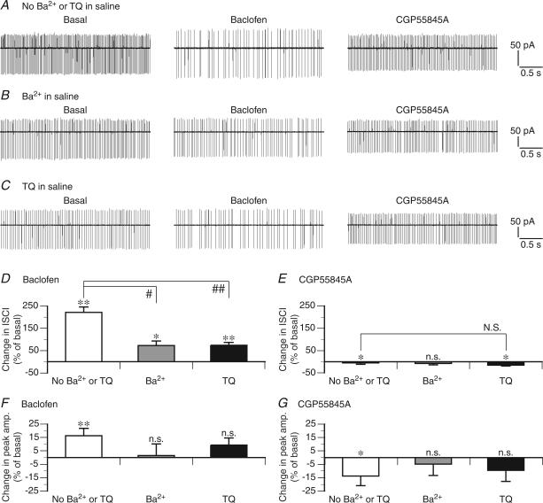
Finally, we examined whether Ibacl can function in situ using Purkinje cells in cerebellar slice preparations. We tried to measure the effects of baclofen on events observed under current clamp in a ruptured-patch whole-cell configuration. However, this attempt was hampered because the effects of baclofen and the intrinsic excitability of the examined cells severely ran down during a prolonged recording in this configuration presumably due to cytoplasmic disturbance (n = 3, data not illustrated). Instead, we used a noninvasive recording technique (cell-attached technique), and evaluated the effects of baclofen on spontaneous firing. In a cell-attached configuration, the depolarizing phase of an action potential can be observed as a transient inward current (spike current). In the normal saline, most of the examined cells displayed regular spontaneous firing with an ISCI of 21–93 ms (55.2 ± 8.0 ms, n = 7). Inclusion of CNQX and bicuculline, antagonists against ionotropic glutamate and GABA receptors in the saline, did not stop or slow down spontaneous firing (ISCI, 18–37 ms, n = 4; data not illustrated), suggesting that the intrinsic mechanisms but not excitatory or inhibitory synaptic inputs initiate spontaneous firing. In the regularly firing cells, baclofen significantly increased the ISCI by 221.8 ± 23.6% (n = 7, Fig. 8A and D). Subsequent application of 2 μm CGP55845A, a GABABR-selective antagonist, reversed this effect of baclofen (n = 7, Fig. 8A–C and E). Baclofen significantly increased the ISCI even in the continuous presence of 0.1 mm BaCl2 (by 73.4 ± 19.7%, n = 4) or 150–300 nm TQ (by 74.6 ± 12.3, n = 7) in the saline (Fig. 8B–D). However, the extent of the effect with BaCl2 or TQ was significantly smaller that that without these agents (Fig. 8D, # and ##). The baclofen-induced increase in the ISCI was accompanied with augmentation of spike currents (Fig. 8A and F). This effect was significant only in the absence of 0.1 mm BaCl2 or 150–300 nm TQ (Fig. 8A–C and F), and reversed by 2 μm CGP55845 (Fig. 8A and G). The results shown in Fig. 8 demonstrate that Ibacl can function in situ and influence the intrinsic excitability of Purkinje cells.
Figure 8. Actions of Ibacl on the intrinsic excitability in situ.
Ibacl reduces the rate of spontaneous firing of Purkinje cells in cerebellar slices presumably by hyperpolarizing the Erest. A–C, sample spike currents (see Methods) recorded from single Purkinje cells consecutively before (Basal) and during baclofen application (10 μm, at 5–8 min of the application onset), and during CGP55845A application (a GABABR-selective antagonist, 2 μm, at 5–8 min of the application onset) in the BaCl2-/TQ-free (A), 0.1 mm BaCl2-containing (B) and 300 nm TQ-containing (C) saline. The recordings were made in a cell-attached mode. D–G, mean changes in inter-spike current interval (ISCI; D and E) and the peak amplitude (F and G) of spike currents following baclofen and CGP55845A application without (n = 7) or with 0.1 mm BaCl2 (n = 4) or 150–300 nm TQ (n = 7) measured as in A–C. Error bars, ± s.e.m. For each condition, the effects of baclofen (difference between the basal and baclofen records) and CGP55845A (difference between the basal and CGP55845A records) were tested by rank sum test (n.s., P > 0.05; *P < 0.05; **P < 0.01). When judged significant by the previous test, the effects were further compared between the conditions by rank sum test (N.S., P > 0.05; #P < 0.05; ##P < 0.01).
Discussion
Functional identification of a GABABR-operated inwardly rectifying current in cerebellar Purkinje cells
In this study, we characterized an inwardly rectifying current activated by the GABABR-selective agonist baclofen (Ibacl) in cultured cerebellar Purkinje cells, using a ruptured-patch whole-cell voltage-clamp technique (Figs 1–5). In central neurones, inwardly rectifying currents may be produced by Na+-/K+-permeable channels (HCN channels; Robinson & Siegelbaum, 2003) and/or the supergene family of K+-selective channels (Kir channels; Bichet et al. 2003). Cerebellar Purkinje cells possess Ih (Crepel & Penit-Soria, 1986; Li et al. 1993), which is produced by HCN channels (Robinson & Siegelbaum, 2003), and they appear to express GIRK2–4 (Kir3.2–3.4, see below), IRK1,3 (Kir2.1,2.3) (Falk et al. 1995; Miyashita & Kubo, 1997), and Kir7.1 (Krapivinsky et al. 1998). In this section, we compare the physiological properties of Ibacl (Figs 1, 2, 3, 4, 5) with these channels.
The Erev of Ibacl was close to the EK, and shifted depending on the extracellular concentration of K+ but not that of Na+ (Figs 1 and 3). These results demonstrate that Ibacl is produced by Kir channels but not HCN channels. Uncoupling Gi/o-proteins from GABABR almost completely suppressed Ibacl (Fig. 2). This result indicates that Ibacl is operated by GABABR via Gi/o-proteins and is produced by GIRK channels which are highly dependent on Gi/o-proteins compared with other Kir channels (Isomoto et al. 1997; Bichet et al. 2003; Sadja et al. 2003). Both Ba2+ and Cs+, at millimolar levels, completely blocked Ibacl (Fig. 4A–C). Ibacl shares these metal ion sensitivities with GIRK (Hommers et al. 2003) and IRK (Isomoto et al. 1997) channels. By contrast, HCN channels (Ih) are resistant to Ba2+ (Crepel & Penit-Soria, 1986; Tabata & Ishida, 1996; Robinson & Siegelbaum, 2003), and Kir7.1 channels are resistant to both Ba2+ and Cs+ (Krapivinsky et al. 1998). TQ at 30 nm blocked a large fraction of Ibacl (Fig. 4D and E). The degree of blockade is consistent with the reported affinity of TQ for GIRK channels (Ki, 13.3 nm) but not that for IRK channels (no significant effect even at 1 μm) (Jin & Lu, 1999). Upon the onset of a hyperpolarizing voltage step, Ibacl displayed the activation phase consisting of rapid and slow components with much different time constants (Fig. 5, Table 1). The activation phases of GIRK currents in cerebellar granule neurones (Slesinger et al. 1997) and GIRK-transfected heterologous cells (Wischmeyer et al. 1997) also consist of rapid and slow components. Ibacl is distinguished from Kir7.1 current, which is instantaneously activated to the maximal level (Krapivinsky et al. 1998), and Ih, which displays single-exponential activation phase with a time constant (50–100 ms; Crepel & Penit-Soria, 1986; Tabata & Ishida, 1996) much longer than that of the main activation component of Ibacl (∼0.8 ms, Table 1). During a 100 ms hyperpolarization, Ibacl sustained without inactivation (Fig. 5). Ibacl is distinguished from IRK1 currents, which display apparent inactivation due to open channel block by external Na+ (Kubo et al. 1993). Taken together, our voltage-clamp measurements (Figs 1–5) demonstrate that cerebellar Purkinje cells are equipped with a GABABR-operated inwardly rectifying current most likely produced by GIRK channels.
The subunit composition of GIRK channels mediating Ibacl remains to be elucidated in future studies. Some insitu hybridization studies show that Purkinje cells express the mRNAs of GIRK2 (Lauritzen et al. 1997), GIRK3 (Karschin et al. 1996) and GIRK4 (Karschin et al. 1996; Iizuka et al. 1997). Some immunohistochemical studies show that GIRK2 protein is present in Purkinje cells (Lauritzen et al. 1997) (albeit there are opposing reports; Liao et al. 1996; Murer et al. 1997), and GIRK4 protein localizes to the proximal dendrites, somata and proximal axonal segments of Purkinje cells (Iizuka et al. 1997; Murer et al. 1997). Thus, Purkinje cells may express homo- and/or heteromeric channels consisting of GIRK2–4. In certain heterologous expression systems, GIRK4 form functional channels alone or together with GIRK3 (Iizuka et al. 1995; Wischmeyer et al. 1997). These channels could be candidates for the Ibacl-mediating channels. On the other hand, Purkinje cells do not have detectable levels of the mRNA (Karschin et al. 1996) and protein of GIRK1 (Ponce et al. 1996; Miyashita & Kubo, 1997). Thus, Purkinje cells express virtually no GIRK1/2 heteromeric channel, which is the predominant type in many other central neurones (Jan & Jan, 1997). In the heterologous expression system, GIRK1-free channels produce smaller macroscopic currents than GIRK1-containing channels (Wischmeyer et al. 1997). Lack of GIRK1 may explain that the amplitude of Ibacl in Purkinje cells is comparable with that in much smaller neurones (e.g. cerebellar granule cells; Slesinger et al. 1997).
Possible physiological roles of a GABABR-operated inwardly rectifying current in cerebellar Purkinje cells
Using a perforated-patch whole-cell current-clamp technique, we assessed how Ibacl influence the postsynaptic excitability of cultured cerebellar Purkinje cells (Figs 6 and 7). GABABR in Purkinje cells is reported to respond to not only GABA released from the innervating interneurones, but also to GABA spillover from neighbouring inhibitory synapses between other neurones in situ (Hirono et al. 2001). One study (Dittman & Regehr, 1997) estimates that the spillover elevates the extracellular GABA concentration around Purkinje cells to ≤10 μm. GABA and baclofen have similar binding affinities (∼60 nm) and potencies for GABABR (Bowery, 1993). Thus, the events observed with 3 μm baclofen in Figs 6 and 7 may occur under physiological conditions when cerebellar cortical GABAergic neurones are active.
Baclofen (3 μm) did not much affect the waveform of an action potential evoked by a suprathreshold stimulus (Fig. 6A). This result indicates that Ibacl may not limit the contribution of strong excitatory synaptic inputs (e.g. climbing fibre inputs) to the generation of action potentials, whereas Ibacl may affect weak excitatory inputs (e.g. parallel fibre inputs) in a manner mentioned below. In some cells, baclofen raised the peak level achieved by the action potentials (see Results; Fig. 6A, inset). In general, such augmentation could result from the increased activity of the voltage-gated Na+ channels producing the depolarizing phase of an action potential. One possibility is that the GABABR–Gi/o-protein signalling cascade suppresses activation of cyclic adenosine monophosphate-dependent kinase, which is reported to reduce the peak current of the voltage-gated Na+ channels (Catterall, 1999). Another possibility is that a hyperpolarizing shift in the Erest caused by Ibacl (Figs 6A and C) facilitates disinactivation of the Na+ channels (i.e. recovery from depolarization-induced inactivation; for review see Hayashida & Ishida, 2004). The latter possibility is consistent with the observation that the peak level depended on the Em prior to the action potential (Fig. 6D).
In hippocampal pyramidal neurones, activation of GIRK channels results in attenuation of GEPs initiated in the dendrites (Seeger & Alzheimer, 2001), presumably because GIRK channels effectively reduce the input resistance at the stimulated site (Koch, 1999). In cerebellar Purkinje cells, by contrast, such attenuation was not observed (Fig. 7B). Some immunohistochemical studies (Iizuka et al. 1997; Murer et al. 1997) suggest condensation of GIRK subunits around the soma. Thus, it could be possible that K+ conductance through the Ibacl-producing channels is relatively small in the dendrites, and does not effectively reduce the input resistance for GEPs. However, baclofen significantly hyperpolarized the Erest (Fig. 6A and C) and the peak level achieved by GEPs in Purkinje cells (Fig. 7B and D). These effects are due largely to Ibacl because they were not observed in the continuous presence of extracellular Ba2+ (Figs 6B and C, and 7C and D). These results indicate that when activated, Ibacl effectively increases K+ conductance around the soma, and may impede excitatory synaptic inputs from driving the Em towards the firing threshold. The coincidence of synaptic inputs from parallel fibres and a climbing fibre in the Purkinje cell is a key step to induce cerebellar long-term depression (LTD), a form of synaptic plasticity crucial for cerebellar motor learning (for review see Ito, 2002). GABAergic activation of Ibacl might influence induction of LTD by preferentially limiting the contribution of relatively weak parallel fibre inputs to postsynaptic excitation.
The cell-attached recordings from Purkinje cells in cerebellar slices showed that baclofen decreases the rate of spontaneous firing (Fig. 8A and D), which is initiated by the intrinsic mechanisms of Purkinje cells but not synaptic inputs (see Results). Ibacl activated via the GABABR is thought to be the primary mechanism enabling this effect of baclofen because the GABABR-selective antagonist CGP55845A reversed it (Fig. 8A and E), and extracellular Ba2+ and TQ largely eliminated it (Fig. 8B–D). Ibacl could exert the effect on the firing rate by hyperpolarizing the Erest (Fig. 6A and C). This notion is consistent with the observation that baclofen augmented the peak amplitude of spike currents (Fig. 8A and F); such augmentation is expected to occur with the hyperpolarized Erest (see above; Fig. 6A and D). These results suggest that Ibacl reduces the intrinsic excitability of Purkinje cells, and strongly support the notion Ibacl can function in Purkinje cells in situ.
In conclusion, we have functionally identified a GABABR-operated inwardly rectifying K+ current most likely produced by GIRK channels, and demonstrated its possible inhibitory actions on the postsynaptic and intrinsic excitability of cerebellar Purkinje cells. These findings give a new insight into the physiological roles of GABABR signalling in cerebellar cortex.
Acknowledgments
We thank Dr K. Hashimoto for his advice, Ms A. Imai for cell preparation, and Professor K. Yamada, Department of Pharmacy and Health Science, Graduate School of Natural Science and Technology, Kanazawa University, for the opportunity to pursue this project. This work was supported by the Grants-in-Aid for Scientific Research, and the Special Coordination Funds for Promoting Science and Technology from the Ministry of Education, Science, Sports, Culture and Technology of Japan, and a grant from the Cell Science Research Foundation.
References
- Bichet D, Haass FA, Jan LY. Merging functional studies with structures of inward-rectifier K+ channels. Nat Rev Neurosci. 2003;4:957–967. doi: 10.1038/nrn1244. [DOI] [PubMed] [Google Scholar]
- Bowery NG. GABAB receptor pharmacology. Annu Rev Pharmacol Toxicol. 1993;33:109–147. doi: 10.1146/annurev.pa.33.040193.000545. [DOI] [PubMed] [Google Scholar]
- Catterall WA. Molecular properties of brain sodium channels: an important target for anticonvulsant drugs. Adv Neurol. 1999;79:441–456. [PubMed] [Google Scholar]
- Crepel F, Penit-Soria J. Inward rectification and low threshold calcium conductance in rat cerebellar Purkinje cells. An in vitro study. J Physiol. 1986;372:1–23. doi: 10.1113/jphysiol.1986.sp015993. [DOI] [PMC free article] [PubMed] [Google Scholar]
- Dittman JS, Regehr WG. Contributions of calcium-dependent and calcium-independent mechanisms to presynaptic inhibition at a cerebellar synapse. J Neurosci. 1996;16:1623–1633. doi: 10.1523/JNEUROSCI.16-05-01623.1996. [DOI] [PMC free article] [PubMed] [Google Scholar]
- Dittman JS, Regehr WG. Mechanism and kinetics of heterosynaptic depression at a cerebellar synapse. J Neurosci. 1997;17:9048–9059. doi: 10.1523/JNEUROSCI.17-23-09048.1997. [DOI] [PMC free article] [PubMed] [Google Scholar]
- Falk T, Meyerhof W, Corrette BJ, Schafer J, Bauer CK, Schwarz JR, Richiter D. Cloning, functional expression and mRNA distribution of an inwardly rectifying potassium channel protein. FEBS Lett. 1995;367:127–131. doi: 10.1016/0014-5793(95)00527-g. 10.1016/0014-5793(95)00527-G. [DOI] [PubMed] [Google Scholar]
- Fritschy J-M, Meskenaite V, Weinmann O, Honer M, Benke D, Mohler H. GABAB-receptor splice variants GB1a and GB1b in rat brain: developmental regulation, cellular distribution and extrasynaptic localization. Eur J Neurosci. 1999;11:761–768. doi: 10.1046/j.1460-9568.1999.00481.x. 10.1046/j.1460-9568.1999.00481.x. [DOI] [PubMed] [Google Scholar]
- Hamill OP, Marty A, Neher E, Sakmann B, Sigworth FJ. Improved patch-clamp techniques for high-resolution current recording from cells and cell-free membrane patches. Pflugers Arch. 1981;391:85–100. doi: 10.1007/BF00656997. 10.1007/BF00656997. [DOI] [PubMed] [Google Scholar]
- Hayashida Y, Ishida AT. Dopamine receptor activation can reduce voltage-gated Na+ current by modulating both entry into and recovery from inactivation. J Neurophysiol. 2004;92:3134–3141. doi: 10.1152/jn.00526.2004. 10.1152/jn.00526.2004. [DOI] [PMC free article] [PubMed] [Google Scholar]
- Hirono M, Yoshioka T, Konishi S. GABAB receptor activation enhances mGluR-mediated responses at cerebellar excitatory synapses. Nat Neurosci. 2001;4:1207–1216. doi: 10.1038/nn764. 10.1038/nn764. [DOI] [PubMed] [Google Scholar]
- Hommers LG, Lohse MJ, Bunemann M. Regulation of the inward rectifying properties of G-protein-activated inwardly rectifying K+ (GIRK) channels by Gbetagamma subunits. J Biol Chem. 2003;278:1037–1043. doi: 10.1074/jbc.M205325200. 10.1074/jbc.M205325200. [DOI] [PubMed] [Google Scholar]
- Ige AO, Bolam JP, Billinton A, White JH, Marshall FH, Emson PC. Cellular and sub-cellular localisation of GABAB1 and GABAB2 receptor proteins in the rat cerebellum. Brain Res Mol Brain Res. 2000;83:72–80. doi: 10.1016/s0169-328x(00)00199-6. 10.1016/S0169-328X(00)00199-6. [DOI] [PubMed] [Google Scholar]
- Iizuka M, Kubo Y, Tsunenari I, Pan CX, Akiba I, Kono T. Functional characterization and localization of a cardiac-type inwardly rectifying K+ channel. Receptors Channels. 1995;3:299–315. [PubMed] [Google Scholar]
- Iizuka M, Tsunenari I, Momota Y, Akiba I, Kono T. Localization of a G-protein-coupled inwardly rectifying K+ channel, CIR, in the rat brain. Neuroscience. 1997;77:1–13. doi: 10.1016/s0306-4522(96)00460-5. 10.1016/S0306-4522(96)00460-5. [DOI] [PubMed] [Google Scholar]
- Isomoto S, Kondo C, Kurachi Y. Inwardly rectifying potassium channels: their molecular heterogeneity and function. Jpn J Physiol. 1997;47:11–39. doi: 10.2170/jjphysiol.47.11. 10.2170/jjphysiol.47.11. [DOI] [PubMed] [Google Scholar]
- Ito M. The molecular organization of cerebellar long-term depression. Nat Rev Neurosci. 2002;3:896–902. doi: 10.1038/nrn962. 10.1038/nrn962. [DOI] [PubMed] [Google Scholar]
- Jan LY, Jan YN. Voltage-gated and inwardly rectifying potassium channels. J Physiol. 1997;505:267–282. doi: 10.1111/j.1469-7793.1997.267bb.x. 10.1111/j.1469-7793.1997.267bb.x. [DOI] [PMC free article] [PubMed] [Google Scholar]
- Jin W, Lu Z. Synthesis of a stable form of tertiapin: a high-affinity inhibitor for inward-rectifier K+ channels. Biochemistry. 1999;38:14286–14293. doi: 10.1021/bi991205r. [DOI] [PubMed] [Google Scholar]
- Jones KA, Borowsky B, Tamm JA, Craig DA, Durkin MM, Dai M, Yao WJ, Johnson M, Gunwaldsen C, Huang LY, Tang C, Shen Q, Salon JA, Morse K, Laz T, Smith KE, Nagarathnam D, Noble SA, Branchek TA, Gerald C. GABAB receptors function as a heteromeric assembly of the subunits GABABR1 and GABABR2. Nature. 1998;396:674–679. doi: 10.1038/25348. 10.1038/25348. [DOI] [PubMed] [Google Scholar]
- Kano M, Hashimoto K, Chen C, Albeliovich A, Aiba A, Kurihara H, Watanabe M, Inoue Y, Tonegawa S. Impaired synapse elimination during cerebellar development in PKC gamma mutant mice. Cell. 1995;83:1223. doi: 10.1016/0092-8674(95)90147-7. [DOI] [PubMed] [Google Scholar]
- Kano M, Rexhausen U, Dreessen J, Konnerth A. Synaptic excitation produces a long-lasting rebound potentiation of inhibitory synaptic signals in cerebellar Purkinje cells. Nature. 1992;356:601–604. doi: 10.1038/356601a0. 10.1038/356601a0. [DOI] [PubMed] [Google Scholar]
- Karschin C, Dissmann E, Stuhmer W, Karschin A. IRK (1–3) and GIRK (1–4) inwardly rectifying K+ channel mRNAs are differentially expressed in the adult rat brain. J Neurosci. 1996;16:3559–3570. doi: 10.1523/JNEUROSCI.16-11-03559.1996. [DOI] [PMC free article] [PubMed] [Google Scholar]
- Kaupmann K, Malitschek B, Schuler V, Heid J, Froestl W, Beck P, Mosbacher J, Bischoff S, Kulik A, Shigemoto R, Karschin A, Bettler B. GABAB-receptor subtypes assemble into functional heteromeric complexes. Nature. 1998;396:683–687. doi: 10.1038/25360. [DOI] [PubMed] [Google Scholar]
- Kawaguchi S, Hirano T. Suppression of inhibitory synaptic potentiation by presynaptic activity through postsynaptic GABAB receptors in a Purkinje neuron. Neuron. 2000;27:399–347. doi: 10.1016/s0896-6273(00)00041-6. 10.1016/S0896-6273(00)00046-5. [DOI] [PubMed] [Google Scholar]
- Kawato M, Kuroda T, Imamizu H, Nakano E, Miyauchi S, Yoshioka T. Internal forward models in the cerebellum: fMRI study on grip force and load force coupling. Prog Brain Res. 2003;142:171–188. doi: 10.1016/S0079-6123(03)42013-X. [DOI] [PubMed] [Google Scholar]
- Koch C. Biophysics of Computation: Information Processing in Single Neurons. New York: Oxford University Press; 1999. [Google Scholar]
- Krapivinsky G, Medina I, Krapivinsky L, Yang Y, Clapham DE. A novel inward rectifier K+ channel with unique properties. Neuron. 1998;20:995–1005. doi: 10.1016/s0896-6273(00)80480-8. 10.1016/S0896-6273(00)80480-8. [DOI] [PubMed] [Google Scholar]
- Kubo Y, Baldwin TJ, Jan YN, Jan LY. Primary structure and functional expression of a mouse inward rectifier potassium channel. Nature. 1993;362:127–133. doi: 10.1038/362127a0. 10.1038/362127a0. [DOI] [PubMed] [Google Scholar]
- Kulik A, Nakadate K, Nyiri G, Notomi T, Malitschek B, Bettler B, Shigemoto R. Distinct localization of GABAB receptors relative to synaptic sites in the rat cerebellum and ventrobasal thalamus. Eur J Neurosci. 2002;15:291–307. doi: 10.1046/j.0953-816x.2001.01855.x. 10.1046/j.0953-816x.2001.01855.x. [DOI] [PubMed] [Google Scholar]
- Kuner R, Kohr G, Grunewald S, Eisenhardt G, Bach A, Kornau HC. Role of heteromer formation in GABAB receptor function. Science. 1999;283:74–77. doi: 10.1126/science.283.5398.74. 10.1126/science.283.5398.74. [DOI] [PubMed] [Google Scholar]
- Lauritzen I, De Weille J, Adelbrecht C, Lesage F, Murer G, Raisman-Vozari R, Lazdunski M. Comparative expression of the inward rectifier K+ channel GIRK2 in the cerebellum of normal and weaver mutant mice. Brain Res. 1997;753:8–17. doi: 10.1016/s0006-8993(96)01491-6. 10.1016/S0006-8993(96)01491-6. [DOI] [PubMed] [Google Scholar]
- Li S-J, Wang Y, Strahlendorf HK, Strahlendorf JC. Serotonin alters an inwardly rectifying current (Ih) in rat cerebellar Purkinje cells under voltage clamp. Brain Res. 1993;617:87–95. doi: 10.1016/0006-8993(93)90617-v. 10.1016/0006-8993(93)90617-V. [DOI] [PubMed] [Google Scholar]
- Liao YJ, Jan YN, Jan LY. Heteromultimerization of G-protein-gated inwardly rectifying K+ channel proteins GIRK1 and GIRK2 and their altered expression in weaver brain. J Neurosci. 1996;16:7137–7150. doi: 10.1523/JNEUROSCI.16-22-07137.1996. [DOI] [PMC free article] [PubMed] [Google Scholar]
- Liesi P, Stewart RR, Wright JM. Involvement of GIRK2 in postnatal development of the weaver cerebellum. J Neurosci Res. 2000;60:164–173. doi: 10.1002/(SICI)1097-4547(20000415)60:2<164::AID-JNR5>3.0.CO;2-M. 10.1002/(SICI)1097-4547(20000415)60:2<164::AID-JNR5>3.0.CO;2-M. [DOI] [PubMed] [Google Scholar]
- Llano I, Marty A, Armstrong CM, Konnerth A. Synaptic- and agonist-induced excitatory currents of Purkinje cells in rat cerebellar slices. J Physiol. 1991;434:183–213. doi: 10.1113/jphysiol.1991.sp018465. [DOI] [PMC free article] [PubMed] [Google Scholar]
- Llinas RR, Walton KD, Lang EJ. Cerebellum. In: Shepherd GM, editor. The Synaptic Organization of the Brain. 5. New York: Oxford University Press; 2003. pp. 271–309. [Google Scholar]
- Llinas R, Welsh JP. On the cerebellum and motor learning. Curr Opin Neurobiol. 1993;3:958–965. doi: 10.1016/0959-4388(93)90168-x. 10.1016/0959-4388(93)90168-X. [DOI] [PubMed] [Google Scholar]
- Mark MD, Herlitze S. G-protein mediated gating of inward-rectifier K+ channels. Eur J Biochem. 2000;267:5830–5836. doi: 10.1046/j.1432-1327.2000.01670.x. 10.1046/j.1432-1327.2000.01670.x. [DOI] [PubMed] [Google Scholar]
- Marty A, Neher E. Tight-seal whole-cell recording. In: Sakmann B, Neher E, editors. Single-Channel Recording. 2. New York: Plenum; 1995. pp. 31–52. [Google Scholar]
- Miyashita T, Kubo Y. Localization and developmental changes of the expression of two inward rectifying K+-channel proteins in the rat brain. Brain Res. 1997;750:251–263. doi: 10.1016/s0006-8993(96)01365-0. 10.1016/S0006-8993(96)01365-0. [DOI] [PubMed] [Google Scholar]
- Murer G, Adelbrecht C, Lauritzen I, Lesage F, Lazdunski M, Agid Y, Raisman-Vozari R. An immunocytochemical study on the distribution of two G-protein-gated inward rectifier potassium channels (GIRK2 and GIRK4) in the adult rat brain. Neuroscience. 1997;80:345–357. doi: 10.1016/s0306-4522(97)00001-8. 10.1016/S0306-4522(97)00001-8. [DOI] [PubMed] [Google Scholar]
- Ohyama T, Nores WL, Murphy M, Mauk MD. What the cerebellum computes. Trends Neurosci. 2003;26:222–227. doi: 10.1016/S0166-2236(03)00054-7. 10.1016/S0166-2236(03)00054-7. [DOI] [PubMed] [Google Scholar]
- Palay SL, Chan-Palay V. Cerebellar Cortex: Cytology and Organization. Berlin: Springer-Verlag; 1974. [Google Scholar]
- Ponce A, Bueno E, Kentros C, Vega-Saenz de Miera E, Chow A, Hillman D, Chen S, Zhu L, Wu MB, Wu X, Rudy B, Thornhill WB. G-protein-gated inward rectifier K+ channel proteins (GIRK1) are present in the soma and dendrites as well as in nerve terminals of specific neurons in the brain. J Neurosci. 1996;16:1990–2001. doi: 10.1523/JNEUROSCI.16-06-01990.1996. [DOI] [PMC free article] [PubMed] [Google Scholar]
- Robinson RB, Siegelbaum SA. Hyperpolarization-activated cation currents: from molecules to physiological function. Annu Rev Physiol. 2003;65:453–480. doi: 10.1146/annurev.physiol.65.092101.142734. 10.1146/annurev.physiol.65.092101.142734. [DOI] [PubMed] [Google Scholar]
- Sadja R, Alagem N, Reuveny E. Gating of GIRK channels: details of an intricate, membrane-delimited signaling complex. Neuron. 2003;39:9–12. doi: 10.1016/s0896-6273(03)00402-1. 10.1016/S0896-6273(03)00402-1. [DOI] [PubMed] [Google Scholar]
- Seeger T, Alzheimer C. Muscarinic activation of inwardly rectifying K+ conductance reduces EPSPs in rat hippocampal CA1 pyramidal cells. J Physiol. 2001;535:383–396. doi: 10.1111/j.1469-7793.2001.00383.x. 10.1111/j.1469-7793.2001.00383.x. [DOI] [PMC free article] [PubMed] [Google Scholar]
- Slesinger PA, Stoffel M, Jan YN, Jan LY. Defective gamma-aminobutyric acid type B receptor-activated inwardly rectifying K+ currents in cerebellar granule cells isolated from weaver and Girk2 null mutant mice. Proc Natl Acad Sci U S A. 1997;94:12210–12217. doi: 10.1073/pnas.94.22.12210. 10.1073/pnas.94.22.12210. [DOI] [PMC free article] [PubMed] [Google Scholar]
- Tabata T, Araishi K, Hashimoto K, Hashimotodani Y, Van der Putten H, Bettler B, Kano M. Ca2+ activity at GABAB receptor constitutively promotes metabotropic glutamate signaling in the absence of GABA. Proc Natl Acad Sci U S A. 2004;101:16952–16957. doi: 10.1073/pnas.0405387101. 10.1073/pnas.0405387101. [DOI] [PMC free article] [PubMed] [Google Scholar]
- Tabata T, Ishida AT. Transient and sustained depolarization of retinal ganglion cells by Ih. J Neurophysiol. 1996;75:1932–1943. doi: 10.1152/jn.1996.75.5.1932. [DOI] [PubMed] [Google Scholar]
- Tabata T, Sawada S, Araki K, Bono Y, Furuya S, Kano M. A reliable method for culture of dissociated mouse cerebellar cells enriched for Purkinje neurons. J Neurosci Methods. 2000;104:45–53. doi: 10.1016/s0165-0270(00)00323-x. 10.1016/S0165-0270(00)00323-X. [DOI] [PubMed] [Google Scholar]
- Thach WT, Goodkin HP, Keating JG. The cerebellum and the adaptive coordination of movement. Annu Rev Neurosci. 1992;15:403–442. doi: 10.1146/annurev.ne.15.030192.002155. 10.1146/annurev.ne.15.030192.002155. [DOI] [PubMed] [Google Scholar]
- Wischmeyer E, Doring F, Spauschus A, Thomzig A, Veh R, Karschin A. Subunit interactions in the assembly of neuronal Kir3.0 inwardly rectifying K+ channels. Mol Cell Neurosci. 1997;9:194–206. doi: 10.1006/mcne.1997.0614. 10.1006/mcne.1997.0614. [DOI] [PubMed] [Google Scholar]


