Abstract
To get some insight into the regulatory mechanisms controlling the sterol branch of the mevalonate pathway, tobacco (Nicotiana tabacum cv Bright Yellow-2) cell suspensions were treated with squalestatin-1 and terbinafine, two specific inhibitors of squalene synthase (SQS) and squalene epoxidase, respectively. These two enzymes catalyze the first two steps involved in sterol biosynthesis. In highly dividing cells, SQS was actively expressed concomitantly with 3-hydroxy-3-methylglutaryl coenzyme A reductase and both sterol methyltransferases. At nanomolar concentrations, squalestatin was found to inhibit efficiently sterol biosynthesis as attested by the rapid decrease in SQS activity and [14C]radioactivity from acetate incorporated into sterols. A parallel dose-dependent accumulation of farnesol, the dephosphorylated form of the SQS substrate, was observed without affecting farnesyl diphosphate synthase steady-state mRNA levels. Treatment of tobacco cells with terbinafine is also shown to inhibit sterol synthesis. In addition, this inhibitor induced an impressive accumulation of squalene and a dose-dependent stimulation of the triacylglycerol content and synthesis, suggesting the occurrence of regulatory relationships between sterol and triacylglycerol biosynthetic pathways. We demonstrate that squalene was stored in cytosolic lipid particles, but could be redirected toward sterol synthesis if required. Inhibition of either SQS or squalene epoxidase was found to trigger a severalfold increase in enzyme activity of 3-hydroxy-3-methylglutaryl coenzyme A reductase, giving first evidence for a positive feedback regulation of this key enzyme in response to a selective depletion of endogenous sterols. At the same time, no compensatory responses mediated by SQS were observed, in sharp contrast to the situation in mammalian cells.
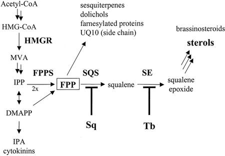
In higher plants, two distinct pathways have been shown to operate concomitantly for synthesizing isopentenyl diphosphate, the common precursor for all isoprenoids. Plastid isoprenoids such as carotenoids, mono- and diterpenes, or the prenyl chains of chlorophylls and plastoquinones are formed from 2-C-methyl-d-erythritol 4-phosphate, which itself arises from the initial condensation of pyruvate with glyceraldehyde 3-phosphate (for review, see Lichtenthaler, 1999; Rohmer, 1999). In the cytosol, isoprenoids are synthesized via the classical acetate/mevalonate (MVA) pathway, in which 3-hydroxy-3-methylglutaryl coenzyme A (CoA) reductase (HMGR) plays a key role. This enzyme is encoded by a multigene family (Bach et al., 1991; Stermer et al., 1994). In this pathway, farnesyl diphosphate (FPP) occupies a central position from which specific cis- and trans-prenyltransferases dispatch isoprene units to either sterols or non-sterol isoprenoids as represented by sesquiterpenes, ubiquinone, heme a, polyprenols, or prenylated proteins (Fig. 1). It has been recently proposed that specific classes of isoprenoids might be produced within distinct metabolic channels or metabolons, probably involving individual HMGR isoforms (Chappell, 1995; Weissenborn et al., 1995). Sterols represent the major end products of this multibranched pathway, but what controls the whole pathway is still far from being understood. Such a control might either concern only branch point enzymes or involve coordinated functioning of distinct metabolic channels, each one being regulated independently from one another (Chappell, 1995).
Figure 1.
Cytosolic isoprenoid biosynthetic pathway. Cytosolic isoprenoids are synthesized from acetyl CoA via the intermediate formation of MVA. IPP, Isopentenyl diphosphate; DMAPP, dimethylallyl diphosphate; IPA, isopentenyl adenine; and UQ, ubiquinone. Sq and Tb inhibit SQS and SE, respectively.
The first step committed to the sterol branch of the isoprenoid pathway is catalyzed by the squalene synthase (SQS), which mediates the reductive head-to-head condensation of two molecules of FPP to form squalene via presqualene diphosphate (Poulter, 1990). This reaction takes place in membranes of endoplasmic reticulum, as do all subsequent steps involved in sterol biosynthesis. Because of its particular position at the interface between hydrophilic and hydrophobic intermediates, SQS might constitute a major control point for regulating the sterol branch in directing FPP molecules into either sterols or non-sterol isoprenoids in response to changing cellular requirements. The sequence of reactions needed to convert squalene into end products is now well known (Benveniste, 1986), and the “state of art” on relevant enzymes and genes has been just reviewed (Bach and Benveniste, 1997). In contrast to animal and fungal cells, higher plants synthesize a mixture of sterols in which sitosterol, stigmasterol, and 24-methylcholesterol often predominate. These compounds play an essential role as membrane components in regulating the fluidity and permeability of membranes and the activity of membrane-associated proteins (Hartmann, 1998), but some sterols or biosynthetic intermediates might also serve as signal molecules during plant growth and development (Clouse, 2000). Despite the critical importance of sterols, mechanisms responsible for sterol homeostasis in higher plants are still largely unknown. Recent reports have pointed out that HMGR could regulate the flux of intermediates toward the sterol branch (Gondet et al., 1994; Chappell et al., 1995; Schaller et al., 1995), but whether or not this enzyme is able to respond to a depletion of sterol end products had not yet been investigated until now.
To get some insight into the regulatory mechanisms controlling the sterol biosynthetic pathway, the present study was focused on the role played by the plant SQS. Early work with plant cell suspension cultures has already emphasized its involvement in plant defense reactions. A very fast inhibition of SQS activity was observed after addition of fungal elicitors. The resulting arrest of sterol biosynthesis has been interpreted as a means for the cell to either redirect metabolic intermediates, especially FPP, toward the synthesis of sesquiterpene phytoalexins (Threlfall and Whitehead, 1988; Vögeli and Chappell, 1988; Zook and Kuc′, 1991) or simply to leave “house-keeping” metabolism pending better conditions (Haudenschild and Hartmann, 1995). This work was aimed at probing the effects of a direct inhibition of SQS in tobacco (Nicotiana tabacum cv Bright Yellow-2 [TBY-2]) cell suspension cultures. The recent availability of squalestatins (also called zaragozic acids), which are highly potent and specific inhibitors of SQS (Baxter et al., 1992; Bergstrom et al., 1993), gives the opportunity to investigate whether or not compensatory responses take place in the case of a depletion of only squalene-derived products. In particular, we wanted to check the possibility for HMGR to be a target for positive feedback regulation by endogenous sterols. Our study also comprises an investigation of responses of TBY-2 cells to an inhibition of squalene epoxidase (SE), the next committed enzyme to the sterol pathway. This enzyme catalyzes the stereospecific conversion of squalene to (3S)-2,3-oxidosqualene in the presence of molecular oxygen. It is the first oxygen-requiring step in the sterol biosynthetic pathway and, thus, might constitute a secondary regulatory level. SE can be specifically inhibited by compounds belonging to the class of allylamines (Ryder, 1991).
Here, we show that both squalestatin-1 (Sq) and terbinafine (Tb) are able to inhibit efficiently sterol biosynthesis in TBY-2 cells and, thus, to induce a decrease in the sterol cell content. In the case of Tb treatment, this depletion was accompanied by a concomitant accumulation of squalene and by an increase in the content and the rate of synthesis of triacylglycerols (TAG). Squalene was found to accumulate in lipid droplets but could be redirected toward sterol biosynthesis if necessary. Our results demonstrate that inhibition of either SQS or SE triggers an increase in HMGR enzyme activity, giving evidence for a positive feedback regulation of this key enzyme in response to a selective depletion of endogenous sterols. Surprisingly, we observed no compensatory responses mediated by SQS, in sharp contrast to the situation in animal systems.
RESULTS
Sterol Enzyme Activities and Free Sterol Levels during a Growth Cycle of TBY-2 Cells
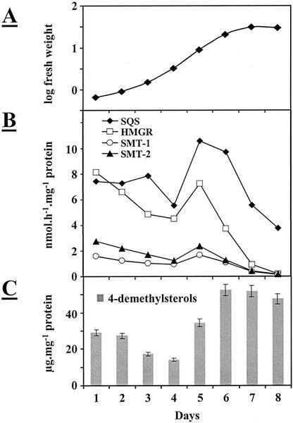
Figure 2 shows a typical growth curve of a TBY-2 cell culture and the corresponding changes in four sterol enzyme activities and in free sterol levels. Microsomal fractions were prepared from cultures collected on the designated days after subculturing and used for both enzymatic assays and sterol determinations. We monitored SQS activity and the activities of HMGR and sterol methyltransferases SMT1 and SMT2 throughout the growth cycle. HMGR, an upstream enzyme, is involved in the synthesis of both sterols and non-sterol isoprenoids. Sterol methyltransferases catalyze two distinct steps downstream of squalene: the S-adenosyl-l-Met-dependent methylation of cycloartenol (SMT1) and of 24-methylenelophenol (SMT2) to give 24-methylenecycloartanol and 24-ethylidenelophenol, respectively.
Figure 2.
Sterol biosynthesis in proliferating TBY-2 cells: changes in cell mass (A), sterol enzyme activities (B), and free 4-demethylsterol levels (C) as a function of time after subculturing. Microsomal membranes were prepared from cultures collected on the designated days after subculturing and used for both measurements of sterol enzyme activities (the values are the means of two replicates and are representative of a standard experiment) and free 4-demethylsterol contents (±se). SMT1, S-adenosyl Met-cycloartenol methyltransferase; and SMT2, S-adenosyl Met-24-methylenelophenol methyltransferase.
One or 2 d after subculturing, the tobacco cell culture entered into a rapid growth phase, as attested by a large increase in fresh cell mass, and reached a stationary phase after 7 d (Fig. 2A). Activities of the four enzymes exhibited very similar changes over the course of a culture cycle, with a maximum found on d 5 and a sharp decrease in the stationary phase, suggesting a coordinated expression of all the enzymes of the sterol pathway in proliferating TBY-2 cells (Fig. 2B). In Figure 2C, changes in the sterol levels over the growth cycle are given. Only free Δ5-sterols, the end products of the pathway, were quantified (as micrograms per milligram of protein). As expected, these compounds mainly accumulated in the stationary phase.
The sterol composition of 3-d-old TBY-2 cells is given in Table I. Stigmasterol, 24-methylcholesterol and sitosterol were by far the predominant sterols. The other sterols were isofucosterol, 24-methylenecholesterol, cholesterol, and Δ7-cholesterol. Other compounds (about 1.2% of total sterols) represented the usual sterol biosynthetic intermediates: 4α-methylsterols (obtusifoliol, cycloeucalenol, 24-methylene-, and 24-ethylidenelophenol) and 4,4-dimethylsterols (cycloartenol and 24-methylenecycloartanol). The relative proportions of the different end products were found not to change over the growth cycle, with the exception of a slight increase (from 1.55 to 1.82) in the ratio of stigmasterol to sitosterol (data not shown).
Table I.
Free sterol composition of TBY-2 cells
| Sterol | Composition of TBY-2 Cells |
|---|---|
| % of total sterols | |
| 4-Demethylsterols | |
| Cholesterol | 1.3 |
| Δ7-Cholesterol | 0.4 |
| 24-Methylenecholesterol | 9.9 |
| 24-Methylcholesterol | 35.2 |
| Stigmasterol | 24.5 |
| Sitosterol | 13.4 |
| Isofucosterol | 11.3 |
| 4α-Methylsterols | |
| Obtusifoliol | 0.2 |
| 24-Methylenelophenol | 1.2 |
| Cycloeucalenol | 0.1 |
| 4,4-Dimethylsterols | |
| Cycloartanol | 0.7 |
| Cycloartenol | 1.1 |
| 24-Methylenecycloartanol | 0.5 |
| Squalene | 0.3 |
| Total sterol (mg g−1 dry wt) | 1.9 ± 0.1 |
Sterols were extracted from 3-d-old cells as described in “Materials and Methods.” After acetylation, they were identified by GC-mass spectroscopy and quantified by GC.
Effects of Sq
Cell Growth, Sterol Content, and Biosynthesis
Three-day-old TBY-2 cells were treated with concentrations of Sq ranging from 0 to 0.5 μm for 24 h and incubated with radioactive sodium acetate for 2 h just before cell harvesting. TBY-2 cells were collected by filtration and analyzed for their sterol composition as described in “Materials and Methods.” Sterols (as the sum of free and esterified forms) were quantified by gas chromatography (GC) as acetate derivatives. Table II shows that Sq severely impaired cell growth as attested by a 50% decrease in the cell mass after a treatment with 0.5 μm. A parallel dose-dependent decrease in the sterol cell content was found. The high sensitivity of TBY-2 cells toward the inhibitor is illustrated by the very fast depletion of the pool of 4,4-dimethylsterols, the early precursors of sterols, which was reduced by 88% after a treatment with only 50 nm Sq (data not shown). The efficiency of Sq as an inhibitor of the sterol pathway was confirmed by measurements of radioactivity from [14C]acetate incorporated into end products. After addition of 50 nm Sq, radioactivity associated with 4-demethylsterols accounted for only 4% compared with that of sterols from control cells (Table II). At the same time, a dose-dependent accumulation of a labeled compound was observed. This compound, which was recovered in the band corresponding to 4α-methylsterols, was identified as farnesol. It might originate from hydrolysis of radioactive FPP molecules not used for sterol synthesis.
Table II.
Effect of Sq on cell growth, sterol biosynthesis, and SQS activity
| Sq | fwt | 4-Demethylsterols | SQS Activity | ||
|---|---|---|---|---|---|
| nm | g | mg g−1 dwt | cpm g−1 dwt | % | nmol h−1 mg−1 protein |
| 0 | 6.5 | 1.7 | 124,050 | 100 | 10.5 ± 1 |
| 50 | 5.0 | 1.0 | 4,980 | 4.0 | 0.25 ± 0.1 |
| 75 | 4.2 | 0.8 | 2,550 | 2.0 | 0.20 ± 0.1 |
| 100 | 3.7 | 0.7 | 2,580 | 2.1 | 0.1 ± 0.1 |
| 500 | 3.2 | 0.6 | 540 | 0.4 | 0.1 ± 0.1 |
Three-day-old TBY-2 cells were treated with various concentrations (from 0 to 500 nm) of Sq for 24 h, then incubated with sodium [1-14C]acetate for 2 h just before cell harvesting. Total sterols were quantified by GC as acetate derivatives. SQS activity was measured in the presence of [1-3H]FPP and 10 μg of microsomal protein. fwt, Fresh weight; dwt, dry weight.
SQS Activity
The target of Sq is SQS as illustrated in Table II. A dose as low as 50 nm Sq was shown to be sufficient to inhibit almost completely SQS activity in microsomal fractions. Such an inhibition was found to take place in a few hours, with an IC50 value of 5.5 nm, indicating the very potent inhibitory power of Sq. This compound, which partially mimics the structure of FPP and/or presqualene diphosphate, has been described as a competitive inhibitor of SQS (Bergstrom et al., 1993).
Attempts were made to recover SQS activity in microsomal membranes from Sq-treated cells. However, neither extensive washes of intact cells nor dilution and additional centrifugations of microsomes were successful, suggesting that Sq also acts as a mechanism-based irreversible inactivator of plant SQS (Lindsey and Harwood, 1995).
SQS, FPP Synthase (FPPS), and HMGR Expression
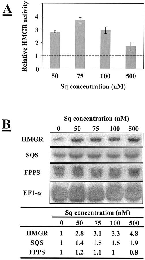
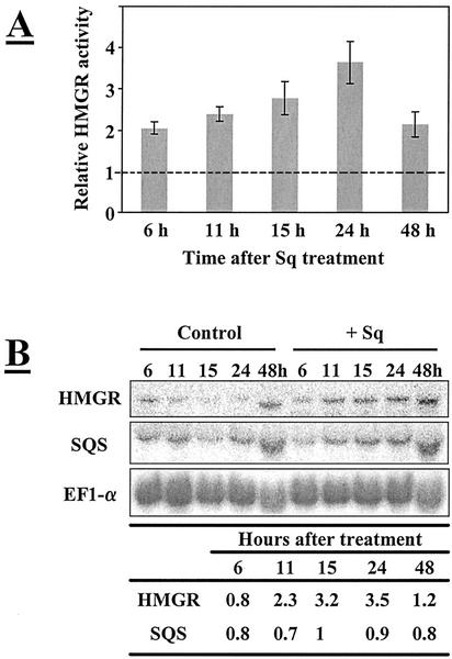
To investigate whether SQS transcription could be affected by the inhibition of SQS activity, total RNA was isolated from TBY-2 cells treated with different Sq concentrations. A full-length SQS cDNA from Nicotiana benthamiana (Hanley et al., 1996), which has 98% identity with the corresponding N. tabacum cDNA, was used as a probe. After hybridization with this probe, transcripts with a size of 1.6 kb could be detected. As shown in Figure 3B, the levels of SQS transcripts were not found to change significantly, despite an almost complete inhibition of SQS activity. A time-course study (from 0 to 48 h) of effects induced by 0.5 μm Sq also showed no differences between SQS mRNA levels from control and treated cells, whatever the period of time (Fig. 4B). Because SQS inhibition possibly induces an increased amount of FPP molecules in the cytosol, we checked the effects of Sq treatments on the transcription of FPPS. Northern-blot experiments were performed with a partial FPPS cDNA from TBY-2 cells as a probe. The corresponding mRNA levels, with a size of about 1.7 kb, remained constant whatever the dose of Sq used, suggesting that FPPS transcription was neither affected by SQS inhibition, nor by the excess of farnesol and FPP.
Figure 3.
Effects of Sq on HMGR activity (A) and HMGR, SQS, and FPPS mRNA levels (B). Three-day-old TBY-2 cells were treated with various concentrations of Sq (from 0 to 500 nm) and used to isolate both microsomal fractions and total RNA. A, HMGR activity was measured in the presence of 30 μm R,S-[3-14C]HMG-CoA and of 30 μg of microsomal protein. Control value (5.5 nmol h−1 mg−1) was set at 1. Enzyme activities were expressed as relative values to the control. Results are from two independent experiments ± sd. B, Total RNA (30 μg) was loaded per lane and transferred to a nylon membrane. Hybridizations were performed with 32P-labeled probes. All of the hybridizations were performed on the same membrane. Relative intensities obtained by PhosphorImage analysis are given in the table after correction for background and normalization relatively to EF1-α mRNA content. Control value was set at 1.
Figure 4.
Time course of Sq effects on HMGR activity (A) and HMGR and SQS mRNA levels (B). Three-day-old TBY-2 cells were treated during various times (from 0 to 48 h) by Sq (500 nm) and were used to isolate both microsomal fractions and total RNA. A, HMGR activity was measured as indicated in Figure 3. For each time, enzyme activities were expressed as relative values to the corresponding control (set at 1). Results are from two independent experiments ± sd. B, Total RNA (30 μg) was loaded per lane and transferred to a nylon membrane. Hybridizations were performed with 32P-labeled probes. All the hybridizations were performed on the same membrane. Relative intensities obtained by PhosphorImage analysis are given in the table after correction for background and normalization relatively to EF1-α mRNA content and compared with the corresponding control (set at 1).
It is well known that HMGR constitutes the major limiting-step in cholesterol biosynthesis in mammalian cells (Goldstein and Brown, 1990). The inhibition of SQS and the resulting depletion of squalene-derived products were shown to induce compensatory responses mediated by HMGR (Ness et al., 1994; Lopez et al., 1998). It seemed interesting to us to investigate whether similar responses could also take place in TBY-2 cells. Three-day-old TBY-2 cells were treated for 24 h with Sq concentrations varying from 0 to 0.5 μm and were used to isolate both total RNA and microsomal fractions, for HMGR activity measurements. As shown in Figure 3A, microsomal fractions from Sq-treated TBY-2 cells were found to exhibit 2- to 4-fold increased HMGR enzyme activities compared with control cells. The highest stimulation rate was observed at 75 nm. Such a stimulation of HMGR activity could be detected as early as 6 h after Sq administration and progressively increased until 24 h (Fig. 4A). For northern-blot experiments, we used as a probe a 1,400-bp fragment corresponding to the C-terminal part (catalytic site) of a Nicotiana sylvestris HMGR cDNA (Genschik et al., 1992). HMGR transcripts with a size of about 2.5 kb could be detected. Figure 3B shows a dose-dependent increase in HMGR mRNA levels from Sq-treated TBY-2 cells. A 5-fold increase was found in cells treated with 500 nm Sq. At this concentration, the highest level of transcripts was observed 24 h after Sq administration (Fig. 4B).
Taken together, these results indicate that the inhibition of SQS by Sq triggered a stimulation of both HMGR steady-state mRNA and enzyme activity, suggesting that the arrest of sterol biosynthesis and the resulting depletion of squalene-derived products exerted a positive feedback regulatory effect on the transcription of HMGR. Surprisingly, this inhibition did not activate SQS transcription.
Effect of Tb
In Vivo Sterol Biosynthesis
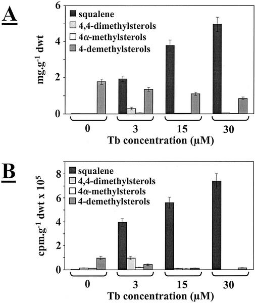
Three-day-old TBY-2 cells were treated with concentrations of Tb ranging from 0 to 30 μm for 30 h, then collected by filtration and analyzed for their free sterol composition as described in “Materials and Methods.” Squalene and free sterols (as acetate derivatives) were quantified by GC. Treatment of TBY-2 cells with Tb was found to induce a dose-dependent accumulation of squalene and a progressive decrease in the content of end products (Fig. 5A). After treatment with 30 μm Tb, the squalene content amounted to 5 mg g−1 dry weight, whereas it was barely detectable in control cells (about 20 μg g−1 dry weight). At the same time, the remaining Δ5-sterols accounted for only 0.85 mg g−1 dry weight compared with 1.8 mg g−1 for control cells, corresponding approximately to a 50% decrease in the usual free sterol content. Such an accumulation of squalene clearly indicates an inhibition of SE by Tb, leading to an arrest of end product biosynthesis. No change in relative proportions of Δ5-sterols was found (data not shown). It is remarkable to see that TBY-2 cells can accommodate such high squalene intracellular concentrations because no inhibition of cell growth or cell death was observed (data not shown).
Figure 5.
Effects of Tb on sterol composition (A) and biosynthesis (B). Three-day-old TBY-2 cells were treated with concentrations of Tb ranging from 0 to 30 μm for 24 h and incubated with sodium [1-14C]acetate for 2 h before cell harvesting. Sterols were extracted as indicated in “Materials and Methods.” A, Squalene and free sterols (acetate derivatives) were quantified by GC. Sterols amounts are expressed in milligrams per gram dry weight. B, Radioactivities incorporated into sterols and precursors are expressed as 105 cpm g−1 dry weight (dwt).
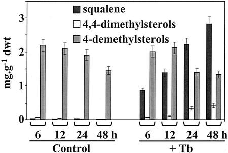
Figure 6 shows a time course of squalene accumulation in TBY-2 cells treated with 3 μm Tb. A significant increase in the squalene content occurred as soon as 6 h after administration of the inhibitor and continued up to 48 h. After 96 h, no additional accumulation of squalene was observed (data not shown).
Figure 6.
Time course of squalene accumulation after Tb administration. Three-day-old TBY-2 cells were treated by 30 μm of Tb for different periods of time in parallel with a control. Sterols were extracted as indicated in “Materials and Methods.” Squalene and free sterols (acetate derivatives) were quantified by GC. Sterol amounts are expressed in milligrams per gram dry weight (dwt).
Further evidence for the SE inhibition was obtained by feeding control and Tb-treated TBY-2 cells with [14C]acetate for 2 h before cell harvesting. In control cells, most of the acetate radioactivity was incorporated into free sterols, whereas in Tb-treated cells, squalene was by far the most labeled compound (Fig. 5B). After treatment with 3 μm Tb, about 71% of the radioactivity recovered in the sterol branch was already associated with squalene. At this concentration, a time-course study of squalene biosynthesis demonstrated that the highest rate of synthesis was obtained after 24 h of treatment (data not shown). After treatment with 30 μm, 97% of the radioactivity was present in squalene and only 2% in the end products.
Cycloartenol Accumulation at Low Tb Concentration
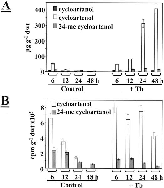
As shown in Figure 5, treatment of TBY-2 cells with 3 μm Tb was found to induce a significant accumulation of 4,4-dimethylsterols. These compounds were mainly represented by cycloartenol and 24-methylenecycloartanol (Table I). GC analysis of the corresponding acetate derivatives indicated a specific increase in the cycloartenol content (data not shown), suggesting that SMT1 involved in the methylation of cycloartenol to give 24-methylenecycloartanol might be down-regulated. To get more insight into this process, TBY-2 cells were treated with 3 μm Tb for periods of time from 0 to 48 h. A 2-h pulse of radioactive acetate was given just before cell harvesting. After lipid extraction, the fraction of 4,4-dimethylsterols was analyzed in more detail. The acetate derivatives were separated on AgNO3-impregnated thin-layer chromatography (TLC) plates. Cycloartenol and 24-methylenecycloartanol were eluted and quantified by GC, and their radioactivity was measured by scintillation counting. As shown in Figure 7A, the cycloartenol content of treated cells was found to increase progressively in function of the duration of contact with the inhibitor. In contrast, cycloartenol did not accumulate in control cells (Fig. 7A). As a consequence, the radioactivity associated with cycloartenol declined rapidly in control cells, whereas in treated cells, cycloartenol continued to be actively synthesized for as long as 24 h (Fig. 7B). Whatever the period of time, only low amounts of radioactivity were detected in 24-methylenecycloartanol.
Figure 7.
Time course of content (A) and biosynthesis (B) of 4,4-dimethylsterols after administration of 3 μm Tb. Three-day-old TBY-2 cells were treated with 3 μm Tb for different periods of time and incubated with sodium [1-14C]acetate for 2 h before cell harvesting. A, Levels of the three major 4,4-dimethylsterols were quantified by GC (acetate derivatives) and expressed in micrograms per gram dry weight. B, Radioactivities incorporated into cycloartenol and 24-methylenecycloartanol were expressed as 105 cpm g−1 dry weight (dwt). 24-me cycloartanol, 24-Methylenecycloartanol.
To investigate whether the methylation of cycloartenol by the SMT1 could be inhibited in TBY-2 cells treated by low Tb concentrations, microsomal fractions were isolated from control and Tb-treated cells and tested for their SMT1 enzyme activities. Similar enzyme activities were found (data not shown), indicating that the SMT1 protein remained active despite the Tb treatment of tobacco cells. We also checked that 30 μm Tb had no direct inhibitory effect on SMT1 activity. Thus, these data indicate that the inhibition of the methylation reaction occurs only in intact treated cells and could result from a secondary regulatory effect.
SQS and HMGR Expression
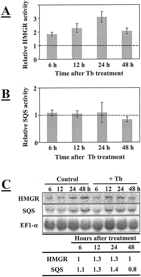
As stated above, treatment of TBY-2 cells with Tb triggered the accumulation of impressive amounts of squalene, the product of the reaction catalyzed by SQS. In control cells, endogenous squalene was barely detectable. Despite such large increases of squalene, SQS was found to exhibit a constant enzyme activity, similar to that of control cells and whatever the duration of the Tb treatment (Fig. 8B). At the same time, no significant change in the SQS steady-state mRNA levels was observed (Fig. 8C), indicating no negative regulatory effect on the gene transcription by squalene.
Figure 8.
Time course of Tb effects on HMGR (A) and SQS (B) activities and mRNA levels (C). Three-day-old TBY-2 cells were treated for various time periods (from 0 to 48 h) with 30 μm Tb and used to isolate both microsomal fractions and total RNA. A, HMGR activity was measured as indicated in Figure 3. For each point, enzyme activities were expressed as relative values to the corresponding control (set at 1). Results are from three independent experiments ± sd. B, SQS activity was measured in the presence of [1-3H]FPP and of 10 μg of microsomal protein. For each time, enzyme activities were expressed as relative values to the corresponding control (set at 1). Results are from three independent experiments ± sd. C, Total RNA (30 μg) was loaded per lane and transferred to a nylon membrane. Hybridizations were performed with 32P-labeled probes. All the hybridizations were performed on the same membrane. Relative intensities obtained by PhosphorImage analysis are given in the table after correction for background and normalization relative to EF1-α mRNA content and compared with the corresponding control (set at 1).
We also investigated effects of Tb treatments on HMGR expression. As shown in Figure 8A, a 2- to 4-fold increase in the HMGR enzyme activity was observed in TBY-2 cells treated with 30 μm Tb. The stimulation occurred already after 6 h and reached a maximum after 24 h. However, this increase in enzyme activity was not correlated with significant modifications of the corresponding mRNA levels (Fig. 8C).
TAG Synthesis
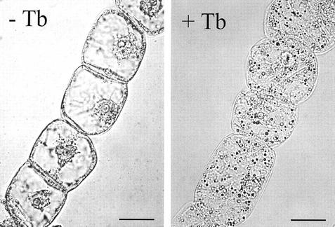
Besides the accumulation of squalene, Tb treatment of TBY-2 cells also triggered a significant dose-dependent increase in the TAG content, with an 8-fold enhancement of the mean value measured for control cells after administration of 30 μm (Table III). This increase directly resulted from a stimulation of their de novo biosynthetic rate, as indicated by the parallel increase in their [14C]radioactivity (Table III). A concomitant accumulation of lipid droplets in the cytosol of treated cells was found to take place. Light microscopy observations showed the presence of many orange spheres after staining of cells with Sudan IV, a lipid-specific dye (Fig. 9). Very few or none of these lipid droplets were seen in control cells. These results suggest that relationships between sterol and TAG biosynthetic pathways might occur in vivo.
Table III.
Effects of Tb on TAG content and biosynthesis
| TAG | Tb Concentration
|
|||
|---|---|---|---|---|
| 0 | 3 | 15 | 30 | |
| μm | ||||
| μmol g−1 dwt | 0.5 ± 0.1 | 1.6 ± 0.2 | ND | 4.1 ± 0.6 |
| cpm g−1 dwt × 103 | 21 | 73 | 92 | 118 |
Three-day-old TBY-2 cells were treated for 24 h with different concentrations (from 0 to 30 μm) of Tb, then incubated with sodium [1-14C]acetate for 2 h just before cell harvesting. TAGs were extracted and quantified as described in “Materials and Methods.” ND, Not determined; dwt, dry weight.
Figure 9.
Lipid particles in the cytosol of TBY-2 cells treated with Tb. Three-day-old TBY-2 cells were treated for 24 h with Tb 30 μm and then observed in optical microscopy after staining by Sudan IV, in parallel with a control. Lipid particles appear as spherical orange droplets. Bar = 20 μm.
Squalene Intracellular Localization
To address the question of the intracellular localization of the overproduced squalene, TBY-2 cells were treated with 30 μm Tb for 24 h before being used for isolation of a microsomal fraction. After sedimentation at 100,000g, lipid particles appeared as a fluffy lipid layer at the surface of the corresponding supernatant. Both fractions were then analyzed for their squalene content. In control cells, squalene could be not detected in the supernatant. In contrast, most (i.e. higher than 90%) of the squalene from treated cells was recovered in the lipid droplets. The remaining 10% were associated with the microsomal fraction.
It seemed to us interesting to investigate whether or not the squalene stored in these lipid particles could be reused as a precursor for sterol biosynthesis. TBY-2 cells were first treated with 30 μm Tb for 24 h and fed with radioactive acetate for 2 h just before cell harvesting. They were then extensively washed and resuspended in a Murashige and Skoog medium containing 0.5 μm Sq to block the synthesis of endogenous squalene. These cells were allowed to grow for 12, 24, and 48 h, respectively, collected by filtration, and analyzed for their content in squalene and in free sterols and their precursors (4,4-dimethyl- and 4α-methylsterols). Radioactivity associated with each class of compounds was also measured. A sample of TBY-2 cells treated with Tb for 24 h was taken as a control (t0h). As shown in Table IV, TBY-2 cells collected after 12 and 24 h exhibited dramatic decreases in both the content and radioactivity of squalene, whereas concomitant and similar increases were found for the sterol end products. A 2-fold increase in the level of 4-demethylsterols occurred after 24 h, despite the inhibition of SQS by Sq, and these compounds contained the most radioactivity initially associated with squalene. In the further 24 h (t48h), their amount was reduced by 50% because no more squalene was available. The data from Table IV also indicate a transient increase in both the contents and radioactivities of the sterol biosynthetic precursors, giving evidence for a restart of an active sterol biosynthesis from the pool of radioactive squalene, triggered by the Tb removal. Finally, it should be emphasized that such a restoration of the sterol biosynthesis was accompanied by a parallel decrease in the content and radioactivity of TAG, indicating once more likely relationships between both pathways. Taken together, these results clearly demonstrate that squalene, which was previously stored in cytoplasmic lipid particles, could be remobilized for an active sterol biosynthesis. Even if the fate of TAG molecules remains to be investigated, it appears that these lipid particles actually do constitute a pool of available metabolic intermediates.
Table IV.
Redirection of squalene from lipid particles to the sterol pathway
| Squalene
|
4-Demethylsterols
|
4,4-Dimethylsterols
|
4α-Methylsterols
|
TAG
|
||||
|---|---|---|---|---|---|---|---|---|
| mg g−1 dwt | Radioactivity | mg g−1 dwt | Radioactivity | Radioactivity | Radioactivity | μmol g−1 dwt | cpm g−1 dwt × 105 | |
| % | % | % | % | |||||
| T0h | 5.2 | 98.2 | 0.9 | 0.8 | 0.2 | 0.8 | 5.1 | 5.8 |
| T12h | 0.7 | 18.6 | 1.4 | 37.6 | 31.6 | 12.1 | 1.2 | 1.5 |
| T24h | 0.2 | 4.3 | 1.6 | 92.5 | 1.5 | 1.6 | 0.4 | 0.1 |
| T48h | 0.05 | 0.3 | 0.8 | 99.7 | ND | ND | ND | ND |
TBY-2 cells were first treated with 30 μm Tb for 24 h and then incubated with sodium [1-14C]acetate for 2 h. Cells were extensively washed to remove the inhibitor and non-incorporated radioactivity. Cells were resuspended in Murashige and Skoog medium (T0h) and allowed to grow with 0.5 μm Sq. Aliquots of cells were taken after 0 (T0h), 12 (T12h), 24 (T24h), and 48 h (T48h). All samples were analyzed for their squalene, sterol, and TAG contents and radioactivities. dwt, Dry weight; ND, not determined.
DISCUSSION
In contrast to the situation in animal cells, much less is known regarding regulation of the sterol pathway in plants. In that context, we planned to investigate the potential regulatory role played by the plant SQS for the following reasons: (a) SQS is the first committed enzyme to the sterol branch of the isoprenoid pathway and as such, may play a critical role in directing FPP molecules in either sterol or non-sterol isoprenoids in response to changing cellular requirements (Fig. 1); (b) because sterols are major isoprenoid end products, most part of precursors goes through SQS; (c) SQS has been known to participate in plant defense reactions against pathogens (Threlfall and Whitehead, 1988; Vögeli and Chappell, 1988; Zook and Kuć, 1991; Haudenschild and Hartmann, 1995); and (d) in mammalian cells, SQS is coordinately regulated with HMGR by a sterol feedback mechanism (Sakakura et al., 2001). Moreover, inhibition of SQS would not deprive the cell of important non-sterol compounds such as isoprenylated proteins, dolichol, heme a, or ubiquinone. As a plant material, we used tobacco BY-2 cell suspension cultures. This cell line was originally selected for its very short cell cycle (about 15 h; Nagata et al., 1992). Such a suspension was, therefore, particularly suitable for metabolic studies and labeling experiments. TBY-2 cells were first checked for their ability for sterol synthesis. As shown in Figure 2, SQS and SMT1 and SMT2 enzyme activities were expressed during the entire cell growth, with a maximum at 5 d after subculturing. HMGR, which is involved in the synthesis of both sterols and non-sterol isoprenoids, exhibited a very similar expression profile, suggesting that all the enzymes of the sterol pathway were coordinately regulated, to sustain an active synthesis of membranes in rapidly dividing cells.
To investigate the regulatory response of SQS to depletion of sterols, TBY-2 cells were treated with Sq. This inhibitor belongs to the class of Sqs, a family of fungal metabolites recently discovered. These compounds, which are analogs of FPP and presqualene diphosphate, are potent competitive inhibitors of mammalian SQS (Baxter et al., 1992; Bergstrom et al., 1993). We show here that Sq is also a strong inhibitor of the tobacco SQS. The high efficiency of Sq as an inhibitor of sterol synthesis was revealed by the rapid and dramatic decrease in the radioactivity from [14C]acetate incorporated into sterols after treatment with nanomolar concentrations of Sq, resulting in a decrease in the sterol cell content (Table II). At the same time, SQS activity rapidly became barely detectable. Sq was found to inhibit tobacco SQS with an IC50 of 5.5 nm (data not shown), a value similar to that obtained for mammalian SQS (Lindsey and Harwood, 1995). Sq was also found to rapidly impair cell growth of TBY-2 cells (Table II). Such an effect on cell growth probably resulted from an inhibition of cell division. When given to synchronized TBY-2 cells, this inhibitor triggers an arrest of the cell cycle specifically in the G1/G0 phase, but without inducing cytotoxicity or cell death (Hemmerlin et al., 2000).
An intriguing question is related to the fate of FPP molecules, which do not contribute to the build-up of sterols. First, the increase in cytosolic FPP resulting from Sq treatments does not appear to regulate negatively FPPS expression because no changes in corresponding steady-state mRNA levels occurred, whatever the Sq concentration (Fig. 3). However, FPP might have an effect on MVA kinase, an enzyme upstream in the pathway because it has been shown to be a competitive inhibitor of this enzyme with respect to ATP (Schulte et al., 2000). As already stated (Fig. 1), FPP serves as a substrate for a variety of non-sterol isoprenoids. As a consequence, a redirection of FPP toward such pathways could appear to be likely. We have previously shown that exogenous farnesol could be incorporated into sterols but also into the prenyl side chain of ubiquinone Q10 and proteins from TBY-2 cells (Hartmann and Bach, 2001). When radioactive farnesol was given to tobacco cells in the presence of 0.5 μm Sq, no increase in the label of ubiquinone and proteins occurred, indicating that no additional FPP molecules were redirected toward these compounds (M.-A. Hartmann, unpublished data). These results are similar to those from Crick et al. (1995) obtained with brain cells. Under the same conditions, these authors also demonstrated that Sq had no effect on the synthesis of dolichol-phosphate.
Our labeling experiments showed that a significant part of FPP molecules were hydrolyzed in response to Sq treatment, as attested by the dose-dependent accumulation of radioactive farnesol (data not presented). Similar observations were made in the case of Sq-treated mammalian cells (Bergstrom et al., 1993; Lopez et al., 1998). Such a hydrolysis might be catalyzed by a FPP diphosphatase, which could be induced by the stress caused by the Sq treatment (Nah et al., 2001). However, it should be pointed out that farnesol is also known to have deleterious effects. In particular, when added to TBY-2 cells at a concentration higher than 20 μm, farnesol induces cell death (Hemmerlin and Bach, 2000). As a consequence, its level in the cytosol has to be closely controlled. However, the possibility of a conversion of FPP or farnesol into other metabolites should not be excluded.
In sharp contrast to SQS inhibition by Sq, treatment of TBY-2 cells by Tb, an inhibitor of SE, did not affect cell growth. Tb belongs to the class of compounds termed allylamines, which have significant impact as antifungal drugs (Ryder, 1991). Tb is a reversible, noncompetitive inhibitor of SE (Ryder, 1991). We show here that Tb is also active in plant systems, as attested by a dose-dependent decrease in the free sterol content and by a concomitant accumulation of squalene. These data are in agreement with previous results obtained with celery (Apium graveolens) cell suspension cultures (Yates et al., 1991) or wheat (Triticum aestivum) seedlings (Simmen and Gisi, 1995). However, under our conditions, no accumulation of Δ5,7-sterols could be observed, and the absence of unusual intermediates like Δ8- or Δ8,14-sterols suggests that Tb had no secondary target in TBY-2 cells (Yates et al., 1992). Such high amounts of squalene seemed not to be toxic for the cell. We observed that inhibition of SE by Tb in TBY-2 cells was accompanied by a proliferation of cytosolic lipid droplets (Fig. 9), in which squalene accumulated. Our results provide evidence that squalene could be remobilized for an active sterol biosynthesis in response to a depletion of end products (Table IV). Therefore, lipid particles actually do constitute a pool of available biosynthetic intermediates and not only a metabolically inactive storage compartment, in agreement with recent data in yeast (Saccharomyces cerevisiae) from Milla et al. (2002). Concomitant to the squalene accumulation, a dose-dependent increase in the TAG content and rate of synthesis was observed (Table III). These TAG probably also accumulated in lipid droplets because they could not be detected in microsomes. The mechanisms whereby the inhibition of SE induces the TAG synthesis still remain to be elucidated. However, it should be emphasized that a stimulation of TAG synthesis was also observed in leek (Allium porrum) seedlings treated with fenpropimorph, another inhibitor of sterol biosynthesis (M.-A. Hartmann, A.-M. Perret, J.-P. Carde, C. Cassagne, and P. Moreau, unpublished data), suggesting that some regulatory relationships between sterol and TAG biosynthetic pathways might occur in plants.
Because Sq and Tb treatments of TBY-2 cells were found to induce significant decreases in the sterol cell content, it was interesting to investigate whether compensatory responses could be mediated by HMGR. In mammals, HMGR is the major rate-limiting enzyme in the cholesterol biosynthetic pathway. This enzyme is encoded by a single gene. It is well established that reductase activity is controlled through multivalent feedback regulation, involving both sterols and non-sterol compounds derived from MVA (Goldstein and Brown, 1990). In sharp contrast to animal systems, the occurrence of multiple genes encoding HMGR is a general feature of higher plants. Individual genes have been shown to exhibit different expression patterns in response to physiological and environmental stimuli such as light, plant growth regulators, wounding, or pathogen attack (Bach et al., 1991; Stermer et al., 1994). It has been proposed that the different HMGR isoforms might play distinct roles associated with the production of specific isoprenoid compounds (Chappell, 1995; Weissenborn et al., 1995).
Previous work had given evidence for an involvement of plant HMGR in controlling the flux of intermediates toward sterol biosynthesis (Gondet et al., 1994; Chappell et al., 1995; Schaller et al., 1995). Tobacco plants overexpressing either the HMGR1 gene from Hevea brasiliensis (Schaller et al., 1995) or a truncated HMGR gene from guinea pig (Chappell et al., 1995) synthesize higher amounts of sterols and sterol precursors, which accumulate as steryl esters in cytosolic lipid bodies (Gondet et al., 1994). However, no change in the free sterol content was observed. Thus, sterol acylation appears as a means for the cell to maintain sterol homeostasis. We show here for the first time, to our knowledge, that tobacco HMGR is able to respond to a selective depletion of endogenous sterols. Decreases in squalene and squalene-derived compounds resulting from treatments of TBY-2 cells with Sq or Tb triggered 2- to 4-fold increases in HMGR enzyme activity (Figs. 3A, 4A, and 8A). In the case of SQS inhibition by Sq, an enhancement of corresponding mRNA levels was observed, giving evidence for an activation of HMGR gene transcription. In rat liver, a similar stimulation of HMGR transcription was previously reported in response to Sq (Ness et al., 1994; Lopez et al., 1998). On the other hand, SE inhibition by Tb was not accompanied by changes in the HMGR transcripts, suggesting that the regulatory response mediated by HMGR could be exerted at a translational or posttranslational (i.e. catalytic efficiency or protein degradation) level. To our knowledge, whereas plant HMGR appears to be mainly transcriptionally controlled (Weissenborn et al., 1995), such a feedback regulatory effect occurring at the HMGR protein level in response to a depletion of end products has not yet been reported in plants. Thus, our results show differential regulatory responses induced by both inhibitors.
Jiang et al. (1993) and Keller et al. (1993) have presented convincing evidence that the mammalian SQS genes are regulated by their transcription rate in response to exogenous sterols and inhibitors of sterol synthesis. Surprisingly, our results indicate that tobacco SQS exhibits a different behavior. We show that SQS mRNA levels were not altered after treatment of TBY-2 cells with either Sq or Tb. Thus, the expression pattern of SQS appears to be insensitive to both the inhibition of SQS activity and the accumulation of squalene, the product of the reaction. Such an impressive accumulation of squalene, which was induced by the inhibition of SE by Tb, can likely be attributed to the enhancement of HMGR expression, leading to a greater amount of enzyme protein and, thus, to a higher synthesis of early intermediates. At the same time, no change in the SQS enzyme activity occurred, indicating that SQS would not be a limiting step for sterol synthesis in tobacco cells. However, as described above, squalene did not accumulate in endoplasmic reticulum membranes but in lipid droplets. Thus, SQS could not “sense” the excess of squalene. In the same context, no changes in SQS activity were observed when TBY-2 cells were treated with Lab 170250F, an inhibitor of obtusifoliol 14-demethylase (Taton et al., 1988), an enzyme downstream of squalene, or with mevinolin, an inhibitor of HMGR (L. Wentzinger and M.-A. Hartmann, unpublished data). Taken together, these data indicate that SQS is regulated differently in TBY-2 cells and in mammalian cells (Tansey and Shechter, 2000). The next step will be to check whether similar regulatory mechanisms for SQS also take place in intact plants.
Genes encoding enzymes making up a specific metabolon must have similar transcriptional networks to coordinate expression of the metabolic unit. In mammals, the cholesterol feedback system is mediated by a family of membrane-bound transcription factors known as sterol regulatory element (SRE)-binding proteins, which recognize a 10-bp sequence (SRE) within the target genes (Brown and Goldstein, 1997). It has been just reported that such a SRE-binding protein activation mechanism concerns every step of the cholesterol biosynthetic pathway (Sakakura et al., 2001). In the promoters of plant HMGR genes, no consensus sequences similar to these SRE or other cis regulatory elements from animal sterol-regulated genes have been found so far (Enjuto et al., 1995). Moreover, many enzymes involved in plant sterol biosynthesis are encoded by multiple genes (Bach and Benveniste, 1997). For instance, this is the case for SQS (Kribii et al., 1997; Devarenne et al., 1998) and SE (Schäfer et al., 1999). The biological significance of such a plethora of genes remains to be elucidated. Higher plants have probably evolved specific mechanisms for regulating their complex isoprenoid pathway, mechanisms that remain to be discovered. In this challenging context, we are currently investigating in more detail sterol homeostasis in plant cells.
MATERIALS AND METHODS
Chemicals
All chemicals were purchased from Sigma (St Louis). Sodium [1-14C]acetate (54 Ci mol−1), [3-14C]HMG-CoA and S-adenosyl l-[methyl-3H]Met were from Amersham (Buckinghamshire, UK). [1-3H]FPP was from Isotopchim (Ganagobie-Peyruis, France). Sq was obtained from Glaxo (Greenford, Middelsex, UK) and dissolved in 0.1 m Tris-HCl (pH 7.4) to give a 0.1 mm. stock solution. Tb was kindly supplied by Dr. N.S. Ryder (Vienna) and dissolved in dimethyl sulfoxide at a concentration of 50 mm. Final dimethyl sulfoxide concentrations did not exceed 0.05% (v/v).
Plant Material
Cell suspension cultures of tobacco (Nicotiana tabacum cv Bright Yellow-2 [TBY-2]) were usually grown in 250-mL Erlenmeyer flasks containing 80 mL of modified Murashige and Skoog medium at 26°C in the dark and subcultured weekly as reported (Nagata et al., 1992). Cells were harvested by filtration. In all cases, sterol inhibitors were added to 3-d-old cell cultures.
In Vivo Labeling Experiments
Cells were usually incubated with sodium [1-14C]acetate (5 μCi, 0.2 mm) for 2 h just before cell harvesting. Unincorporated radioactivity was removed by washing cells on the filter.
Isolation of Microsomes
Frozen cells were ground in a mortar in the presence of liquid N2. The powder was resuspended in a medium consisting of 0.25 m Suc, 4 mm EDTA, 100 mm potassium fluoride, 40 mm sodium ascorbate, and 0.2% (w/v) bovine serum albumin in 0.1 m Tris-HCl (pH 8.0; 10 mL g−1 fresh wt). After filtration through a nylon blutex, the homogenate was centrifuged at 10,000g for 25 min. The resulting supernatant was centrifuged at 100,000g for 60 min. The microsomal pellet was resuspended in 0.1 m Tris-HCl (pH 7.5) containing 1.5 mm dithioerythritol and 20% (w/v) glycerol and stored at −80°C until use. Protein concentrations were determined according to Bradford (1976) with bovine serum albumin as a standard.
Isolation of Lipid Particles
Frozen cells were ground in a mortar in the presence of liquid N2. The powder was homogenized in 0.1 m Tris-HCl (pH 8.0) containing 1 mm EDTA. After filtration through a nylon blutex, the homogenate was centrifuged at 10,000g for 25 min. The resulting supernatant was centrifuged at 100,000g for 60 min. The white fat pad at the top of the tube was collected and lyophilized for lipid analyses. The microsomal fraction was resuspended in 0.1 m Tris-HCl (pH 8.0) and 1 mm EDTA, and centrifuged at 100,000g for 60 min. The microsomal pellet was freeze-dried before lipid analysis.
Lipid Analyses
Freeze-dried material was ground and extracted by refluxing twice with dichloromethane:methanol (2:1, v/v) for 2 h. Extracts were combined, dried under reduced pressure, and thoroughly washed at room temperature with hexane to recover squalene, free sterols, and TAG.
Sterols were isolated and quantified as previously reported (Hartmann and Benveniste, 1987). After recovering from total lipids with hexane, sterols were loaded on TLC plates, which were developed in dichloromethane (two runs). Purified sterols were then eluted and acetylated before being analyzed by GC on a glass capillary column (30 m long, 0.25 mm i.d., coated with DB-1). The temperature program used includes a fast rise from 60°C to 230°C (30°C min−1), then a slow rise from 230°C to 280°C (2°C min−1). A cholesterol standard was added to the samples before analysis. Sterols were identified by GC-mass spectroscopy (Rahier and Benveniste, 1989). Squalene and steryl esters were separated by TLC with cyclohexane:toluene (95:5, v/v) as the solvent. Squalene (RF 0.5) was eluted and quantified by GC. The radioactivity was measured by liquid scintillation spectrometer. TAG were quantified using the colorimetric assay from Sigma (kit 336-10).
Assays for Enzyme Activities
HMGR and SQS activities were measured according to Bach et al. (1986) and Haudenschild and Hartmann (1995), respectively. SMT activities were measured in the presence of 0.1 m Tris-HCl (pH 7.5) containing 100 μm [3H-methyl]Ado-Met (1 μCi), 0.1% (w/v) Tween 80, 1 mm 2-mercaptoethanol, 50 to 100 μg of microsomal membranes, and 100 μm cycloartenol (SMT1) or 50 μm 24-methylenelophenol (SMT2), in a total volume of 100 μL. Incubations were carried out at 30°C for 1 h and stopped by adding 12% (w/v) KOH in ethanol. The neutral lipids were extracted with hexane and loaded on TLC plates. The bands of 4,4-dimethyl sterols (SMT1) or 4α-methylsterols (SMT2) were scrapped off the plate and their radioactivities were measured by liquid scintillation counting.
Northern Blots
Total RNA was isolated from TBY-2 cells using the guanidine thiocyanate-phenol-chloroform method (Chomczynski and Sacchi, 1987). It was analyzed (30 μg) by formaldehyde-agarose gel electrophoresis and blotted onto Hybond-N membranes (Amersham). Radiolabeled cDNA probes were prepared by a random priming method (Sambrook et al., 1989). The nylon membranes were hybridized overnight with a 32P-labeled probe (106 cpm mL−1) in a solution containing 5× Denhardt solution, 6× SSC, 0.5% (w/v) SDS, and 5 mg mL−1 denatured salmon sperm DNA, under low stringency conditions (55°C). Membranes were washed twice with 2× SSC and 0.1% (w/v) SDS at room temperature, twice with 0.2× SSC and 0.1% (w/v) SDS at 45°C for 30 min. Transcript levels were quantified from the blots using a PhosphorImager (Molecular Dynamics, Sunnyvale, CA), after an overnight exposure. The membranes were boiled for 3 min in 0.1% (w/v) SDS to remove the bound probe and to reuse them for other hybridizations. Data were normalized for EF1-α mRNA content and corrected for background. The probes, which were used, are the following: (a) HMGR, a 1.4-kb fragment corresponding to the catalytic part of a Nicotiana sylvestris HMGR cDNA (Genschik et al., 1992); (b) SQS, a 1.1-kb XhoI/SpeI fragment of a Nicotiana benthamiana SQS cDNA (Hanley et al., 1996); (c) EF1-α, a 1.7-kb full length of an Arabidopsis EF1-α cDNA; and (d) FPPS, a 390-bp cDNA fragment obtained by PCR amplification of a TBY-2 cDNA library with degenerate oligonucleotides: B1 [5′-(T/C) TT(T/C)(T/C) TIGTII(C/T) IGA(T/C) GA(T/C) ATIAATTGA] and E1 [5′-TA(A/G) TC(A/G) TC(T/C) TGIAT(T/C) GA(A/G) AA] as described by Attucci et al. (1995).
Optical Observations
TBY-2 cells were stained with Sudan IV (70% [v/v] ethanol). Lipid droplets appear as orange spherical granules on light microscopy.
ACKNOWLEDGMENTS
We thank Dr. Neil S. Ryder (Novartis Research Institute, Vienna) for providing us a sample of Tb. Sq was kindly supplied by Glaxo Group research. We thank Drs. Elisabeth Jamet and Nicole Chaubet-Gigot (Institut de Biologie Moléculaire des Plantes [IBMP]) for the HMGR and EF1-α cDNA probes, respectively, and Dr. Kathleen M. Hanley (Biosource Technologies, Vacaville, CA) for the SQS cDNA from N. benthamiana. FPP was a generous gift from Prof. Bilal Camara (Université Louis Pasteur, Strasbourg, France). Finally, we thank Dr. Wen-Hui Shen (IBMP) for providing us his cDNA library prepared from 3-d-old TBY-2 cells and Dr. Pierrette Bouvier-Navé (IBMP) for samples of cycloartenol and 24-methylenelophenol.
Footnotes
Article, publication date, and citation information can be found at www.plantphysiol.org/cgi/doi/10.1104/pp.004655.
LITERATURE CITED
- Attucci S, Aitken SM, Gulick PJ, Ibrahim RK. Farnesyl pyrophosphate synthase from white lupin: molecular cloning, expression, and purification of the expressed protein. Arch Biochem Biophys. 1995;321:493–500. doi: 10.1006/abbi.1995.1422. [DOI] [PubMed] [Google Scholar]
- Bach TJ, Benveniste P. Cloning of cDNAs or genes encoding enzymes of sterol biosynthesis from plants and other eukaryotes: heterologous expression and complementation analysis of mutations for functional characterization. Prog Lipid Res. 1997;36:197–226. doi: 10.1016/s0163-7827(97)00009-x. [DOI] [PubMed] [Google Scholar]
- Bach TJ, Boronat A, Caelles C, Ferrer A, Weber T, Wettstein A. Aspects related to mevalonate biosynthesis in plants. Lipids. 1991;26:637–648. doi: 10.1007/BF02536429. [DOI] [PubMed] [Google Scholar]
- Bach TJ, Rogers DH, Rudney H. Detergent-solubilization, purification, and characterization of membrane-bound 3-hydroxy-3-methylglutaryl coenzyme A reductase from radish seedlings. Eur J Biochem. 1986;154:103–111. doi: 10.1111/j.1432-1033.1986.tb09364.x. [DOI] [PubMed] [Google Scholar]
- Baxter A, Fitzgerald BJ, Hutson JL, McCarty AD, Motteram JM, Ross BC, Sapra M, Snowden MA, Watson NS, Williams RJ et al. Squalestatin 1, a potent inhibitor of squalene synthase, which lowers serum cholesterol in vivo. J Biol Chem. 1992;267:11705–11708. [PubMed] [Google Scholar]
- Benveniste P. Sterol biosynthesis. Annu Rev Plant Physiol. 1986;37:275–307. [Google Scholar]
- Bergstrom JD, Kurtz MM, Rew DJ, Amend AM, Karkas JD, Bostedor RG, Bansal VS, Dufresne C, Van Middlesworth FL, Hensens OD et al. Zaragozic acids: a family of fungal metabolites that are picomolar competitive inhibitors of squalene synthase. Proc Natl Acad Sci USA. 1993;90:80–84. doi: 10.1073/pnas.90.1.80. [DOI] [PMC free article] [PubMed] [Google Scholar]
- Bradford MM. A rapid and sensitive method for the quantitation of microgram quantities of protein utilizing the principle of protein-dye binding. Anal Biochem. 1976;72:248–254. doi: 10.1016/0003-2697(76)90527-3. [DOI] [PubMed] [Google Scholar]
- Brown MS, Goldstein JL. The SREBP pathway: regulation of cholesterol metabolism by proteolysis of a membrane-bound transcription factor. Cell. 1997;89:331–340. doi: 10.1016/s0092-8674(00)80213-5. [DOI] [PubMed] [Google Scholar]
- Chappell J. Biochemistry and molecular biology of the isoprenoid biosynthetic pathway in plants. Annu Rev Plant Physiol Plant Mol Biol. 1995;46:521–547. [Google Scholar]
- Chappell J, Wolf F, Proulx J, Cuellar RE, Saunders C. Is the reaction catalyzed by 3-hydroxy-3-methylglutaryl coenzyme A reductase a rate-limiting step for isoprenoid biosynthesis in plants? Plant Physiol. 1995;109:1337–1343. doi: 10.1104/pp.109.4.1337. [DOI] [PMC free article] [PubMed] [Google Scholar]
- Chomczynski P, Sacchi N. Single step method of RNA isolation by acid guanidium thiocyanate-phenol-chlorophorm extraction. Anal Biol. 1987;162:156–159. doi: 10.1006/abio.1987.9999. [DOI] [PubMed] [Google Scholar]
- Clouse SD. Plant development: a role for sterols in embryogenesis. Curr Biol. 2000;10:R601–R604. doi: 10.1016/s0960-9822(00)00639-4. [DOI] [PubMed] [Google Scholar]
- Crick DC, Suders J, Kluthe CM, Andres DA, Waechter CJ. Selective inhibition of cholesterol biosynthesis in brain cells by squalestatin 1. J Neurochem. 1995;65:1365–1373. doi: 10.1046/j.1471-4159.1995.65031365.x. [DOI] [PubMed] [Google Scholar]
- Devarenne TP, Shin DH, Back K, Yin S, Chappell J. Molecular characterization of tobacco squalene synthase and regulation in response to fungal elicitor. Arch Biochem Biophys. 1998;349:205–215. doi: 10.1006/abbi.1997.0463. [DOI] [PubMed] [Google Scholar]
- Enjuto M, Lumbreras V, Marin C, Boronat A. Expression of the Arabidopsis HMG2 gene, encoding 3-hydroxy-3-methylglutaryl coenzyme A reductase, is restricted to meristematic and floral tissues. Plant Cell. 1995;7:517–527. doi: 10.1105/tpc.7.5.517. [DOI] [PMC free article] [PubMed] [Google Scholar]
- Genschik P, Criqui MC, Parmentier Y, Marbach J, Durr A, Fleck J, Jamet E. Isolation and characterization of a cDNA encoding a 3-hydroxy-3-methylglutaryl coenzyme A reductase from Nicotiana sylvestris. Plant Mol Biol. 1992;20:337–341. doi: 10.1007/BF00014504. [DOI] [PubMed] [Google Scholar]
- Goldstein JL, Brown MS. Regulation of the mevalonate pathway. Nature. 1990;343:425–430. doi: 10.1038/343425a0. [DOI] [PubMed] [Google Scholar]
- Gondet L, Bronner R, Benveniste P. Regulation of sterol content in membranes by subcellular compartmentation of steryl esters accumulating in a sterol-overproducing tobacco mutant. Plant Physiol. 1994;105:509–518. doi: 10.1104/pp.105.2.509. [DOI] [PMC free article] [PubMed] [Google Scholar]
- Hanley KM, Nicolas O, Donaldson TB, Smith-Monroy C, Robinson GW, Hellmann GM. Molecular cloning, in vitro expression and characterization of a plant squalene synthetase cDNA. Plant Mol Biol. 1996;30:1139–1151. doi: 10.1007/BF00019548. [DOI] [PubMed] [Google Scholar]
- Hartmann M-A. Plant sterols and the membrane environment. Trends Plant Sci. 1998;3:170–175. [Google Scholar]
- Hartmann M-A, Bach TJ. Incorporation of all-trans-farnesol into sterols and ubiquinone in Nicotiana tabacum L. cv Bright Yellow-2 cell cultures. Tetrahedron Lett. 2001;42:655–657. [Google Scholar]
- Hartmann M-A, Benveniste P. Plant membrane sterols: isolation, identification, and biosynthesis. Methods Enzymol. 1987;148:632–650. [Google Scholar]
- Haudenschild C, Hartmann M-A. Inhibition of sterol biosynthesis during elicitor-induced accumulation of furanocoumarins in parsley cell suspension cultures. Phytochemistry. 1995;40:1117–1124. [Google Scholar]
- Hemmerlin A, Bach TJ. Farnesol-induced cell death and stimulation of 3-hydroxy-3-methylglutaryl-coenzyme A reductase activity in tobacco cv Bright Yellow-2 cells. Plant Physiol. 2000;123:1257–1268. doi: 10.1104/pp.123.4.1257. [DOI] [PMC free article] [PubMed] [Google Scholar]
- Hemmerlin A, Fischt I, Bach TJ. Differential interaction of branch-specific inhibitors of isoprenoid biosynthesis with cell cycle progression in tobacco BY-2 cells. Physiol Plant. 2000;110:342–349. [Google Scholar]
- Jiang G, McKenzie TL, Conrad DG, Shechter I. Transcriptional regulation by lovastatin and 25-hydroxycholesterol in HepG2 cells and molecular cloning and expression of the cDNA for the human hepatic squalene synthase. J Biol Chem. 1993;268:12818–12824. [PubMed] [Google Scholar]
- Keller RK, Cannons A, Vilsaint F, Zhao Z, Ness GC. Identification and regulation of rat squalene synthetase mRNA. Arch Biochem Biophys. 1993;302:304–306. doi: 10.1006/abbi.1993.1215. [DOI] [PubMed] [Google Scholar]
- Kribii R, Arro M, Del Arco A, Gonzalez V, Balcells L, Delourme D, Ferrer A, Karst F, Boronat A. Cloning and characterization of the Arabidopsis thaliana SQS1 gene encoding squalene synthase: involvement of the C-terminal region of the enzyme in the channeling of squalene through the sterol pathway. Eur J Biochem. 1997;249:61–69. doi: 10.1111/j.1432-1033.1997.00061.x. [DOI] [PubMed] [Google Scholar]
- Lichtenthaler HK. The 1-deoxy-d-xylulose-5-phosphate pathway of isoprenoid biosynthesis in plants. Annu Rev Plant Physiol Plant Mol Biol. 1999;50:47–65. doi: 10.1146/annurev.arplant.50.1.47. [DOI] [PubMed] [Google Scholar]
- Lindsey S, Harwood HJ. Inhibition of mammalian squalene synthetase activity by zaragozic acid A is a result of competitive inhibition followed by mechanism-based irreversible inactivation. J Biol Chem. 1995;270:9083–9096. doi: 10.1074/jbc.270.16.9083. [DOI] [PubMed] [Google Scholar]
- Lopez D, Chambers CM, Keller RK, Ness GC. Compensatory responses to inhibition of hepatic squalene synthase. Arch Biochem Biophys. 1998;351:159–166. doi: 10.1006/abbi.1997.0556. [DOI] [PubMed] [Google Scholar]
- Milla P, Athenstaedt K, Viola F, Oliaro-Bosso S, Kohlwein SD, Daum G, Balliano G. Yeast oxidosqualene cyclase (Erg7p) is a major component of lipid particles. J Biol Chem. 2002;277:2406–2412. doi: 10.1074/jbc.M104195200. [DOI] [PubMed] [Google Scholar]
- Nagata T, Nemoto Y, Hasezawa S. Tobacco BY-2 cell line as the “Hela” cell in the cell biology of higher plants. Int Rev Cytol. 1992;132:1–30. [Google Scholar]
- Nah J, Song SJ, Back K. Partial characterization of farnesyl and geranylgeranyl diphosphatases induced in rice seedlings by UV-C irradiation. Plant Cell Physiol. 2001;42:864–867. doi: 10.1093/pcp/pce102. [DOI] [PubMed] [Google Scholar]
- Ness GC, Eales S, Lopez D, Zhao Z. Regulation of 3-hydroxy-3-methylglutaryl coenzyme A reductase gene expression by sterols and nonsterols in rat liver. Arch Biochem Biophys. 1994;308:420–425. doi: 10.1006/abbi.1994.1059. [DOI] [PubMed] [Google Scholar]
- Poulter CD. Biosynthesis of nonhead-to-tail terpenes: formation of 1′-1 and 1′-3 linkages. Acc Chem Res. 1990;23:70–77. [Google Scholar]
- Rahier A, Benveniste P. Mass spectral identification of phytosterols. In: Nes WD, Parish E, editors. Analysis of Sterols and Other Significant Steroids. New York: Academic Press; 1989. pp. 223–250. [Google Scholar]
- Rohmer M. The discovery of a mevalonate-independent pathway for isoprenoid biosynthesis in bacteria, algae and higher plants. Nat Prod Rep. 1999;16:565–574. doi: 10.1039/a709175c. [DOI] [PubMed] [Google Scholar]
- Ryder NS. Squalene epoxidase as a target for the allylamines. Biochem Soc Trans. 1991;19:774–777. doi: 10.1042/bst0190774. [DOI] [PubMed] [Google Scholar]
- Sakakura Y, Shimano H, Sone H, Takahashi A, Inoue K, Toyoshima H, Suzuki S, Yamada N. Sterol regulatory element-binding proteins induce an entire pathway of cholesterol synthesis. Biochem Biophys Res Comm. 2001;286:176–183. doi: 10.1006/bbrc.2001.5375. [DOI] [PubMed] [Google Scholar]
- Sambrook J, Fritsch EF, Maniatis T. Molecular Cloning: A Laboratory Manual. Ed 2. Cold Spring Harbor, NY: Cold Spring Harbor Laboratory Press; 1989. [Google Scholar]
- Schäfer UA, Reed DW, Hunter DG, Yao K, Weninger AM, Tsang EWT, Reaney MJT, McKenzie SL, Covello PS. An example of intron junctional sliding in the gene families encoding squalene monooxygenase homologues in Arabidopsis thaliana and Brassica napus. Plant Mol Biol. 1999;39:721–728. doi: 10.1023/a:1006172120929. [DOI] [PubMed] [Google Scholar]
- Schaller H, Grausem B, Benveniste P, Chye ML, Tan CT, Song YH, Chua NH. Expression of the Hevea brasiliensis 3-hydroxy-3-methylglutaryl coenzyme A reductase in tobacco results in sterol overproduction. Plant Physiol. 1995;109:761–770. doi: 10.1104/pp.109.3.761. [DOI] [PMC free article] [PubMed] [Google Scholar]
- Schulte AE, van der Heijden R, Verpoorte R. Purification and characterization of mevalonate kinase from suspension-cultured cells of Catharanthus roseus (L.) G. Don Arch Biochem Biophys. 2000;378:287–298. doi: 10.1006/abbi.2000.1779. [DOI] [PubMed] [Google Scholar]
- Simmen U, Gisi U. Effects of seed treatment with SAN 789F, a homopropargylamine fungicide, on germination and contents of squalene and sterols of wheat seedlings. Pestic Biochem Physiol. 1995;52:25–32. [Google Scholar]
- Stermer BA, Bianchini GM, Korth KL. Regulation of HMG-CoA reductase activity in plants. J Lipid Res. 1994;35:1133–1140. [PubMed] [Google Scholar]
- Tansey TR, Shechter I. Structure and regulation of mammalian squalene synthase. Biochim Biophys Acta. 2000;1529:49–62. doi: 10.1016/s1388-1981(00)00137-2. [DOI] [PubMed] [Google Scholar]
- Taton M, Ullmann P, Benveniste P, Rahier A. Interaction of triazole fungicides and plant growth regulators with microsomal cytochrome P-450-dependent obtusifoliol 14α-methyl demethylase. Pestic Biochem Physiol. 1988;30:178–189. [Google Scholar]
- Threlfall DR, Whitehead IM. Coordinated inhibition of squalene synthetase and induction of enzymes of sesquiterpenoids phytoalexin biosynthesis in cultures of Nicotiana tabacum. Phytochemistry. 1988;27:2567–2580. [Google Scholar]
- Vögeli U, Chappell J. Induction of sesquiterpene cyclase and suppression of squalene synthetase activities in plant cell cultures treated with fungal elicitor. Plant Physiol. 1988;88:1291–1296. doi: 10.1104/pp.88.4.1291. [DOI] [PMC free article] [PubMed] [Google Scholar]
- Weissenborn DL, Denbow CJ, Laine M, Lång SS, Yang Z, Yu X, Cramer CL. HMG-CoA reductase and terpenoid phytoalexins: molecular specialization within a complex pathway. Physiol Plant. 1995;93:393–400. [Google Scholar]
- Yates PJ, Haughan PA, Lenton JR, Goad LJ. Effects of terbinafine on growth, squalene, and steryl ester content of a celery cell suspension culture. Pestic Biochem Physiol. 1991;40:221–226. [Google Scholar]
- Yates PJ, Haughan PA, Lenton JR, Goad LJ. Four Δ5,7-sterols from terbinafine treated celery cell suspension cultures. Phytochemistry. 1992;31:3051–3058. [Google Scholar]
- Zook MN, Kuć JA. Induction of sesquiterpene cyclase and suppression of squalene synthase activity in elicitor-treated or fungal-infected potato tuber tissue. Physiol Mol Plant Pathol. 1991;39:377–390. [Google Scholar]


