Figure 8.—
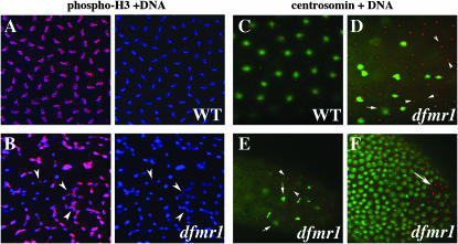
Mitotic asynchrony in early dfmr13 embryos. Wild-type and dfmr13 embryos were stained with DNA dye, Hoechst (imaged in blue), and phospho-histone H3 (pH 3) antibody (imaged in red). (A) A close-up of a wild-type syncytial blastoderm embryo showing synchronously dividing, evenly spaced nuclei with pH 3. (B, left and right) A close-up of a similar age syncytial blastoderm dfmr1 embryo showing unevenly spaced and asynchronously dividing nuclei. (Arrowheads in B) Asynchronously dividing or incompletely separated nuclei. Note that only a subset of the nuclei in the dfmr1 embryo are positive for pH 3. (C–F) Defective centrosomal assembly in dfmr13 embryos. (C) Wild-type and (D–F) dfmr13 embryos stained with centrosomin (red) antibodies and DNA dye, Hoechst (green). In the wild-type embryo in C, there appear to be two centrosomes associated with each nucleus. (D and E) A subset of nuclei appear to be undergoing normal mitosis; however, a number of abnormalities are also evident. These include “orphan” centrosome pairs that are not associated with DNA/nuclei (arrowheads in D and E). Some of the duplicated centrosomes remain in close proximity (arrows in D and E). Similar abnormalities are seen in the stage 5 embryo in F.

