Abstract
Recently, the possibility of using virus vectors to immunize cattle against selected bovine viral diarrhea virus (BVDV) genes has gained widespread interest. However, when we attempted to express the E2 protein from type 2 (890 strain) BVDV in a bovine herpesvirus 1 (BHV1) vector, we observed that expression was poor. This often happens when genes from a cytoplasmic virus are expressed in the cell nucleus. To counter this effect, we attempted to enhance expression by a strategy employed by viruses. RNAs of retroviruses and hepadnaviruses contain cis-acting elements that facilitate expression of RNAs that otherwise are degraded or retained within the nucleus. In Mason-Pfizer monkey virus, the required RNA sequence element is known as a constitutive transport element (CTE). A related element from woodchuck hepatitis virus is known as the woodchuck posttranscriptional regulatory element (WPRE). We tested the ability of the CTE, the WPRE, and introns to enhance expression of E2. All three elements stimulated expression of E2 from plasmids. The combination of the WPRE and an intron yielded the highest level of E2 expression in plasmids. However, when E2 was expressed from a BHV1 vector, the presence of an intron was inhibitory. In contrast, the WPRE was very efficient at stimulating E2 expression from a BHV1 vector. This result represents the first expression of a type 2 BVDV E2 protein from a mammalian virus vector and raises the possibility that the WPRE may provide a general method of enhancing foreign gene expression from BHV1 and other herpesvirus vectors.
Bovine viral diarrhea virus (BVDV) is a ubiquitous pathogen of cattle with a worldwide distribution. It is classified as a pestivirus within the Flaviviridae family (25) and is divided into genotypes 1 and 2 (39, 46). In addition, 11 BVDV subtypes of genotype 1 (3, 24, 54, 55) and 2 subtypes of genotype 2 have been described (5).
The majority of BVDV infections in susceptible, immunocompetent cattle result in clinically inapparent or mild disease. Beginning in 1989, however, reports of hypervirulent strains of BVDV capable of inducing severe disease during uncomplicated acute infection of immunocompetent cattle began to appear (9, 14, 39, 43, 46). The new BVDV strains could induce thrombocytopenia in infected animals, resulting in a hemorrhagic syndrome characterized by high mortality rates. To date, BVDV strains isolated from hypervirulent outbreaks belong primarily to genotype 2 (24). Although vaccination programs employing either inactivated or modified live vaccines are used extensively to protect cattle against the consequences of infection (8), these vaccines are not considered optimal for controlling BVDV infection (52).
Recently, the possibility of using recombinant virus vectors to immunize cattle against selected BVDV genes has gained widespread interest (4, 16-18, 23, 27, 44, 50). Among the virus vectors tested, bovine herpesvirus 1 (BHV1) has arguably the best potential for use as an expression vector for genes from BVDV and other bovine pathogens (see Discussion). However, BHV1 is a nuclear virus and its use as a vector for genes from cytoplasmic viruses has seldom been successful. For example, the vesicular stomatitis virus G gene is expressed poorly, and expression of the bovine respiratory syncytial virus G and F genes and the bovine parainfluenza virus 3 HN and F genes is undetectable in BHV1 vectors (W. C. Lawrence, J. C. Whitbeck, and L. J. Bello, unpublished data). The reason is not always clear but may involve features of the foreign gene transcript that render it nonfunctional when expressed in the nucleus. Possible mRNA problems include RNA instability, the presence of cryptic splice sites and, for intronless transcripts, the absence of signals for nucleocytoplasmic transport. One strategy used to cope with this problem in the past has been to chemically synthesize a new version of the gene in which the defect is relieved if not eliminated. This strategy was used successfully to express the G protein of bovine respiratory syncytial virus (BRSV) and the E2 protein of type 1 BVDV (C86 strain) in BHV1 recombinants (26, 50). With respect to the BVDV E2 protein, however, it should be noted that the unmodified, genomic form of the E2 open reading frame (ORF) from type 1 (NADL strain) BVDV has been successfully expressed in a BHV1 vector (27). Our laboratory has recently confirmed this observation (56).
In the study described in this paper we attempted to express the genomic form of the E2 ORF from the 890 strain of BVDV in a BHV1 vector and observed that expression was poor compared to expression of the NADL strain E2 ORF. The 890 strain, in contrast to the NADL strain, is a type 2 BVDV (39, 46). Since the specific mRNA defect responsible for poor expression of the 890 E2 ORF was not known, we reasoned that construction of a synthetic ORF capable of improved E2 expression could not be carried out in a rational way. As an alternative, we attempted to enhance expression by using a strategy employed by some viruses. The RNAs of viruses, such as retroviruses and hepadnaviruses, contain cis-acting elements that stabilize and facilitate the expression of RNAs that otherwise would be degraded or retained within the nucleus. In simple retroviruses, such as Mason-Pfizer monkey virus (M-PMV), the required RNA sequence element is known as a constitutive transport element (CTE) (11, 20). A related element from the woodchuck hepatitis virus, a hepadnavirus, is known as the woodchuck posttranscriptional regulatory element (WPRE) (15).
We have tested the ability of the CTE and the WPRE to enhance synthesis of the 890 strain E2 protein. The initial tests were carried out in plasmids and indicated that although both elements could enhance E2 synthesis, the WPRE was more effective. We then tested the effect of the WPRE on E2 synthesis in a BHV1 recombinant containing a cloned copy of the E2 ORF. We found the WPRE to be as effective in stimulating E2 synthesis from a BHV1 recombinant as it was in plasmids. In the presence of the WPRE, the 890 E2 ORF was expressed from a BHV1 vector as well as the NADL ORF. We also tested the effect of an intron on E2 synthesis but found that in BHV1 recombinants it resulted in a decrease in E2 synthesis.
In addition to representing the first expression of a type 2 BVDV E2 ORF in a mammalian virus vector, our studies raise the possibility that the WPRE may provide a generic solution to the problem of expressing foreign genes in BHV1 vectors.
MATERIALS AND METHODS
Virus and cell culture.
The Colorado 1 strain of BHV1 was obtained from the American Type Culture Collection. The 890 strain of BVDV was a gift of Christopher C. L. Chase. Viruses were propagated in Madin-Darby bovine kidney (MDBK) cells as previously described (28), except that 10% horse serum was used in place of calf serum. MDBK cells and bovine turbinate (BT) cells were obtained from the American Type Culture Collection.
Plasmid constructions.
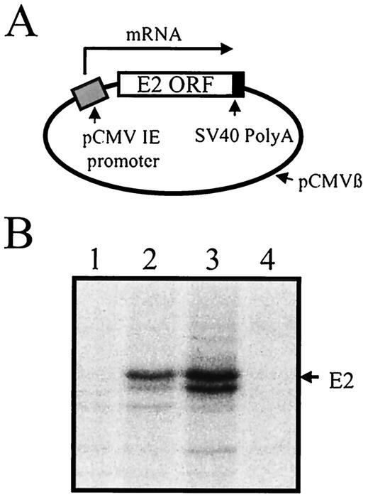
To construct pEC106, we used a cDNA clone of the E2 ORF from the type 2, 890 strain of BVDV (9). The clone contains nucleotides 2372 to 3457 of the BVDV (890) sequence from GenBank accession no. U18059 and was previously amplified from BVDV RNA by reverse transcription-PCR and inserted into pVL1393 (Invitrogen) to form a plasmid designated 159-1. The cloned BVDV region encodes the signal sequence and extracellular domain of the E2 protein but not the transmembrane anchor region. To provide an anchor, we joined the above 1,086-bp sequence, in frame, to a 177-bp fragment from pBVDV1 (56). The latter fragment contains nucleotides 3464 to 3640 of the BVDV (NADL) sequence from GenBank accession no. M31182 encoding the anchor region of E2. Upstream of the 1,263-bp E2 fusion sequence we inserted the sequence GTCGACtctagaggatccaccATG by PCR. This provided an ATG start codon as well as an upstream SalI site (shown in uppercase letters). Downstream of the E2 sequence we inserted the sequence ctcTAGaGCGGCCGC. This provided a TAG stop codon and a downstream NotI site. The 1.3-kb SalI-NotI E2 sequence was digested with SalI and NotI and cloned into XhoI-NotI-digested pCMVβ (Clontech). In the resulting construct, designated pEC106, expression of the E2 ORF is regulated by the cytomegalovirus (CMV) immediate-early (IE) promoter and simian virus 40 (SV40) late poly(A) signal present in pCMVβ (Fig. 1A). It should be noted that digestion with XhoI and NotI removed the intron and β-galactosidase gene normally present in pCMVβ.
FIG. 1.
Immunoprecipitation and SDS-PAGE analysis of E2 protein from cells transfected with pEC106 or infected with BVDV. BT cells were transfected with 2 μg of pEC106, and MDBK cells were infected with BVDV at 1 TCID50/cell. Control, transfected, and infected cells were labeled with [35S]methionine-cysteine for 4 h beginning 16 h posttransfection or infection. Cell extracts were prepared and analyzed by immunoprecipitation with an E2-specific monoclonal antibody and then by SDS-PAGE and autoradiography of dried gels. (A) Structure of pEC106. (B) 35S-labeled proteins from BT control cells (lane 1), pEC106-transfected cells (lane 2), BVDV-infected cells (lane 3), and MDBK control cells (lane 4).
To generate the CTE used to construct pEC89, a 292-bp fragment from a molecular clone of the M-PMV, kindly provided by Michael Malim, was amplified by PCR using primers that generated NotI sites at both ends of the CTE PCR product. The amplified fragment contains nucleotides 8003 to 8294 of the M-PMV sequence from GenBank accession no. M12349 and includes the region (nucleotides 8022 to 8175) that encodes the CTE of M-PMV (20). The PCR product was digested with NotI and cloned into the NotI site of pEC106 to generate pEC89.
To construct pEC107, the 592-bp woodchuck hepatitis virus WPRE element (15) was excised from pWPRE B11 by digesting with HindIII and HincII. The plasmid, kindly provided by Thomas J. Hope, contained woodchuck hepatitis virus nucleotides 1093 to 1684 from GenBank accession no. J04514 cloned into the ClaI site of pBluescript II SK(+) (Stratagene). The isolated fragment was converted to blunt ends by filling (Klenow) and ligated to NotI linkers. The fragment was then digested with NotI and cloned into the NotI site of pEC106 to generate pEC107.
To construct pEC121, the 1.3-kb E2 SalI-NotI fragment described above in the construction of pEC106 was used. The SalI site of the fragment was blunted by filling (Klenow) and ligated to a NotI linker. The fragment was then digested with NotI and cloned into NotI-cut pCMVβ to generate pEC121. Note that pEC121 is similar to pEC106 except that the intron of pCMVβ remains.
To construct pEC122, the 1.1-kb EcoRI-KpnI fragment from pEC121 was ligated to the 4.5-kb KpnI-EcoRI fragment of pEC107. Thus, pEC122 contains the intron from pEC121 and the WPRE from pEC107.
Construction of v40A and v40B.
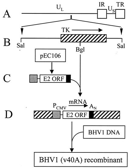
To express the E2 ORF in a BHV1 recombinant, it was first necessary to transfer the expression cassette consisting of the CMV promoter, the E2 ORF, and the SV40 poly(A) signal from pEC106 to a BHV1 insertion vector. To this end, the 2.2-kb EcoRI-SalI fragment containing the expression cassette from pEC106 was isolated, converted to blunt ends by filling, and cloned into the unique BglII site (blunted) of pBH95X (6) (Fig. 2). Plasmid pBH95X is a pUC18 plasmid containing the 2.7-kb SalI BHV1 fragment that encodes the BHV1 tk gene (Fig. 2B). Because insertion of the expression cassette into pBH95X was by blunt-end ligation, constructs containing the expression cassette in both possible orientations relative to the tk gene were isolated. In construct pIV40A the expression cassette is in the same orientation as tk, and in pIV40B the orientation is opposite to tk. To construct a BHV1 recombinant containing the E2 expression cassette within the tk gene, BT cells were cotransfected with mixtures of infectious BHV1 DNA and either pIV40A or pIV40B. Procedures for preparation of infectious DNA and cotransfection of cells have been described in detail elsewhere (7, 28, 33). Recombinant viruses containing the cassette were isolated by selecting large plaques in the presence of 200 μg of 5-bromo-2-deoxyuridine using the TK-negative MDBK(BU100) cell line previously described (7). To confirm the presence of an E2 expression cassette in the selected viruses, extracts were prepared from cells infected with each potential recombinant. DNA from the extracts was then analyzed by PCR amplification of the region spanning the insertion site. Recombinant viruses with confirmed inserts were then subjected to three rounds of plaque purification. The plaque-purified BHV1 recombinant resulting from cotransfection with pIV40A (Fig. 2D) was designated v40A. Cotransfection with pIV40B yielded the BHV1 recombinant designated v40B.
FIG. 2.
Construction of the v40A BHV1 recombinant. The E2 expression cassette consisting of the CMV promoter, the E2 ORF, and the SV40 late poly(A) signal was excised from pEC106, cloned into the unique BglII site of the BHV1 tk gene (contained in pBH95X), and inserted into the BHV1 genome by homologous recombination with infectious BHV1 DNA (see Methods and Materials for details). (A) Organization of the BHV1 genome showing the unique long (UL) and short (US) regions and the internal (IR) and terminal (TR) repeat regions of the genome. (B) Expanded structure of the 2.7-kb BHV1 SalI fragment contained in pBH95X and the position of the unique BglII site within it. The location of the fragment within the BHV1 genome and the position and orientation of the tk gene are indicated. (C) E2 expression cassette excised from pEC106. (D) Position and orientation of the E2 expression cassette within the tk gene of the pIV40A insertion vector. The orientation is the same in the BHV1 v40A recombinant.
Construction of v41B, v46B, and v47B.
Construction of the v41B, v46B, and v47B BHV1 recombinants was carried out essentially as described above with minor variations. For v41B, the 2.8-kb EcoRI-SalI fragment containing the expression cassette from pEC107 was used. For v46B, the 2.4-kb EcoRI-SalI fragment containing the expression cassette from pEC121 was used. For v47B, the 3.0-kb EcoRI-SalI fragment containing the expression cassette from pEC122 was used. In each case the fragment was blunted and cloned into the BglII site (blunted) of pBH95X to form the desired insertion vector. The orientation of the expression cassette in v41B, v46B, and v47B in each case was in the antisense direction relative to the tk gene.
Preparation of 35S-labeled protein extracts from transfected cells.
BT cells, in 60 to 70% confluent 60-mm plates, were transfected with 2 μg of plasmid DNA using Lipofectamine Plus (Invitrogen) according to the manufacturer's instructions. Fifteen hours posttransfection, complete growth medium (Dulbecco's modified Eagle's medium, 10% fetal bovine serum) was replaced with serum-free, methionine-free, and cysteine-free medium, and the transfected cells were incubated for an additional hour at 37°C. At 16 h posttransfection, the medium was replaced with 1.5 ml of fresh methionine-free and cysteine-free Dulbecco's modified Eagle's medium containing 100 μCi of [35S]methionine-cysteine mix (NEN)/ml. Cells were incubated for 4 h at 37°C, and cell extracts were prepared as described previously (56). The concentration of protein in the cell extracts was determined with the DC protein assay kit (Bio-Rad).
Preparation of 35S-labeled protein extracts from infected cells.
Confluent cell monolayers in 60-mm plates were infected with wild-type BHV1 and the BHV1 recombinants at a multiplicity of infection of 5 PFU/cell or with BVDV at 1 50% tissue culture infective dose (TCID50)/cell. At 16 h postinfection, cells were labeled with 50 μCi of [35S]methionine-cysteine and cell extracts were prepared as described above for transfected cells.
Immunoprecipitations.
Labeled cell extracts (50 μl) were mixed with 1 μl of E2-specific monoclonal antibody, BZ-53 for BVDV (890) or D89 for BVDV (NADL), and mixed overnight at 4°C by rocking. Fifteen microliters of protein G beads (Sigma) was added, and mixing was continued for 1 h at 4°C. Immune complexes were collected and subjected to sodium dodecyl sulfate-polyacrylamide gel electrophoresis (SDS-PAGE) as described previously (56). Labeled proteins were visualized by autoradiography of dried gels (2).
Quantitation of E2 expression.
Relative radioactivity in E2 bands of dried gels was quantitated using a GS-525 Molecular Imager system (Bio-Rad).
RESULTS
Expression of the BVDV E2 ORF in a plasmid vector.
The BVDV genome is normally expressed in the cytoplasm of a cell. Therefore, before attempting to express the E2 ORF of type 2 BVDV (890 strain) in a BHV1 recombinant, we first determined whether expression of the genomic version of the E2 ORF could be achieved in the nucleus of a cell. To this end, a cDNA clone of the E2 ORF was ligated into a modified pCMVβ expression plasmid vector to form pEC106 (Fig. 1A). pCMVβ had been modified to eliminate the intron and β-galactosidase gene normally present in this vector. In pEC106 the expression of E2 is under the control of the CMV IE promoter and the SV40 late poly(A) signal.
pEC106 was transfected into BT cells, and the cells were labeled with a mixture of [35S]methionine-cysteine. As a control, MDBK cells were infected with BVDV (890) and similarly labeled with [35S]methionine-cysteine. Labeled protein from the transfected and BVDV-infected cells was immunoprecipitated using a monoclonal antibody specific for the E2 protein, and proteins were separated on an SDS-polyacrylamide gel (Fig. 1B). A prominent band with an apparent molecular mass of 62 kDa was present in the transfected cell lane (lane 2). The band corresponded in position to that of E2 in the infected cell lane (lane 3), indicating that the E2 ORF had been successfully expressed in transfected cells.
Insertion of the E2 ORF into the BHV1 genome.
The above results indicated that the genomic version of the E2 ORF could be expressed from a plasmid vector. It seemed likely, therefore, that the same ORF could be expressed in a BHV1 recombinant (Fig. 2). To confirm this, the E2 expression cassette consisting of the CMV promoter, the E2 ORF, and the SV40 poly(A) signal was excised from pEC106 and cloned into the BglII site of plasmid pBH95X (6). The latter plasmid contains a 2.7-kb BHV1 SalI fragment containing the entire BHV1 tk gene (Fig. 2B). The BglII site of pBH95X lies within the tk gene. The resultant plasmid, designated pIV40A, served as an insertion vector to introduce the E2 expression cassette into the tk locus of BHV1 by homologous recombination (Fig. 2D). To this end, BT cells were cotransfected with a mixture of infectious BHV1 DNA and pIV40A DNA and a BHV1 recombinant virus, designated v40A, was isolated.
E2 synthesis by v40A.
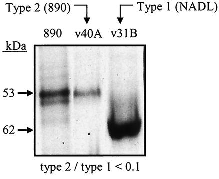
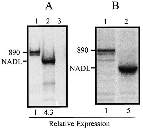
To evaluate E2 expression by the v40A recombinant, BT cells were infected with the virus, labeled with a mixture of [35S]methionine-cysteine, and analyzed by immunoprecipitation and SDS-PAGE (Fig. 3). To assess the efficiency of expression, we compared E2 synthesis by v40A with the synthesis of E2 by a BHV1 recombinant (v31B) expressing the type 1 BVDV (NADL strain) E2 ORF. The latter recombinant was previously constructed in our laboratory (56) and, like v40A, contains an E2 expression cassette inserted into the tk locus of BHV1. However, in contrast to v40A, the E2 expression cassette in v31B is transcribed in an antisense orientation relative to tk. It can be seen in Fig. 3 that v40A-infected cells produce less than 10% as much E2 protein as v31B-infected cells. The fact that the E2 protein from the NADL and 890 strains of BVDV migrate at different rates in SDS-PAGE has been reported previously (46).
FIG. 3.
Comparison of E2 synthesis in cells infected with BHV1 recombinants v40A or v31B. BT cells were infected with v40A or v31B at a multiplicity of infection of 5 PFU/cell or with BVDV (890) at 1 TCID50/cell. Infected cells were labeled with [35S]methionine-cysteine for 4 h beginning at 16 h postinfection. Cell extracts were prepared and analyzed by immunoprecipitation with E2-specific monoclonal antibodies and then by SDS-PAGE and autoradiography of dried gels.
Effect of orientation.
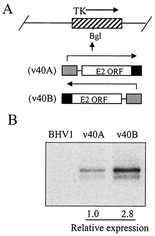
In view of the above results, we considered the possibility that the orientation of the E2 expression cassette relative to transcription of the tk gene might have an effect on E2 expression. To test this, we constructed a BHV1 recombinant similar to v40A but with the type 2 E2 expression cassette transcribed in the opposite direction. Cells were infected with the new recombinant, designated v40B, and E2 expression was evaluated. It can be seen that orientation did, in fact, affect expression (Fig. 4). Insertion of the E2 ORF into the tk locus in an antisense orientation resulted in almost 3 times more E2 expression than that obtained in the sense orientation. However, even in the optimal orientation used for the v40B recombinant, expression of the type 2 ORF was still significantly less than that of the type 1 homologue in v31B (Fig. 5A).
FIG. 4.
Effect of E2 orientation on expression. BHV1 recombinants containing the E2 expression cassette in opposite orientations relative to the tk gene were used to infect BT cells at a multiplicity of infection of 5 PFU/cell. Infected cells were labeled with [35S]methionine-cysteine, and labeled protein was analyzed as described in the legend for Fig. 3. (A) Orientation of the E2 cassette in v40A and v40B. (B) 35S-labeled proteins from cells infected with BHV1, v40A, and v40B.
FIG. 5.
Expression of the E2 ORF from type 1 and type 2 BVDV in BHV1 recombinants and plasmids. (A) BT cells were infected with BHV1 recombinants v40B containing the E2 ORF from type 2 (890) BVDV (lane 1) or v31B containing the E2 ORF from type 1 (NADL) BVDV (lane 2) or with wild-type BHV1 (lane 3). Infected cells were labeled with [35S]methionine-cysteine, and labeled protein was analyzed as described in the legend for Fig. 3. (B) BT cells were transfected with plasmids pEC106 containing the E2 ORF from type 2 (890) BVDV (lane1) or pEC55 containing the E2 ORF from type 1 (NADL) BVDV (lane 2). Transfected cells were labeled with [35S]methionine-cysteine, and labeled protein was analyzed as described in the legend for Fig. 1. The positions of the 890 and NADL E2 bands are indicated.
To determine whether the difference in type 1 versus type 2 E2 expression was specifically related to expression from a BHV1 vector, we compared expression of type 1 and type 2 E2 from E2 expression cassettes cloned in plasmids (Fig. 5B). Plasmids pEC106 (Fig. 1A) and pEC55 are similar constructs containing cassettes that express the type 2 and type 1 E2 ORFs, respectively. It can be seen that when expressed from a plasmid, relative expression of the type 2 E2 protein was no better than when expressed from a BHV1 vector. This suggests that the low level of type 2 E2 protein synthesis observed with the v40B recombinant is not specifically related to BHV1 expression but may reflect an inherent problem with nuclear expression of the E2 ORF.
Effect of cis-acting elements on E2 expression in plasmids.
We have used the limited expression of the type 2 E2 ORF as a way of evaluating possible methods of enhancing foreign gene expression in BHV1 recombinants. The methods tested all involved addition to the pre-mRNA transcript of cis-acting regulatory elements known to stabilize and promote expression of RNAs that otherwise are degraded or retained within the nucleus. The elements tested were an intron, the CTE from M-PMV (11, 20), and the WPRE from the woodchuck hepatitis virus (15). In each case the element was inserted into an untranslated region of the expressed mRNA. The CTE is an element that facilitates transport of unspliced, intron-containing mRNA to the cytoplasm. It, therefore, has the potential to minimize the effect of cryptic splice sites and promote nucleocytoplasmic transport of mRNA. The WPRE is a similar element that facilitates the transport of intronless RNAs, spliced RNAs, and unspliced RNAs containing an intron (15, 57). It, like the CTE, has the potential to minimize the effect of cryptic splice sites and promote nucleocytoplasmic transport of mRNA. Introns were tested because they are known to be required for the efficient processing and transport of many pre-mRNAs (for reviews, see references 32, 37, and 45). We initially avoided the use of introns in our BHV1 constructs because BHV1, like herpes simplex virus, is an α-herpesvirus and introns are known to interfere with pre-mRNA processing and/or transport in herpes simplex virus-infected cells (48).
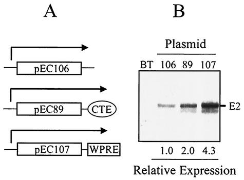
The effects of the CTE and WPRE were first tested in plasmid constructs (Fig. 6). In each case the element was inserted into the 3′-untranslated region of the expressed mRNA. Relative expression of the control construct (pEC106) lacking both a CTE and a WPRE was given a value of 1. The addition of a CTE (pEC89) increased E2 expression twofold. Addition of a WPRE (pEC107) increased expression about fourfold.
FIG. 6.
Effect of the CTE and WPRE on expression of E2 in plasmids. (A) Elements contained in plasmids pEC106, pEC89, and pEC107. (B) BT cells were transfected with plasmids pEC106, pEC89, or pEC107, and transfected cells were labeled with [35S]methionine-cysteine. Labeled protein was analyzed as described in the legend for Fig. 1.
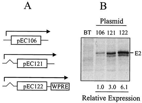
We then tested the effect of inserting an intron into the E2 plasmid construct, either alone or in combination with a WPRE (Fig. 7). The intron was placed in a position 5′ to the E2 ORF. Again, the control construct was given a value of 1. Addition of an intron to the construct increased E2 expression threefold (pEC121). The combination of a WPRE and an intron increased expression sixfold (pEC122).
FIG. 7.
Effect of an intron and the WPRE on E2 expression in plasmids. (A) Structure of tested plasmids. Plasmids pEC121 and pEC122 contain an intron upstream of the E2 ORF. (B) BT cells were transfected with the indicated plasmids, and transfected cells were labeled with [35S]methionine-cysteine. Labeled protein was analyzed as described in the legend for Fig. 1.
Effect of cis-acting elements on E2 expression in BHV1 recombinants.
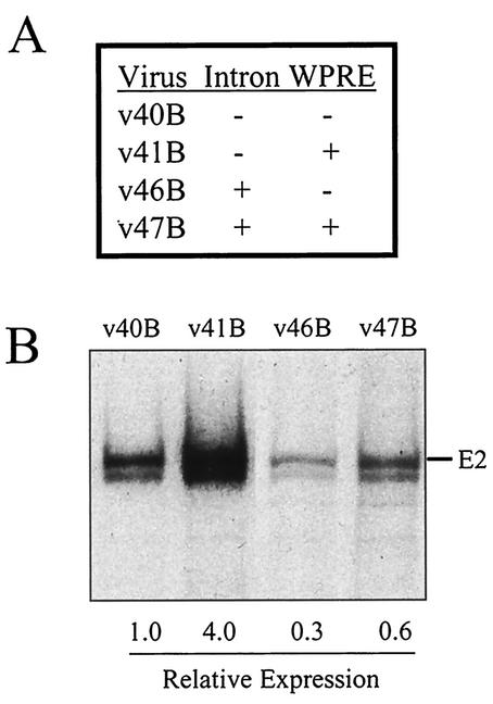
We next analyzed the effect of the WPRE and an intron on E2 expression in BHV1 recombinants. To this end, the expression cassettes from pEC107, pEC121, and pEC122 were excised from the plasmids and cloned into BHV1 as described previously for v40A (Fig. 2). The cassettes, in each case, were inserted into the tk locus of BHV1 in the antisense orientation used for v40B. We confirmed in each case that this was, in fact, the optimal orientation for the new constructs (data not shown). It can be seen in Fig. 8 that the WPRE was as effective in stimulating E2 expression in the BHV1 v41B recombinant as it was in the pEC107 plasmid construct. The intron, in contrast, inhibited expression in BHV1 recombinants whether present alone (v46B) or together with a WPRE (v47B).
FIG. 8.
Effect of an intron and the WPRE on E2 expression in BHV1 recombinants. (A) Elements contained in BHV1 recombinants v40B, v41B, v46B, and v47B. (B) BT cells were infected with the indicated BHV1 recombinants at a multiplicity of infection of 5 PFU/cell. Infected cells were labeled with [35S]methionine-cysteine, and labeled protein was analyzed as described in the legend for Fig. 3.
DISCUSSION
We have successfully expressed the E2 protein of type 2 BVDV (890 strain) from a BHV1 vector containing a cloned copy of the genomic form of the E2 ORF. This represents the first expression of a type 2 BVDV E2 protein from a mammalian virus vector. The E2 ORF was cloned into the thymidine kinase (tk) gene of BHV1 and was expressed under the control of an inserted CMV IE promoter. Best results were achieved when E2 mRNA was transcribed in an antisense orientation relative to transcription of the tk gene. It is possible that this phenomenon reflects the process known as promoter occlusion. The latter process involves transcriptional interference at a downstream promoter caused by transcription of an upstream gene through the promoter. This process has been described extensively in the past (1). However, even when the E2 ORF was in an optimal orientation, expression of the type 2 E2 protein was poor compared to expression of E2 protein from the type 1 BVDV (NADL strain) E2 ORF. By inserting the WPRE into the 3′-untranslated region of the transcribed E2 RNA, we were able to increase expression of the 890 E2 protein to a level comparable to that of the NADL E2.
The E2 ORFs of two type 1 BVDV strains (C86 and NADL) have previously been expressed in BHV1 vectors (27, 50). However, although antiserum directed against type 1 E2 protein neutralizes type 1 BVDV, it generally does not neutralize type 2 BVDV well (10, 13). The construction of a BHV1 recombinant expressing the 890 strain E2 protein, therefore, may provide a viable vaccine candidate for type 2 strains of BVDV.
BVDV represents one of the five viruses that make up the virus component of the bovine respiratory virus complex (BRDC). Despite the widespread use of antibiotics and vaccines, the BRDC continues to be a major problem in cattle production and dairy systems (19). The other viruses involved in this disease complex are BHV1, BRSV, bovine parainfluenza 3 virus (BPI3), and bovine coronavirus. Viral vectors that could safely and effectively be used to express immunodominant proteins from these viruses are actively being sought as vaccine vectors. In addition to BHV1, other viruses evaluated as potential vaccine vectors include adenoviruses (4, 12, 16-18, 34), vaccinia virus (47), and vesicular stomatitis virus (VSV) (23). For economic reasons, a multivalent vector capable of expressing proteins from two or more BRDC viruses simultaneously would be highly desirable. Adenoviruses are unlikely to serve as a multivalent vaccine because of limitations in the amount of foreign DNA that can be incorporated into their genome. Vaccinia virus and VSV have safety issues related to the fact that their host range includes humans. VSV also suffers from the fact that RNA viruses exhibit high rates of mutation. BHV1 does not suffer from any of the above problems and has many advantages as a potential bovine vaccine vector.
First, because cattle are routinely and safely immunized against BHV1, the use of BHV1 as a vector would not pose a new risk to cattle or humans and would obviate the need for an additional vaccination against BHV1 (51, 53). Second, the virus has a narrow host range that limits inappropriate spread to other species (36) and, in particular, to humans. Third, the virus is large and contains many nonessential genes that can be removed. For example, studies from our laboratory have demonstrated that a deletion mutant of BHV1 lacking ORF1, US2, the genes for protein kinase, glycoprotein G, glycoprotein I, and most of glycoprotein E is still replication competent (21). The latter mutant contains 7 kb less BHV1 DNA than wild-type virus. Thus, construction of a multivalent vaccine containing genes from multiple bovine pathogens is feasible and should not be limited by genome size constraints. In fact, since BHV1 is one of the viruses in the BRDC, its use as a vaccine vector immediately results in a multivalent vaccine. Fourth, BHV1 DNA is infectious, and new genes can be easily introduced into the viral genome by homologous recombination with plasmids containing the desired gene (6). Finally, the BHV1 genome has been completely sequenced and an entire library of BHV1 DNA fragments is available, allowing insertion of new genes at many different BHV1 sites.
Despite these advantages, the use of BHV1 as a vector for foreign genes has proven to be a challenge. This is especially true for genes derived from RNA viruses that replicate in the cytoplasm. The expression of the BVDV type 1 E2 gene (27, 50, 56) and, for that matter, even the limited expression of the type 2 E2 gene (in the absence of the WPRE) in a BHV1 recombinant is unusual. Most genes from RNA viruses that have been cloned into BHV1 have not been successfully expressed. Unfortunately, because negative results are usually not published, this fact is not evident from the literature. However, in our own personal studies we have found that copies of the BRSV G and F genes and the BPI3 HN and F genes are not expressed at detectable levels when cloned into BHV1 recombinants. In addition, the VSV G gene is expressed poorly (W. C. Lawrence, J. C. Whitbeck, and L. J. Bello, unpublished data). The important study of Kuhnle et al. (26) was the first published report that one of the above genes (BRSV G) could not be expressed in BHV1. Their report was publishable because they found that a chemically synthesized synthetic version of the G gene could be expressed. The problem of expressing genes from cytoplasmic viruses in BHV1 presumably reflects defects in the foreign gene transcript that only become apparent when the transcript is expressed in the nucleus of a cell. The two most likely problems anticipated in this case are cryptic splice sites that result in inappropriate splicing and the absence of regulatory signals for processing and/or nucleocytoplasmic transport of the mRNA. Unfortunately, the identification of these mRNA defects by examination of the gene sequence is not a simple matter. Sequence motifs for splice donor sites are imprecise and not always easy to recognize (35). Even when present, they do not result in inappropriate splicing unless a splice acceptor and branch point are present. The latter motifs are also not easy to recognize. Finally, defects in processing and nucleocytoplasmic transport, perhaps the most common problems anticipated with intronless mRNAs (29, 31, 32, 41, 45), cannot be predicted by sequence analysis.
For the above reasons we sought a method to enhance expression from BHV1 vectors that could be applied generally and without precise knowledge of the existing RNA defect. Accordingly, we tested the effect of the CTE from M-PMV (11, 20) and the WPRE from the woodchuck hepatitis virus (15) on E2 expression. These cis-acting elements were selected for evaluation because they are known to promote the nuclear export of unspliced mRNA. Therefore, they each have the potential to minimize mRNA problems associated with cryptic splice sites and the absence of regulatory signals for nucleocytoplasmic transport of mRNA.
When tested in plasmid constructs, we found that the WPRE was more effective than the CTE in stimulating E2 expression. Therefore, only the WPRE was studied further. It should be noted, however, that for other genes it is possible that a CTE might function better than the WPRE (49). In BHV1 recombinants the WPRE stimulated E2 expression fourfold. Use of the WPRE, combined with insertion of the E2 ORF in an optimal orientation within the tk locus, has allowed us to increase the overall level of E2 expression more than 10-fold compared with the initial v40A BHV1 recombinant.
As noted by others, the WPRE is a potentially important tool in situations where optimal gene expression is advantageous, since its stimulatory activity may be vector and transgene independent (40, 57). It is particularly useful in circumstances, such as with alphaherpesvirus vectors, where use of an intron may not be feasible (48). The WPRE has been studied extensively in various gene delivery systems (22, 30, 42, 49, 57). In its natural context, the WPRE is located within intronless RNAs. However, when placed in the 3′-untranslated region of a test mRNA, it has been shown to stimulate the expression of spliced, unspliced, and intronless RNAs (15, 57). Although the mechanism of action of the WPRE remains unclear, it has been demonstrated to increase the level of the affected nuclear and cytoplasmic transcripts significantly. The fact that the nucleocytoplasmic ratio of the affected RNAs is not altered (57) appears to indicate, at first sight, that the WPRE does not have a nuclear export role. However, a recent study suggests that the WPRE does, in fact, have an export function that is mediated, in part, by the CRM1-dependent export pathway (40).
Nuclear run-on experiments indicate that the WPRE-induced increase in nuclear RNA is generated by a posttranscriptional mechanism, suggesting that one function of the WPRE may be to increase the efficiency of RNA processing. The increase in RNA levels roughly correlates with the increase in protein expression elicited by the presence of the WPRE (57). The latter result suggests that the translation efficiency of the RNA has not been altered. If this interpretation is correct, the effect of the WPRE differs significantly from that of introns and the homologous (PRE) cis element of the closely related human hepatitis B virus. The latter elements have been reported to greatly enhance the translational yield of the affected mRNAs (29, 31, 38). In conclusion, the present evidence is most consistent with the view that the WPRE exerts its effect on protein expression by enhancing mRNA processing and nucleocytoplasmic transport.
In this study we have shown that the WPRE is clearly beneficial for expressing E2 from a BHV1 vector. This represents the first demonstration that the WPRE is functional in BHV1 and is capable of enhancing the expression of a foreign gene in a BHV1 recombinant. Although the E2 protein was expressed to a significant extent in BHV1, even in the absence of the WPRE, it is likely that a BHV1 recombinant containing the WPRE would elicit a higher E2 antibody response in a vaccinated animal than one lacking the WPRE. However, the use of the WPRE in BHV1 recombinants assumes its greatest importance with respect to foreign genes that are normally expressed at undetectable levels in BHV1 (26). The WPRE has the potential to provide a mechanism for their expression and, in so doing, to furnish a generic solution to the problem of expressing these genes from BHV1 vectors. If this potential is realized, the ability of BHV1, and perhaps other α-herpesviruses, to act as vaccine vectors will be greatly enhanced.
Acknowledgments
Financial support was provided by Commonwealth of Pennsylvania, Department of Agriculture grant ME 440689.
The D89 monoclonal antibody was a gift of Harish C. Minocha. The M-PMV molecular clone was a gift of Michael Malim. The pWPRE B11 plasmid was a gift of Thomas J. Hope.
REFERENCES
- 1.Adhya, S., and M. Gottesman. 1982. Promoter occlusion: transcription through a promoter may inhibit its activity. Cell 29:939-944. [DOI] [PubMed] [Google Scholar]
- 2.Austin, G. E., L. J. Bello, and J. J. Furth. 1973. DNA dependent RNA polymerase of KB cells. I. Isolation of the enzymes and transcription of viral DNA, mammalian DNA and chromatin. Biochim. Biophys. Acta 324:488-500. [DOI] [PubMed] [Google Scholar]
- 3.Baule, C., M. van Vuuren, J. P. Lowings, and S. Belak. 1997. Genetic heterogeneity of bovine viral diarrhoea viruses isolated in Southern Africa. Virus Res. 52:205-220. [DOI] [PubMed] [Google Scholar]
- 4.Baxi, M. K., D. Deregt, J. Robertson, L. A. Babiuk, T. Schlapp, and S. K. Tikoo. 2000. Recombinant bovine adenovirus type 3 expressing bovine viral diarrhea virus glycoprotein E2 induces an immune response in cotton rats. Virology 278:234-243. [DOI] [PubMed] [Google Scholar]
- 5.Becher, P., M. Orlich, A. Kosmidou, M. Konig, M. Baroth, and H. J. Thiel. 1999. Genetic diversity of pestiviruses: identification of novel groups and implications for classification. Virology 262:64-71. [DOI] [PubMed] [Google Scholar]
- 6.Bello, L. J., J. C. Whitbeck, and W. C. Lawrence. 1992. Bovine herpesvirus 1 as a live virus vector for expression of foreign genes. Virology 190:666-673. [DOI] [PubMed] [Google Scholar]
- 7.Bello, L. J., J. C. Whitbeck, and W. C. Lawrence. 1987. Map location of the thymidine kinase gene of bovine herpesvirus 1. J. Virol. 61:4023-4025. [DOI] [PMC free article] [PubMed] [Google Scholar]
- 8.Bolin, S. R. 1995. Control of bovine viral diarrhea infection by use of vaccination. Vet. Clin. N. Am. Food Anim. Pract. 11:615-625. [DOI] [PubMed] [Google Scholar]
- 9.Bolin, S. R., and J. F. Ridpath. 1992. Differences in virulence between two noncytopathic bovine viral diarrhea viruses in calves. Am. J. Vet. Res. 53:2157-2163. [PubMed] [Google Scholar]
- 10.Bolin, S. R., and J. F. Ridpath. 1996. Glycoprotein E2 of bovine viral diarrhea virus expressed in insect cells provides calves limited protection from systemic infection and disease. Arch. Virol. 141:1463-1477. [DOI] [PubMed] [Google Scholar]
- 11.Bray, M., S. Prasad, J. W. Dubay, E. Hunter, K. T. Jeang, D. Rekosh, and M. L. Hammarskjold. 1994. A small element from the Mason-Pfizer monkey virus genome makes human immunodeficiency virus type 1 expression and replication Rev-independent. Proc. Natl. Acad. Sci. USA 91:1256-1260. [DOI] [PMC free article] [PubMed] [Google Scholar]
- 12.Breker-Klassen, M. M., D. Yoo, S. K. Mittal, S. D. Sorden, D. M. Haines, and L. A. Babiuk. 1995. Recombinant type 5 adenoviruses expressing bovine parainfluenza virus type 3 glycoproteins protect Sigmodon hispidus cotton rats from bovine parainfluenza virus type 3 infection. J. Virol. 69:4308-4315. [DOI] [PMC free article] [PubMed] [Google Scholar]
- 13.Bruschke, C. J., R. J. Moormann, J. T. van Oirschot, and P. A. van Rijn. 1997. A subunit vaccine based on glycoprotein E2 of bovine virus diarrhea virus induces fetal protection in sheep against homologous challenge. Vaccine 15:1940-1945. [DOI] [PubMed] [Google Scholar]
- 14.Corapi, W. V., R. D. Elliott, T. W. French, D. G. Arthur, D. M. Bezek, and E. J. Dubovi. 1990. Thrombocytopenia and hemorrhages in veal calves infected with bovine viral diarrhea virus. J. Am. Vet. Med. Assoc. 196:590-596. [PubMed] [Google Scholar]
- 15.Donello, J. E., J. E. Loeb, and T. J. Hope. 1998. Woodchuck hepatitis virus contains a tripartite posttranscriptional regulatory element. J. Virol. 72:5085-5092. [DOI] [PMC free article] [PubMed] [Google Scholar]
- 16.Elahi, S. M., S. H. Shen, S. Harpin, B. G. Talbot, and Y. Elazhary. 1999. Investigation of the immunological properties of the bovine viral diarrhea virus protein NS3 expressed by an adenovirus vector in mice. Arch. Virol. 144:1057-1070. [DOI] [PubMed] [Google Scholar]
- 17.Elahi, S. M., S. H. Shen, B. G. Talbot, B. Massie, S. Harpin, and Y. Elazhary. 1999. Induction of humoral and cellular immune responses against the nucleocapsid of bovine viral diarrhea virus by an adenovirus vector with an inducible promoter. Virology 261:1-7. [DOI] [PubMed] [Google Scholar]
- 18.Elahi, S. M., S. H. Shen, B. G. Talbot, B. Massie, S. Harpin, and Y. Elazhary. 1999. Recombinant adenoviruses expressing the E2 protein of bovine viral diarrhea virus induce humoral and cellular immune responses. FEMS Microbiol. Lett. 177:159-166. [DOI] [PubMed] [Google Scholar]
- 19.Ellis, J. A. 2001. The immunology of the bovine respiratory disease complex. Vet. Clin. N. Am. Food Anim. Pract. 17:535-550, vi-vii. [DOI] [PMC free article] [PubMed]
- 20.Ernst, R. K., M. Bray, D. Rekosh, and M. L. Hammarskjold. 1997. A structured retroviral RNA element that mediates nucleocytoplasmic export of intron-containing RNA. Mol. Cell. Biol. 17:135-144. [DOI] [PMC free article] [PubMed] [Google Scholar]
- 21.Furth, J. J., J. C. Whitbeck, W. C. Lawrence, and L. J. Bello. 1997. Construction of a viable BHV1 mutant lacking most of the short unique region. Arch. Virol. 142:2373-2387. [DOI] [PubMed] [Google Scholar]
- 22.Glover, C. P., A. S. Bienemann, D. J. Heywood, A. S. Cosgrave, and J. B. Uney. 2002. Adenoviral-mediated, high-level, cell-specific transgene expression: a SYN1-WPRE cassette mediates increased transgene expression with no loss of neuron specificity. Mol. Ther. 5:509-516. [DOI] [PubMed] [Google Scholar]
- 23.Grigera, P. R., M. P. Marzocca, A. V. Capozzo, L. Buonocore, R. O. Donis, and J. K. Rose. 2000. Presence of bovine viral diarrhea virus (BVDV) E2 glycoprotein in VSV recombinant particles and induction of neutralizing BVDV antibodies in mice. Virus Res. 69:3-15. [DOI] [PubMed] [Google Scholar]
- 24.Hamers, C., P. Dehan, B. Couvreur, C. Letellier, P. Kerkhofs, and P. P. Pastoret. 2001. Diversity among bovine pestiviruses. Vet. J. 161:112-122. [DOI] [PubMed] [Google Scholar]
- 25.Horzinek, M. C. 1991. Pestiviruses—taxonomic perspectives. Arch. Virol. Suppl. 3:1-5. [PubMed] [Google Scholar]
- 26.Kuhnle, G., A. Heinze, J. Schmitt, K. Giesow, G. Taylor, I. Morrison, F. A. Rijsewijk, J. T. van Oirschot, and G. M. Keil. 1998. The class II membrane glycoprotein G of bovine respiratory syncytial virus, expressed from a synthetic open reading frame, is incorporated into virions of recombinant bovine herpesvirus 1. J. Virol. 72:3804-3811. [DOI] [PMC free article] [PubMed] [Google Scholar]
- 27.Kweon, C. H., S. W. Kang, E. J. Choi, and Y. B. Kang. 1999. Bovine herpes virus expressing envelope protein (E2) of bovine viral diarrhea virus as a vaccine candidate. J. Vet. Med. Sci. 61:395-401. [DOI] [PubMed] [Google Scholar]
- 28.Lawrence, W. C., R. C. D'Urso, C. A. Kundel, J. C. Whitbeck, and L. J. Bello. 1986. Map location of the gene for a 130,000-dalton glycoprotein of bovine herpesvirus 1. J. Virol. 60:405-414. [DOI] [PMC free article] [PubMed] [Google Scholar]
- 29.Le Hir, H., A. Nott, and M. J. Moore. 2003. How introns influence and enhance eukaryotic gene expression. Trends Biochem. Sci. 28:215-220. [DOI] [PubMed] [Google Scholar]
- 30.Loeb, J. E., W. S. Cordier, M. E. Harris, M. D. Weitzman, and T. J. Hope. 1999. Enhanced expression of transgenes from adeno-associated virus vectors with the woodchuck hepatitis virus posttranscriptional regulatory element: implications for gene therapy. Hum. Gene Ther. 10:2295-2305. [DOI] [PubMed] [Google Scholar]
- 31.Lu, S., and B. R. Cullen. 2003. Analysis of the stimulatory effect of splicing on mRNA production and utilization in mammalian cells. RNA 9:618-630. [DOI] [PMC free article] [PubMed] [Google Scholar]
- 32.Luo, M. J., and R. Reed. 1999. Splicing is required for rapid and efficient mRNA export in metazoans. Proc. Natl. Acad. Sci. USA 96:14937-14942. [DOI] [PMC free article] [PubMed] [Google Scholar]
- 33.Miller, J. M., C. A. Whetstone, L. J. Bello, and W. C. Lawrence. 1991. Determination of ability of a thymidine kinase-negative deletion mutant of bovine herpesvirus-1 to cause abortion in cattle. Am. J. Vet. Res. 52:1038-1043. [PubMed] [Google Scholar]
- 34.Mittal, S. K., S. K. Tikoo, J. V. van den Hurk, M. M. Breker-Klassen, D. Yoo, and L. A. Babiuk. 1998. Functional characterization of bovine parainfluenza virus type 3 hemagglutinin-neuraminidase and fusion proteins expressed by adenovirus recombinants. Intervirology 41:253-260. [DOI] [PubMed] [Google Scholar]
- 35.Mount, S. M. 1982. A catalogue of splice junction sequences. Nucleic Acids Res. 10:459-472. [DOI] [PMC free article] [PubMed] [Google Scholar]
- 36.Murphy, F. A., E. P. Gibbs, M. C. Horzinek, and M. J. Studdert. 1999. Veterinary virology, 3rd ed., p. 301-325. Academic Press, New York, N.Y.
- 37.Nakielny, S., U. Fischer, W. M. Michael, and G. Dreyfuss. 1997. RNA transport. Annu. Rev. Neurosci. 20:269-301. [DOI] [PubMed] [Google Scholar]
- 38.Nott, A., S. H. Meislin, and M. J. Moore. 2003. A quantitative analysis of intron effects on mammalian gene expression. RNA 9:607-617. [DOI] [PMC free article] [PubMed] [Google Scholar]
- 39.Pellerin, C., J. van den Hurk, J. Lecomte, and P. Tussen. 1994. Identification of a new group of bovine viral diarrhea virus strains associated with severe outbreaks and high mortalities. Virology 203:260-268. [DOI] [PubMed] [Google Scholar]
- 40.Popa, I., M. E. Harris, J. E. Donello, and T. J. Hope. 2002. CRM1-dependent function of a cis-acting RNA export element. Mol. Cell. Biol. 22:2057-2067. [DOI] [PMC free article] [PubMed] [Google Scholar]
- 41.Proudfoot, N. J., A. Furger, and M. J. Dye. 2002. Integrating mRNA processing with transcription. Cell 108:501-512. [DOI] [PubMed] [Google Scholar]
- 42.Ramezani, A., T. S. Hawley, and R. G. Hawley. 2000. Lentiviral vectors for enhanced gene expression in human hematopoietic cells. Mol. Ther. 2:458-469. [DOI] [PubMed] [Google Scholar]
- 43.Rebhun, W. C., T. W. French, J. A. Perdrizet, E. J. Dubovi, S. G. Dill, and L. F. Karcher. 1989. Thrombocytopenia associated with acute bovine virus diarrhea infection in cattle. J. Vet. Intern. Med. 3:42-46. [DOI] [PubMed] [Google Scholar]
- 44.Reddy, J. R., J. Kwang, V. Varthakavi, K. F. Lechtenberg, and H. C. Minocha. 1999. Semliki forest virus vector carrying the bovine viral diarrhea virus NS3 (p80) cDNA induced immune responses in mice and expressed BVDV protein in mammalian cells. Comp. Immunol. Microbiol. Infect. Dis. 22:231-246. [DOI] [PubMed] [Google Scholar]
- 45.Reed, R., and E. Hurt. 2002. A conserved mRNA export machinery coupled to pre-mRNA splicing. Cell 108:523-531. [DOI] [PubMed] [Google Scholar]
- 46.Ridpath, J. F., S. R. Bolin, and E. J. Dubovi. 1994. Segregation of bovine viral diarrhea virus into genotypes. Virology 205:66-74. [DOI] [PubMed] [Google Scholar]
- 47.Sakai, Y., and H. Shibuta. 1989. Syncytium formation by recombinant vaccinia viruses carrying bovine parainfluenza 3 virus envelope protein genes. J. Virol. 63:3661-3668. [DOI] [PMC free article] [PubMed] [Google Scholar]
- 48.Sandri-Goldin, R. M. 2001. Nuclear export of herpes virus RNA. Curr. Top. Microbiol. Immunol. 259:2-23. [PubMed] [Google Scholar]
- 49.Schambach, A., H. Wodrich, M. Hildinger, J. Bohne, H. G. Krausslich, and C. Baum. 2000. Context dependence of different modules for posttranscriptional enhancement of gene expression from retroviral vectors. Mol. Ther. 2:435-445. [DOI] [PubMed] [Google Scholar]
- 50.Schmitt, J., P. Becher, H. J. Thiel, and G. M. Keil. 1999. Expression of bovine viral diarrhoea virus glycoprotein E2 by bovine herpesvirus-1 from a synthetic ORF and incorporation of E2 into recombinant virions. J. Gen. Virol. 80:2839-2848. [DOI] [PubMed] [Google Scholar]
- 51.van Drunen Littel-van den Hurk, S., S. K. Tikoo, X. Liang, and L. A. Babiuk. 1993. Bovine herpesvirus-1 vaccines. Immunol. Cell Biol. 71:405-420. [DOI] [PubMed] [Google Scholar]
- 52.van Oirschot, J. T., C. J. Bruschke, and P. A. van Rijn. 1999. Vaccination of cattle against bovine viral diarrhoea. Vet. Microbiol. 64:169-183. [DOI] [PubMed] [Google Scholar]
- 53.van Oirschot, J. T., M. J. Kaashoek, and F. A. Rijsewijk. 1996. Advances in the development and evaluation of bovine herpesvirus 1 vaccines. Vet. Microbiol. 53:43-54. [DOI] [PubMed] [Google Scholar]
- 54.van Rijn, P. A., H. G. van Gennip, C. H. Leendertse, C. J. Bruschke, D. J. Paton, R. J. Moormann, and J. T. van Oirschot. 1997. Subdivision of the pestivirus genus based on envelope glycoprotein E2. Virology 237:337-348. [DOI] [PubMed] [Google Scholar]
- 55.Vilcek, S., D. J. Paton, B. Durkovic, L. Strojny, G. Ibata, A. Moussa, A. Loitsch, W. Rossmanith, S. Vega, M. T. Scicluna, and V. Paifi. 2001. Bovine viral diarrhoea virus genotype 1 can be separated into at least eleven genetic groups. Arch. Virol. 146:99-115. [DOI] [PubMed] [Google Scholar]
- 56.Wang, L., J. C. Whitbeck, W. C. Lawrence, D. V. Volgin, and L. J. Bello. Expression of the genomic form of the bovine viral diarrhea virus E2 ORF in a bovine herpesvirus-1 vector. Virus Genes, in press. [DOI] [PubMed]
- 57.Zufferey, R., J. E. Donello, D. Trono, and T. J. Hope. 1999. Woodchuck hepatitis virus posttranscriptional regulatory element enhances expression of transgenes delivered by retroviral vectors. J. Virol. 73:2886-2892. [DOI] [PMC free article] [PubMed] [Google Scholar]


