Abstract
We previously developed a model for studies on hantavirus host adaptation and initiated genetic analysis of Puumala virus variants passaged in colonized bank voles and in cultured Vero E6 cells. With the data presented in this paper, the sequence comparison of the wild-type and Vero E6-adapted variants of Puumala virus, strain Kazan, has been completed. The only amino acid substitution that distinguished the two virus variants was found in the L protein, Ser versus Phe at position 2053. Another mutation found in the L segment, the silent transition C1053U, could result from the selection of a variant with altered L RNA folding. Nucleotide substitutions observed in individual L cDNA clones, most of them A→G and U→C transitions, suggested that the population of L RNA molecules is represented by quasispecies. The mutation frequency in the L segment quasispecies appeared to be similar to the corresponding values for the S and M quasispecies. Analysis of the cDNA clones with the complete S segment sequences from passage 20 confirmed our earlier conclusion that the cell-adapted genotype of the virus is represented mostly by variants with mutated S segment noncoding regions. However, the spectrum of the S segment quasispecies appeared to be changing, suggesting that, after the initial adaptation (passages 1 to 11), the viral population is still being driven by selection for variants with higher fitness.
The genus Hantavirus (family Bunyaviridae) includes more than 20 distinct virus types (10). Hantaviruses are considered prime examples of emerging pathogens, causing hemorrhagic fever with renal syndrome in Eurasia and hantavirus pulmonary syndrome in the Americas, and the number of newly discovered viruses is increasing rapidly (for recent reviews, see references 25 and 36). Hantaviruses are enveloped viruses with a segmented single-stranded RNA genome of negative polarity. The three genomic segments, small (S), medium (M), and large (L), encode the nucleocapsid protein, two surface glycoproteins, G1 and G2, and the RNA polymerase, respectively (10). The 5′ noncoding regions (NCRs) of the three RNA segments are relatively short (30 to 50 nucleotides) and so is the 3′ NCR of the L segment, while the 3′ NCRs of the M segment and, especially, of the S segment, are much longer (up to 700 nucleotides). The first 20 to 22 nucleotides from the 5′ and 3′ termini of every RNA segment are complementary to each other and highly conserved among the different hantavirus types. It is thought that these regions form specific “panhandle” structures involved in the regulation of viral transcription and replication. The functions of other NCRs await further investigation (33, 39).
Why only some hantaviruses are pathogenic for humans remains unclear. Among the factors that might determine or influence the pathogenicity of hantaviruses, the nature of their rodent hosts is considered one of the most important (33, 34). To gain more insight on molecular markers associated with the host specificity and potentially the virulence of hantaviruses, we invented a model based on adaptation of wild-type Puumala virus from its natural rodent host, the bank vole, to primate cells in culture. We showed that when the wild-type Puumala virus passaged in colonized bank voles was passaged serially in Vero E6 cells, its ability to infect bank voles decreased rapidly, i.e., the virus became adapted to primate cells (24).
Sequence analysis of the complete S and the M RNA segments of the wild-type and Vero E6 cell-adapted variants revealed that the adaptation to the new host cells was accompanied by the accumulation of point mutations in the NCRs of the S segment but not the M segment. Notably, S RNA molecules carrying mutations in the 5′ and 3′ NCRs of the S segment (positions 26 and 1577) were accumulating gradually in the genetic (quasispecies) spectrum of the Vero E6 cell-adapted variant, starting from passage 3. At passage 11, the last passage monitored in that study, the mutant S RNA molecules represented the majority of the population. Sequencing of the S segment of another cell-adapted variant, Vero E6-II, obtained in an independent adaptation experiment, revealed a mutation at position 1580, only three nucleotides downstream of the mutation found earlier in cell culture-adapted variants of Puumala virus (24).
Comparative sequence analyses performed in the above-mentioned study did not include the L segment encoding the viral polymerase-replicase. As shown for Hantaan virus (8) and other members of the Bunyaviridae (11, 15), the L segment and the L protein can carry mutations associated with host range restriction and attenuation. To complete the genetic comparison of the wild-type and cell culture-adapted variants of Puumala virus, we present here the results of sequence analysis of their L segments. In addition, we performed a follow-up analysis of the mutations in the 5′ and 3′ NCRs of the S segment found earlier.
MATERIALS AND METHODS
Passaging of virus in Vero E6 cell culture.
Puumala virus strain Kazan E6-I (passage 11) (24) was grown and passaged further on monolayer cultures of Vero E6 cells in Eagle's minimal essential medium supplemented with 2% fetal calf serum, 2 mM l-glutamine, and antibiotics as previously described (24). Virus stocks were kept at −70°C until used.
Sequence analysis of L segment.
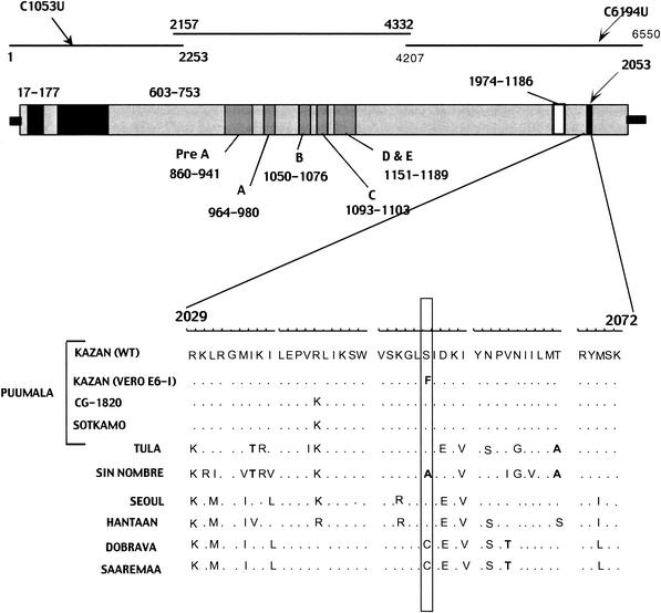
Total RNA was extracted from samples of lung or kidney tissue of infected colonized bank voles (wild-type Puumala virus strain Kazan) or from Puumala virus-infected Vero E6 cells (strain Kazan E6-I), approximately 2 × 10⁶ cells, and purified by acidic guanidine thiocyanate-phenol-chloroform extraction (5). RNA was dissolved in 30 μl of RNase-free water. Half of each RNA preparation was precipitated with ethanol, dissolved in 4 μl of water, and used for RNA ligations. The L segments of both the wild-type and Vero E6-I viruses from passage 11 variants were reverse transcribed and amplified in three overlapping parts of 2,253, 2,176, and 2,344 nucleotides (nucleotides 1 to 2253, 2157 to 4332, and 4207 to 6550, respectively) (Fig. 1).
FIG. 1.
Schematic structure of hantaviral L segment. Black arrows indicate the locations of the differences found between the wild-type (WT) and Vero E6-I variants in the nucleotide and amino acid sequences. The three black lines indicate three fragments of the L segment that were amplified and sequenced separately, and numbers mark the position of each fragment in the nucleotide sequence. The gray bar below represents the L protein and its conserved regions, with numbers showing the location of each region in the amino acid sequence. The black stripe in the C terminus shows the site where the Ser2053Phe mutation resides. Alignment of hantaviral L protein sequences for the region surrounding the amino acid replacement Ser2053Phe is shown below, and nonhomologous amino acid substitutions observed in this region are shown in bold. Hantaviral sequences used for the alignment and retrieved from the GenBank include Hantaan virus strain 76-118 (X55901) (38), Seoul virus strain 80-39 (X56492) (2), Sin Nombre virus strain NM H10 (L37901) (4), Puumala virus strains CG1820 (M63194) (40) and Sotkamo (Z66548) (30), Tula virus strain Tula/Moravia/5302v (AJ005637) (23), Dobrava virus strain Ano-Poroia/9Af/99 (AJ410617) (29), and Saaremaa virus strain Saaremaa/160V (AJ410618) (29).
The primers used for the amplification were chosen based on the L segment sequences of two known Puumala virus strains, Cg1820 (40) and Sotkamo (30): TAGTAGTAGACTCCGAGATAGAG and AGCTTCTTCAGTAAGATT(G/A)CCATG for the first fragment, GGTTGTCTATAAACATTATCGCAG and AGCAGCAAGAACCTCTCTAAATG for the second, and AGCATAGTCACTGCAATGACAATG and TAGTAGTATGCTCCGAGATAAGAG for the third fragment. The PCR products were separated in agarose gels and purified with the QIAquick kit (Qiagen GmbH). Cloning was performed with the pGEM-T cloning kit (Promega). Two clones for each fragment of the wild-type and Vero E6-I variants were subjected to automatic sequencing with the ABI Prism Dye Terminator sequencing kit (Perkin-Elmer Applied Biosystems Division PE/ABI, Foster City, Calif.). Samples were prepared according to the manufacturer's instructions, and reactions were run on an ABI 373 A sequencer (PE/ABI).
Sequencing of S segment.
Cells infected with the Puumala virus Kazan Vero E6-I variant were harvested at passage 20, and reverse transcription (RT)-PCR for the complete S segment was performed as described previously (31). The PCR products were cloned into the pGEM-T plasmid and sequenced automatically as described above.
RNA folding.
RNAstructure program version 3.71 was used for modeling of secondary RNA structures (26). The program is an implementation of the Zuker algorithm to predict RNA secondary structures from the sequence, based on the principle of minimizing free energy.
RESULTS
Comparison of L segment sequences.
Figure 1 shows the schematic structure of the hantavirus L protein. Two black boxes indicate regions identified in the N-terminal part of Bunyaviridae and Arenaviridae polymerases (28). The five motifs common to all RNA-dependent polymerases (pre-A, A, B, C, and D) and motif E, identified in the polymerases of all viruses with a segmented negative-stranded RNA genome (28), are shown as light gray boxes. The white box in the C-terminal part of the protein marks an “acidic” domain found in the sequences of tomato spotted wilt virus (genus Tospovirus) and hantaviruses (4, 23).
Initially, two cDNA clones were sequenced for each of the three overlapping parts of the L segment of the Puumala virus wild-type and Vero E6-I variants. Two mutations that distinguished the cDNA clones of the Vero E6-I variant from those of the wild-type virus were found, a silent transition, C1053U, and a transition, C6194U, that led to a Ser2053Phe substitution in the deduced L protein sequence. Both mutations were further confirmed by sequencing of a third cDNA clone for each of the two variants and independent RT-PCR for nucleotides 539 to 1140 and nucleotides 6112 to 6550, followed by direct sequencing. Thus, the master L segment sequences of the two Puumala virus variants differed at only two positions, and the deduced L protein sequences differed at one position only.
The substitution Ser2053Phe resides in the C-terminal part of the L protein, within a short, moderately conserved region spanning amino acid residues 2029 to 2072 and outside the highly conserved polymerase domains A to E (28). Most of the amino acid substitutions observed between different hantaviruses within this region are homologous, and only four nonhomologous substitutions occurred, at positions 2035 (in Tula virus and Sin Nombre virus), 2053 (in Sin Nombre virus), 2061 (in Dobrava virus and Saaremaa virus), and 2067 (in Tula virus and Sin Nombre virus). The amino acid mutation that accompanied the adaptation of wild-type Puumala virus Kazan to cell culture placed the nonpolar phenylalanine residue at position 2053, where all other hantaviruses except Sin Nombre virus have polar serine or cysteine residues. Since no differences in the sequences of the N protein and the G1-G2 proteins were observed earlier (24), the Ser2053Phe substitution in the L protein is the only amino acid substitution found after complete comparison of all proteins of the Puumala virus wild-type and Vero E6-I variants.
L RNA folding.
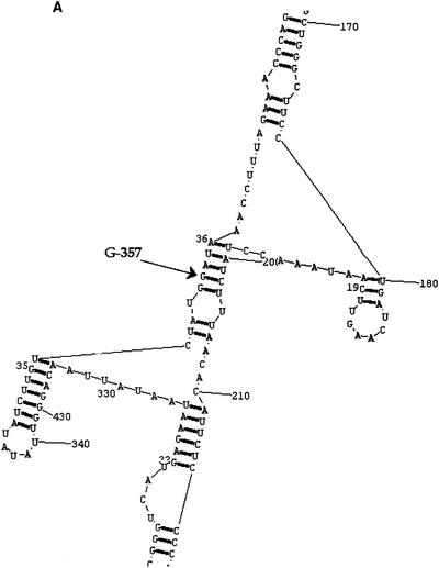
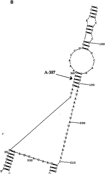
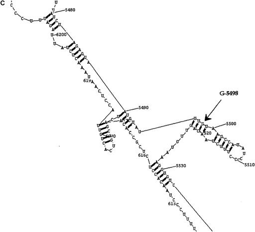
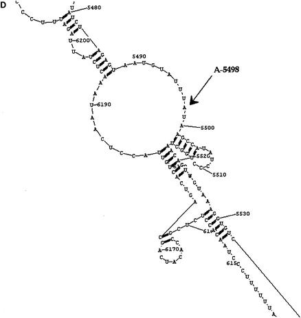
The overall modes of folding of L viral RNA (minus-sense) and cRNA (plus-sense) of the wild-type and Vero E6 cell-adapted Puumala virus variants were similar (data not shown). Most important, both substitutions found in the cell-adapted variant of the virus led to local changes in L viral RNA folding. The C1053U substitution observed in cDNA clones (G5498A, in viral RNA) causes transformation of a double helix region into a loop (Fig. 2C and D). The C6194U substitution (G357A, in viral RNA) induces changes in another small double helix, not disrupting the structure totally but weakening it (Fig. 2A and B). Together, these two events led to a lower level of free energy for the folding of the viral RNA of the cell-adapted variant, −1,263.2 kcal instead of −1,266.9 kcal for the wild-type variant.
FIG. 2.
Folding of L viral RNA of wild-type (A and C) and Vero E6 cell-adapted (B and D) variants of Puumala virus. The positions of the nucleotide differences between the wild-type and Vero E6-I variants are marked by arrows. Note that the C1053U and C6194U substitutions observed in cDNA clones correspond to substitutions G5498A and G357A in the L viral RNA, respectively.
In contrast, modeling of the L cRNA folding gave virtually identical secondary structures for the wild-type and cell-adapted variants of the virus (data not shown). The two nucleotides associated with adaptations are located in separate domains of the molecule, and the two substitutions found in the cell-adapted variant do not seem to disturb the local folding. In both cases, a canonical C:G pair in a double helix is changed to a noncanonical U:G pair; thus, the structure is weakened but preserved. Consequently, the free energy for the folding of the cell-adapted variant becomes lower, −1,532.3 kcal versus −1,536.4 kcal for the wild-type variant.
L segment quasispecies.
In addition to the mutations at position 1053 and 6194, 17 other mutations were observed in individual cDNA clones generated from the wild-type variant and 16 mutations in those generated from the Vero E6-I variant. All positions where the mutations occurred were rechecked with an additional cDNA clone for each variant. As none of the 33 mutations was found in more than one clone, they were considered not relative to the master sequences but instead representing L segment quasispecies, each occupying only a minor portion of the genetic swarm. Similar to what was observed for the S segment quasispecies, mutations in the L segment quasispecies were mostly A→G and U→C transitions (30% and 21%, respectively). The average frequency of nucleotide substitutions observed in the L segment quasispecies, 0.6 × 10⁻³, was comparable to those in the S segment and the M segment quasispecies found earlier (24). As the level of RT-PCR errors determined in the previous study was 0.2 × 10⁻³, some of the mutations in the L segment might have originated from nucleotide misincorporations during the RT or PCR steps. The majority, however, seem to represent “genuine mistakes” of the viral RNA-dependent RNA polymerase.
Follow-up of mutations found in L segment and S segment.
To follow up on the two nucleotide changes observed in the L segment during the adaptation, the corresponding regions of the L segment (nucleotides 6112 to 6550) were also recovered from passages 12 and 20 by RT-PCR and sequenced directly. Both substitutions found in the Vero E6-I variant on passage 11 (positions 1053 and 6194) were seen on the following passages, showing that these mutations are firmly associated with the Vero E6-I geno- and phenotype.
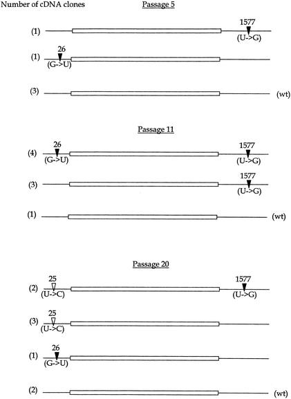
To follow up on the nucleotide changes at positions 26 and 1577 that were observed earlier in the 5′ and 3′ NCRs of the S segment of the Vero E6-I variant (24), cDNA clones containing complete S segment sequences were recovered from passage 20 and analyzed (Fig. 3). It was found that the mutant variants again, as for passage 11, represented a majority of the S segment sequences. However, the structure of the S segment quasispecies seemed to be different from that observed on passage 11. First, the G26U transversion that was previously found in 50% of cDNA clones from the cell-adapted variant (passage 11) now appeared in only one of eight cDNA clones. Instead, a new nucleotide replacement, a U25C transition, was found in five of eight cDNA clones analyzed. Second, only two of the eight cDNA clones carried the U1577G transversion found in the majority of clones (seven of nine) at passage 11. Notably, both cDNA clones with the U1577G mutation carried the U25C transition as well. The likely reasons for the observed differences are discussed below.
FIG. 3.
Mutant spectra of S segment sequences of the Vero E6-I variant. The numbers in parentheses indicate the number of clones with a particular sequence. Solid arrowheads mark mutations in the NCRs identified earlier (24). Open arrowheads mark mutations described in this study. wt, wild type.
DISCUSSION
Adaptation of wild-type viruses to cells in culture usually reduces their ability to infect their natural hosts, animal or human. This phenomenon, attenuation, has been widely used to develop live vaccines as well as to study virus transmissibility and the pathogenesis of different viruses, e.g., rabies virus (3), respiratory syncytial virus (6), Japanese encephalitis virus (9), hepatitis A virus (1), Sendai virus (18), Theiler's virus (19), Sindbis virus (20), measles virus (41), and rotaviruses (42). Sequence comparisons of the wild-type and cell-adapted variants can provide useful markers associated with virus pathogenicity and host restriction.
We have previously developed a model for studies on hantavirus host adaptation and initiated genetic analysis of Puumala virus variants passaged in colonized bank voles and in cultured Vero E6 cells (24). With the data presented in this paper, the sequence comparison of the wild-type and Vero E6-adapted variants of Puumala virus strain Kazan has been completed. No mutations were found in the coding regions of the S and M segments, suggesting that the amino acid sequences of the nucleocapsid protein and surface glycoproteins G1 and G2 were not changed during the adaptation process. The only amino acid substitution that distinguished the two virus variants was found in the L protein, Ser versus Phe at position 2053. Not surprisingly, it was located outside the highly conserved motifs shared by RNA-dependent RNA polymerases of different origins (28, 37). An intriguing feature of this substitution is that the nonpolar phenylalanine appeared at a position where all other known hantaviruses except Sin Nombre virus have polar serines or cysteines (Fig. 1).
Another mutation found in the L segment, the silent transition C1053U, could result from the selection of a variant with altered (and more favorable for Vero E6 cell culture) L RNA folding. Random genetic drift at this position cannot be totally excluded. Notably, both mutations were found at passage 20 as well and thus were firmly associated with the Vero E6-I variant of the virus and considered advantageous for its survival in cell culture. They could lead to local changes in the L viral RNA folding (Fig. 2), increasing the efficiency of transcription and/or replication. The amino acid substitution in the L protein could contribute to more efficient replication in cell culture as well, e.g., by supporting a conformation which works better on the mutated RNA templates and/or in concert with some as yet unknown cellular factor(s).
Our findings of mutations in the polymerase-encoding gene during the cell culture adaptation of Puumala virus are in line with data accumulated earlier for other bunyaviruses (11, 15, 43). As for hantaviruses, it was recently shown that one mutation in the L segment contributes to a better replication of Hantaan virus in suckling mice and thus increases its virulence (8). The authors, however, suggested that a single amino acid substitution observed in the G1 protein was primarily responsible for the altered phenotype of the virus, while the mutation in the L segment was considered less crucial.
The nucleotide substitutions that we observed in individual L cDNA clones, most of them A→G and U→C transitions, suggested that the population of L RNA molecules is represented by quasispecies. Although calculated from a limited number of nucleotides sequenced (approximately 27,000 altogether), the mutation frequency in the L segment quasispecies appeared to be similar to the corresponding values for the S and M quasispecies (and well above the level of RT-PCR errors) (24). Such a similarity confirms their common origin, base misincorporation occurring during virus replication. This is the mechanism known to operate in RNA viruses in general (7, 17) and in hantaviruses in particular (13, 32).
It should be noted that the very terminal sequences of all three genome segments of the wild-type and Vero E6 cell-adapted variants, which were used as annealing sites for primers in reverse transcription and PCR amplification, could not be compared in the same way as the rest of the Puumala virus genome. We therefore performed this comparison by RNA ligation followed by RT-PCR through the ligation point, an approach exploited earlier to recover terminal sequences of other hantaviruses (4, 23). Not surprisingly, we found that all three genome segments of the wild-type and cell-adapted variants had deletions in the 3′ termini, while the 5′ termini were mostly intact. However, none of the 12 cDNA clones representing the termini of the Vero E6-I S segment from passage 11 were shown to carry the G26U mutation that was earlier observed in half of the cDNA clones with complete S segment sequences. Similarly, no mutations at position 25 or 26 were seen in 10 cDNA clones representing termini of the Vero E6-I S segment from passage 20. These observations led to the conclusion that cDNA clones prepared via ligation followed by RT-PCR through the ligation point and those obtained via RT-PCR for the complete S segment represented different (sub)populations of S RNA molecules from the infected cells. It would be of interest to study this phenomenon in greater detail, perhaps exploiting other methodologies as well (see, e.g., reference 27). This, however, is beyond the scope of the current study.
Analysis of the cDNA clones with complete S segment sequences from passage 20 confirmed our earlier conclusion (24) that the cell-adapted genotype of the virus is represented mostly by variants with mutated S segment NCRs. However, the structure of the S segment quasispecies (as can be seen within the modest number of the cDNA clones sequenced) appeared to be changed. The two mutations described earlier for the cell-adapted variant at passage 11 (U1577G and G26U) remained in the population but were seen in only a small portion of the cDNA clones. Instead, S RNA molecules with a U25C mutation became dominant. One can hypothesize that the S RNA molecules with the U25C mutation (and, consequently, the viral particles that carry them) possess a higher fitness for growth in cell culture than the molecules carrying the mutation at position 26. Although no new mutations appeared at passage 20 in the 3′ NCR, the U1577G substitution was found in only two out of eight cDNA clones. A likely reason for the “stepping down” of this mutation might be that the U25C substitution alone produces a better-fit virus that is able to maintain stable growth in cell culture. Alternatively, having only a portion of the S RNA molecules with mutated nucleotide 1577 might be enough for the virus to survive in cell culture. Looking from this angle, it is of interest that both cDNA clones with the U1577G substitution also carried the U25C substitution.
The noncoding regions of many RNA viruses are known to carry important molecular determinants of virulence (reference 21 and references therein). For instance, the poliovirus type 3 vaccine strain consistently reverted to a neurovirulent phenotype by acquiring the same single nucleotide substitution in the 5′ NCR (12). In alphaviruses, modifications of the 3′ and 5′ NCRs had an impact on their virulence, and double mutants carrying mutations in both NCRs were more attenuated than single mutants (22). It was suggested that these mutations could change the viral phenotypes through modification of the secondary structure of genome RNA and/or alteration of binding sites for cell-specific protein factors. In a study of two Hantaan virus variants with different virulence for suckling mice (8), one mutation was found in the 3′ NCR of the L segment. Some heterogeneity of complementary sequences at the 5′ and 3′ termini in the L and M segments was also observed. It has already been mentioned that the authors hold the single amino acid substitution in the G1 protein primarily responsible for the altered phenotype of the virus.
Taken together with our earlier observations (24), the data obtained in this study suggested that the cell-adapted genotype of Puumala virus Kazan is associated with mutations in the L segment and S segment NCRs. These findings naturally led to the question of how functionally sound the observed mutations are. Unfortunately, we were not able to adapt the cell-adapted virus back into bank voles. The few animals that showed the presence of viral nucleocapsid antigen in their lungs (24) failed to produce enough virus for further passaging; our attempts to recover viral sequences from them were not successful either (our unpublished observations). For many RNA viruses, the answer to the question above was found from the fruitful field of reverse genetics (16). Manipulations of the hantavirus genome, however, have proven to be a formidable task, and even minigenomes (which would allow detailed mutational analysis of hantavirus promoters) remained elusive until very recently (14). This hampered functional dissection of individual mutations observed via comparative sequence analyses, such as the one reported here for Puumala virus or elsewhere for the Dobrava and Saaremaa viruses (29). More hope for progress in this direction also rises from recent findings on transfection-driven recombination in cell culture, which yields functionally competent and genetically stable hantavirus (35).
Acknowledgments
We thank Katarina Brus Sjölander for excellent technical assistance.
This work was supported by grants from the Academy of Finland, the Sigrid Jusélius Foundation, Helsinki, Finland, the Swedish Medical Research Council (no. 12177 and 12642), and the European Community (no. QLK2-1999-01119 and QLRT-2001-01358) and by the Nordic Academy for Advanced Study (guest professorship for A.P.).
REFERENCES
- 1.Andre, F. E. 1995. Approaches to a vaccine against hepatitis A: development and manufacture of an inactivated vaccine. J. Infect. Dis. 171(Suppl. 1):S33-S39. [DOI] [PubMed] [Google Scholar]
- 2.Antic, D., B. U. Lim, and C. Y. Kang. 1991. Nucleotide sequence and coding capacity of the large (L) genomic RNA segment of Seoul 80-39 virus, a member of the hantavirus genus. Virus Res. 19:59-66. [DOI] [PubMed] [Google Scholar]
- 3.Bijlenga, C., and E. M. Hernandez-Baumgarten. 1980. Adaptation, attenuation and plaque purification of a rabies virus isolate (V319) from a vampire bat (Desmodus rotundus). Cornell Vet. 70:290-299. [PubMed] [Google Scholar]
- 4.Chizhikov, V. E., C. Spiropoulou, S. P. Morzunov, M. C. Monroe, C. J. Peters, and S. T. Nichol. 1995. Complete genetic characterization and analysis of isolation of Sin Nombre virus. J. Virol. 69:8132-8136. [DOI] [PMC free article] [PubMed] [Google Scholar]
- 5.Chomczynski, P., and N. Sacchi. 1987. Single-step method of RNA isolation by acid guanidinium thiocyanate-phenol-chloroform extraction. Anal. Biochem. 162:156-159. [DOI] [PubMed] [Google Scholar]
- 6.Crowe, J. E., Jr., P. T. Bui, C. Y. Firestone, M. Connors, W. R. Elkins, R. M. Chanock, and B. R. Murphy. 1996. Live subgroup B respiratory syncytial virus vaccines that are attenuated, genetically stable, and immunogenic in rodents and nonhuman primates. J. Infect. Dis. 173:829-839. [DOI] [PubMed] [Google Scholar]
- 7.Domingo, E., and J. J. Holland. 1997. RNA virus mutations and fitness for survival. Annu. Rev. Microbiol. 51:151-178. [DOI] [PubMed] [Google Scholar]
- 8.Ebihara, H., K. Yoshimatsu, M. Ogino, K. Araki, Y. Ami, H. Kariwa, I. Takashima, D. Li, and J. Arikawa. 2000. Pathogenicity of Hantaan virus in newborn mice: genetic reassortant study demonstrating that a single amino acid change in glycoprotein G1 is related to virulence. J. Virol. 74:9245-9255. [DOI] [PMC free article] [PubMed] [Google Scholar]
- 9.Eckels, K. H., Y. X. Yu, D. R. Dubois, N. J. Marchette, D. W. Trent, and A. J. Johnson. 1988. Japanese encephalitis virus live-attenuated vaccine, Chinese strain SA14-14-2; adaptation to primary canine kidney cell cultures and preparation of a vaccine for human use. Vaccine 6:513-518. [DOI] [PubMed] [Google Scholar]
- 10.Elliott, R. M., M. Bouloy, C. H. Calisher, R. Goldbach, J. T. Moyer, S. T. Nichol, R. Pettersson, A. Plyusnin, and C. S. Schmaljohn. 2000. Bunyaviridae, p. 599-621. In M. H. V. van Regenmortel, C. M. Fauquet, D. H. L. Bishop, E. B. Carstens, M. K. Estes, S. M. Lemon, J. Maniloff, M. A. Mayo, D. J. McGeoch, C. R. Pringle, and R. B. Wickner, Virus taxonomy: classification and nomenclature of viruses. Seventh report of the International Committee on Taxonomy of Viruses. Academic Press, San Diego, Calif.
- 11.Endres, M. J., C. Griot, F. Gonzalez-Scarano, and N. Nathanson. 1991. Neuroattenuation of an avirulent bunyavirus variant maps to the L RNA segment. J. Virol. 65:5465-5470. [DOI] [PMC free article] [PubMed] [Google Scholar]
- 12.Evans, D. M., G. Dunn, P. D. Minor, G. C. Schild, A. J. Cann, G. Stanway, J. W. Almond, K. Currey, and J. V. Maizel, Jr. 1985. Increased neurovirulence associated with a single nucleotide change in a noncoding region of the Sabin type 3 polio vaccine genome. Nature 314:548-550. [DOI] [PubMed] [Google Scholar]
- 13.Feuer, R., J. D. Boone, D. Netski, S. P. Morzunov, and S. C. St. Jeor. 1999. Temporal and spatial analysis of Sin Nombre virus quasispecies in naturally infected rodents. J. Virol. 73:9544-9554. [DOI] [PMC free article] [PubMed] [Google Scholar]
- 14.Flick, K., J. Hooper, C. Schmaljohn, R. F. Pettersson, H. Feldmann, and R. Flick. 2003. Rescue of Hantaan virus minigenomes. Virology 306:219-224. [DOI] [PubMed]
- 15.Griot, C., F. Gonzalez-Scarano, and N. Nathanson. 1993. Molecular determinants of the virulence and infectivity of California serogroup bunyaviruses. Annu. Rev. Microbiol. 47:117-138. [DOI] [PubMed] [Google Scholar]
- 16.Hatta, M., P. Gao, P. Halfmann, and Y. Kawaoka. 2001. Molecular basis for high virulence of Hong Kong H5N1 influenza A viruses. Science 293:1840-1842. [DOI] [PubMed] [Google Scholar]
- 17.Holland, J. J., J. C. De La Torre, and D. A. Steinhauer. 1992. RNA virus populations as quasispecies. Curr. Top. Microbiol. Immunol. 176:1-20. [DOI] [PubMed] [Google Scholar]
- 18.Itoh, M., Y. Isegawa, H. Hotta, and M. Homma. 1997. Isolation of an avirulent mutant of Sendai virus with two amino acid mutations from a highly virulent field strain through adaptation to LLC-MK2 cells. J. Gen. Virol. 78:3207-3215. [DOI] [PubMed] [Google Scholar]
- 19.Jnaoui, K., and T. Michiels. 1998. Adaptation of Theiler's virus to L929 cells: mutations in the putative receptor binding site on the capsid map to neutralization sites and modulate viral persistence. Virology 244:397-404. [DOI] [PubMed] [Google Scholar]
- 20.Klimstra, W. B., K. D. Ryman, and R. E. Johnston. 1998. Adaptation of Sindbis virus to BHK cells selects for use of heparan sulfate as an attachment receptor. J. Virol. 72:7357-7366. [DOI] [PMC free article] [PubMed] [Google Scholar]
- 21.Kobiler, D., C. M. Rice, C. Brodie, A. Shahar, J. Dubuisson, M. Halevy, and S. Lustig. 1999. A single nucleotide change in the 5′ noncoding region of Sindbis virus confers neurovirulence in rats. J. Virol. 73:10440-10446. [DOI] [PMC free article] [PubMed] [Google Scholar]
- 22.Kuhn, R. J., D. E. Griffin, H. Zhang, H. G. Niesters, and J. H. Strauss. 1992. Attenuation of Sindbis virus neurovirulence by using defined mutations in nontranslated regions of the genome RNA. J. Virol. 66:7121-7127. [DOI] [PMC free article] [PubMed] [Google Scholar]
- 23.Kukkonen, S. K. J., O. Vapalahti, A. Vaheri, and A. Plyusnin. 1998. Completion of the sequencing of the genome of Tula hantavirus. Heterogeneity found in the 3′ ends of S and L genome RNAs. J. Gen. Virol. 78:2615-2622. [DOI] [PubMed] [Google Scholar]
- 24.Lundkvist, Å., Y. Cheng, K. B. Sjölander, B. Niklasson, A. Vaheri, and A. Plyusnin. 1997. Cell culture adaptation of Puumala hantavirus changes the infectivity for its natural reservoir, Clethrionomys glareolus, and leads to accumulation of mutants with altered genomic RNA S segment. J. Virol. 71:9515-9523. [DOI] [PMC free article] [PubMed] [Google Scholar]
- 25.Lundkvist, Å., and A. Plyusnin. 2002. Molecular epidemiology of hantavirus infections, p. 351-384. In T. Leitner (ed.), The molecular epidemiology of human viruses. Kluwer Academic Publishers, Boston, Mass.
- 26.Mathews, D. H., J. Sabina, M. Zuker, and D. H. Turner. 1999. Expanded sequence dependence of thermodynamic parameters improves prediction of RNA secondary structure. J. Mol. Biol. 288:911-940. [DOI] [PubMed] [Google Scholar]
- 27.Meyer, B. J., and C. S. Schmaljohn. 2000. Persistent hantavirus infections: characteristics and mechanisms. Trends Microbiol. 8:61-67. [DOI] [PubMed] [Google Scholar]
- 28.Müller, R., O. Poch, M. Delarue, D. H. L. Bishop, and M. Bouloy. 1994. Rift Valley fever virus L segment: correction of the sequence and possible functional role of newly identified regions conserved in RNA-dependent polymerases. J. Gen. Virol. 75:1345-1352. [DOI] [PubMed] [Google Scholar]
- 29.Nemirov, K., O. Vapalahti, A. Papa, A. Plyusnina, H. Henttonen, Å. Lundkvist, A. Antoniadis, A. Vaheri, and A. Plyusnin. 2003. Genetic characterization of new Dobrava hantavirus isolate from Greece. J. Med. Virol. 69:408-416. [DOI] [PubMed] [Google Scholar]
- 30.Piiparinen, H., O. Vapalahti, A. Plyusnin, A. Vaheri, and H. Lankinen. 1997. Sequence analysis of Puumala virus Sotkamo strain L segment. Virus Res. 51:1-7. [DOI] [PubMed] [Google Scholar]
- 31.Plyusnin, A., O. Vapalahti, K. Ulfves, H. Lehväslaiho, N. Apekina, I. Gavrilovskaya, V. Blinov, and A. Vaheri. 1994. Sequences of wild Puumala virus genes show a correlation of genetic variation with geographic origin of the strains. J. Gen. Virol. 75:405-409. [DOI] [PubMed] [Google Scholar]
- 32.Plyusnin, A., Y. Cheng, O. Vapalahti, H. Lehväslaiho, and A. Vaheri. 1996. Quasispecies in wild Tula hantavirus populations. J. Virol. 70:9060-9063. [DOI] [PMC free article] [PubMed] [Google Scholar]
- 33.Plyusnin, A., O. Vapalahti, and A. Vaheri. 1996. Hantaviruses: genome structure, expression and evolution. J. Gen. Virol. 77:2677-2687. [DOI] [PubMed] [Google Scholar]
- 34.Plyusnin, A., and S. Morzunov. 2001. Virus evolution and genetic diversity of hantaviruses and their rodent hosts. Curr. Top. Microbiol. Immunol. 256:47-75. [DOI] [PubMed] [Google Scholar]
- 35.Plyusnin, A., S. K. J. Kukkonen, A. Plyusnina, O. Vapalahti, and A. Vaheri. 2002. Transfection-mediated generation of functionally competent tula hantavirus with recombinant S RNA segment. EMBO J. 21:1497-1503. [DOI] [PMC free article] [PubMed] [Google Scholar]
- 36.Plyusnin, A. 2002. Genetics of hantaviruses: implications to taxonomy. Arch. Virol. 147:665-682. [DOI] [PubMed] [Google Scholar]
- 37.Poch, O., I. Sauvaget, M. Delarue, and N. Tordo. 1989. Identification of four conserved motifs among the RNA-dependent polymerase encoding elements. EMBO J. 8:3867-3874. [DOI] [PMC free article] [PubMed] [Google Scholar]
- 38.Schmaljohn, C. S. 1990. Nucleotide sequence of the L genome segment of Hantaan virus. Nucleic Acids Res. 18:6728.. [DOI] [PMC free article] [PubMed] [Google Scholar]
- 39.Schmaljohn, C. S. 1996. Molecular biology of hantaviruses, p. 63-90. In R. M. Elliott (ed.), The Bunyaviridae. Plenum Press, New York, N.Y.
- 40.Stohwasser, R., K. Raab, G. Darai, and E. K. F. Bautz. 1991. Primary structure of the large (L) RNA segment of Nephropatia epidemica virus strain Hällnäs B1 coding for the viral RNA polymerase. Virology 183:386-391. [DOI] [PubMed] [Google Scholar]
- 41.Takeda, M., A. Kato, F. Kobune, H. Sakata, Y. Li, T. Shioda, Y. Sakai, M. Asakawa, and Y. Nagai. 1998. Measles virus attenuation associated with transcriptional impediment and a few amino acid changes in the polymerase and accessory proteins. J. Virol. 72:8690-8696. [DOI] [PMC free article] [PubMed] [Google Scholar]
- 42.Vesikari, T. 1999. Rotavirus vaccines: development and prevention of diarrhoeal disease. Ann. Med. 31:79-85. [DOI] [PubMed] [Google Scholar]
- 43.Vialat, P., R. Muller, T. H. Vu, C. Prehaud, and M. Bouloy. 1997. Mapping of the mutations present in the genome of the Rift Valley fever virus attenuated MP12 strain and their putative role in attenuation. Virus Res. 52:43-50. [DOI] [PubMed] [Google Scholar]


