A common concept is that dermatologists look at a rash, lesion, or even a photograph, and make an instant diagnosis. This can be true, but a clinical history, other sensory modalities, examination of other sites and supporting tests (biopsy, patch tests, etc.) may all be required to make a diagnosis and management plan. An experienced dermatologist typically touches lesions and rashes to accumulate extra information, a striking difference from new students, who rarely do so unless specifically instructed. This article concentrates on palpation, and specifically on quality of scaling, as an additional component of the examination of skin.
WHY PALPATE SKIN?
Touching a patient conveys empathy and reassurance (where appropriate) that the patient's rash is not contagious. Palpation, specifically, is an important but underestimated examination modalitity.1,2 It assesses quality of scale or keratosis, texture changes, and skin temperature or sweating differences. For localized lesions, palpation identifies tenderness, consistency, induration, depth and fixation. Palpation can be essential—small actinic keratoses are much easier to feel than to see, chilblains are described as ‘burning’ but are palpably cold.
Variations include pressure, demonstrating oedema, blanching, or the dermal defects of anetoderma or neurofibromas; stretching, which causes blanching; shearing, for Nikolsky sign in pemphigus; stroking or rubbing, to demonstrate demographism or urtication of mast cell lesions (Darier's sign); and squeezing, for expression of mucin in follicular mucinosis. Picking at scale may cause Auspitz sign (bleeding points after picking off scale, typically in psoriasis but not specific) or demonstrate the follicular plugging of discoid lupus; scratching scale in psoriasis (‘grattage’) makes it more silvery in colour, by introducing light-reflecting air-keratin interfaces. Additionally, skin laxity and relaxation lines for skin surgery are assessed by palpation.
QUALITY OF SCALING—A BIT OF HISTORY
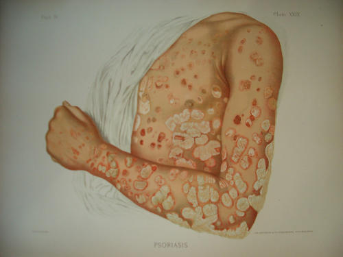
Two centuries of dermatology textbooks identify the quality of scaling in eczema (dermatitis) and psoriasis to be different, the latter having unique hard silvery scale (Figure 1). Fox uses scaling and lesional demarcation to distinguish eczema from psoriasis.3,4 ‘The scales of psoriasis... have been compared to silver and to mother of pearl... in chronic cases they become thick and hard like plates of armour’3 contrasts with the description of eczema squamosum ‘... scaling varies greatly in different cases, ranging from a slight mealiness of the skin to thick whitish masses or irregular flakes of epidermis curling up at the margin’ and (in eczema) ‘When the scales are thick and whitish and the patches are isolated and numerous the appearance... may suggest psoriasis, but the rounded and circumscribed character of psoriatic patches is always lacking in eczema.'4
Figure 1.
Psoriasis. From Taylor RW. A Clinical Atlas of Venereal & Skin Diseases. Philadelphia: Lea Brothers & Co., 1889. Plate XX1X (in colour online)
Similar differences are recorded between other erythrosquamous conditions: ‘Psoriasis... lesions are congested areas covered with masses of silvery scales’, ‘in pityriasis rosea the patches... are covered with fine scales,’ and ‘erythroderma... scaling, which is often profuse, a regular exfoliation.’5
DISEASE IDENTIFICATION BY SCALING
In order to demonstrate that research does not require test tubes, that dermatological thought can be a bit lateral, and that scaling is truly important, I recently performed a simple trial to determine whether I could distinguish psoriasis from eczema by palpation alone. With ethical approval, formal consent, etc., a cohort of 16 adults (five atopic dermatitis, nine psoriasis) were examined using touch alone, the patients being behind a screen and the examining hand being guided to a representative area by a Nurse Practitioner. Sites of predilection, or that might cause embarrassment, were excluded. The diagnosis was correctly made in 14 of 16 cases (χ2, P=0.012). This study shows that, at least in distinguishing between two inflammatory dermatoses with a different scale quality, palpation alone may be sufficient to make the diagnosis. It is unlikely that these diagnoses would not have been made visually, so quality of scale may not have been essential, but the research was to prove a concept rather than to imply that palpation is always necessary.
WHAT ABOUT TELEDERMATOLOGY?
Teledermatology has fans and critics. It precludes discussion of history-taking and discussion of management with the patient unless ‘real-time’ teledermatology is used, and this is about four-fold more costly than a clinic appointment. ‘Store-and-forward’ images reduce this cost but may have problems with image quality, inability to ‘examine’ other body sites (e.g. nails in psoriasis, mouth in lichen planus) and may miss ‘incidental’ lesions of importance. Diagnostic accuracy rates of teledermatology compared with face-to-face diagnosis are consistently lower (about 55-90% correlation in different studies6-8), and there is a higher rate of suggested need for skin biopsy by teledermatology consultation (probably reflecting lower diagnostic certainty). However, this is not always the main issue—the use of teledermatology for triage (is a clinic visit required?, is a biopsy needed?) may answer important patient management questions without needing a fully correct diagnosis.
Some studies have suggested that diagnosis of rashes is less reliable than that of localized lesions.9 Several contributory reasons are discussed above; diagnosis of rashes is often difficult anyhow, but an additional problem with ‘store-and-forward’ teledermatology is that (even if high quality), the submitted picture(s) may not be adequately distant to show the distribution or adequately close to show fine detail. Also, even good quality photos are two-dimensional; raised lesions of urticaria, for example, may be difficult to distinguish from flat lesions of a similar colour, and quality of scaling can only be guessed at. Touching the skin is a modality that is omitted in teledermatology, but there are clearly situations where it can be important. Indeed, the inability to palpate lesions has also been given as a reason for dermatologists being less satisfied than primary care physicians with the results of teledermatology.7 Even enthusiasts admit that this can be a problem.
CONCLUSION
Dermatological diagnosis involves both history and clinical features. Palpation is a modality that most dermatologists automatically do concurrently with visually observing, and, unconsciously or otherwise, the results are incorporated into the diagnostic conclusion. Palpation of lesions or rashes may appear to be something of an orphan part of skin examination but it is an important issue—I hope I have convinced you.
References
- 1.Lawrence CM, Cox NH. Physical Signs in dermatology, 2nd edn. London: Mosby, 2002
- 2.Cox NH, Coulson IH. Diagnosis of Skin Disease. In: Burns DA, Breathnach SM, Cox NH, Griffiths CEM (eds). Rook's Textbook of Dermatology, 7th edn. Oxford: Blackwell Science, 2004: 5.9
- 3.Fox GH. Photographic Illustrations of Skin Diseases, 2nd edn. New York: EB Treat, 1886: 79
- 4.Fox GH. Photographic Atlas of the Diseases of the Skin. Eczema squamosum. Philadelphia: JB Lippincott Company, 1905:late XXII.
- 5.Sequeira JH. Diseases of the Skin, 2nd edn. Philadelphia: P Blakiston's Son & Co, 1915: 11
- 6.Gilmour E, Campbell SM, Loane MA, et al. Comparison of teleconsultations and face-to-face consultations: preliminary results of a United Kingdom multicentre teledermatology study. Br J Dermatol 1998;139: 81-7 [DOI] [PubMed] [Google Scholar]
- 7.Eedy DJ, Wootton R. Teledermatology: a review. Br J Dermatol 2001;144: 606-707 [DOI] [PubMed] [Google Scholar]
- 8.Oakley AMM, Reeves F, Bennett J, Holmes SH, Wickham H. Diagnostic value of written referral and/or images for skin lesions. J Telemed Telecare 2006;12: 151-8 [DOI] [PubMed] [Google Scholar]
- 9.Futral M, Pare AK, Ling M, et al. Poster presentation (cited in Burdick AE, Berman B, Teledermatology). Adv Dermatol 1997;12: 19-45 [PubMed] [Google Scholar]


