Abstract
TALL-1 is a recently identified member of the tumor necrosis factor (TNF) family that costimulates B lymphocyte proliferation. Here we show that B cell maturation protein (BCMA), a member of the TNF receptor family that is expressed only by B lymphocytes, specifically binds to TALL-1. A soluble receptor containing the extracellular domain of BCMA blocks the binding of TALL-1 to its receptor on the plasma membrane and inhibits TALL-1-triggered B lymphocyte costimulation. Overexpression of BCMA activates NF-κB, and this activation is potentiated by TALL-1. Moreover, BCMA-mediated NF-κB activation is inhibited by dominant negative mutants of TNF receptor-associated factor 5 (TRAF5), TRAF6, NF-κB-inducing kinase (NIK), and IκB kinase (IKK). These data indicate that BCMA is a receptor for TALL-1 and BCMA activates NF-κB through a TRAF5-, TRAF6-, NIK-, and IKK-dependent pathway. The identification of BCMA as a NF-κB-activating receptor for TALL-1 suggests molecular targets for drug development against certain immunodeficient or autoimmune diseases.
Members of the tumor necrosis factor (TNF) ligand family play important roles in various physiological and pathological processes, including cell proliferation, differentiation, apoptosis, modulation of immune response, and induction of inflammation (1–6). At least 16 members of the TNF ligand family have been identified. These include TNF, FasL, lymphotoxin-α, lymphotoxin-β, TRAIL/APO-2L, CD27L, CD30L, CD40L, 4–1BBL, OX40L, TRANCE/RANKL, LIGHT, TWEAK, TL1, APRIL/TALL-2, and TALL-1 (1–12). Most TNF family members are synthesized as type II transmembrane precursors. Their extracellular domains can be cleaved by metalloproteinases to form soluble cytokines. The soluble and membrane-bound TNF ligand family members bind to receptors belonging to the TNF receptor family, which are type I transmembrane proteins with characteristic cysteine-rich motifs (1–6).
TALL-1 is a novel TNF family member recently identified by us (13) and subsequently by three other groups (14–16). Unlike most members of the TNF family that are expressed by activated immune cells, TALL-1 is constitutively expressed by monocytes and macrophages (13, 14). Flow cytometry analysis indicates that the receptor for TALL-1 is expressed only by peripheral B lymphocytes or B lymphocyte-derived cell lines, but not by peripheral T lymphocytes, monocytes, and non-B lymphocyte-derived cell lines (14, 15). Functional studies indicate that soluble TALL-1 (sTALL-1) costimulates B lymphocyte proliferation in vitro and administration or overexpression of sTALL-1 causes lymphocytic disorders and autoimmune manifestations in mice (15, 17). These data suggest that TALL-1 plays an important role in monocyte/macrophage-driven B lymphocyte activities.
Here we report that B cell maturation protein (BCMA), a member of the TNF receptor family that is expressed specifically by B lymphocytes, is a receptor for TALL-1. Our findings also suggest that BCMA can activate the transcription factor NF-κB through a TNF receptor-associated factor 5 (TRAF5)-, TRAF6-, NF-κB-inducing kinase (NIK)-, and IκB kinase (IKK)-dependent pathway.
Materials and Methods
Plasmids.
To construct the mammalian secretion expression plasmid for Flag-sTALL-1, a cDNA fragment encoding for amino acids 134–285 of human TALL-1 was amplified from a TALL-1 full-length cDNA clone (6) by PCR with the following primers: 5′-GAAGCTTATGGACTACAAGGACGACGATG-3′ and 5′-AAAGGATCCTACAGACATGGTGTAAGTAG-3′. The PCR product was digested with HindIII and BamHI and inserted into the HindIII and BamHI sites of the pSec-Tag2B plasmid (Invitrogen) to make pSec-Flag-sTALL-1. To construct the mammalian expression plasmid for C-terminal hemagglutinin (HA)-tagged BCMA, a cDNA fragment encoding BCMA and C-terminal HA epitope was amplified by PCR from an expressed sequence tag clone (GenBank accession no. AA259026) with the following primers: 5′-ATAAGCTTTTTGTGATGATGTTG-3′ and 5′-TTGGATCCTTAAGCGTAATCTGGAACATCGTATGGGTACCTAGCAGAAATTGAT-3′. The amplified cDNA fragment was digested with HindIII and BamHI and cloned into the HindIII and BamHI site of a cytomegalovirus (CMV) promoter-based plasmid to make pCMV-BCMA-HA. To construct mammalian secretion expression plasmid for soluble BCMA and Fc fusion protein, a cDNA encoding amino acids 1–62 of human BCMA was amplified by PCR from the expressed sequence tag clone AA259026 with the following primers: 5′-GGGAATTCCATGTTGCAGATGGCTG-3′ and 5′-GGGGATCCAAACAGGTCCAGAG-3′. The PCR product was digested with EcoRI and BamHI and inserted into the EcoRI and BglII sites of the pCMV1-Flag-Fc plasmid (18) to make pCMV1-Flag-sBCMA-Fc.
The mammalian expression plasmid for TRAF5 dominant negative mutant was constructed by insertion of a PCR product encoding amino acids 69–557 of human TRAF5 into the pRK-Flag vector (19, 20).
The expression plasmids for Flag-sTRAIL-R1-Fc and Flag-sTRAIL-R2-Fc (Claudio Vincenz, University of Michigan), CD40, TNF-R2, TRADD(296S), Flag-TRAF2(87–501), Myc-RIP(559–671), Myc-NIK(KK/AA), Flag-IKKα(K44A), and Flag-IKKβ(K44A) (David Goeddel, Tularik, Inc., South San Francisco, CA), Flag-TRAF6(289–523) (Zhaodan Cao, Tularik, Inc.), and the NF-κB-luciferase (Gary Johnson, National Jewish Center) and IRF-luciferase (Uli Schindler, Tularik, Inc.) reporter plasmids have been provided by the indicated investigators.
Protein Purification.
To purify Flag-sTALL-1, 500 ml of conditioned medium from the 293 stable cell line expressing Flag-sTALL-1 was collected and supplemented with 10 mM Tris (pH 7.5) and 100 mM NaCl. Two milliliters of anti-Flag antibody affinity chromatography column (Sigma) was prewashed with three sequential 5-ml aliquots of 0.1 M glycine (pH 3.5) and followed by three sequential 5-ml aliquots of TBS buffer (50 mM Tris/150 mM NaCl, pH 7.4). The medium was passed through the column, and the column was washed with 12-ml aliquots of TBS three times. The proteins bound to the column were eluted with 100 μg/ml Flag peptide (Sigma).
Immunoprecipitation and Western Blot Experiments.
To detect Flag-sTALL-1 expression, 293 cells (3 × 10⁶/100-mm dish) were transfected with 10 μg of pSec-Flag-sTALL-1 or the empty control pSec-TaqB2 plasmid by Ca3(PO4)2 precipitation (19, 20). Twenty-four hours after transfection, cell culture medium was collected. A 1-ml aliquot of the medium was incubated with 0.5 μg of anti-Flag mAb (Sigma) or 0.5 μg of control mouse IgG, 25 μl of a 1:1 slurry of GammaBind G Plus Sepharose (Amersham Pharmacia) at 4°C for 3 h. The Sepharose beads were washed three times with 1 ml of lysis buffer. The precipitates were fractionated on SDS/PAGE, and Western blot analysis was performed with anti-Flag antibody. To detect interaction between Flag-sTALL-1 and BCMA-HA, 293 cells (3 × 10⁶/100-mm dish) were transfected with 10 μg of the expression plasmid for BCMA-HA or an empty control plasmid. Twenty-four hours after transfection, cells were lysed in 1.0 ml of lysis buffer (20 mM Tris, pH 7.5/150 mM NaCl/1% Triton/1 mM EDTA/10 μg/ml aprotinin/10 μg/ml leupeptin/1 mM PMSF). The lysates were mixed with 5 ml of control conditioned medium (cell culture medium from the empty pSec-TagB2 plasmid-transfected 293 cells), Flag-sTALL-1 containing medium, or control conditioned medium plus 0.1 μg of recombinant Flag-sTRAIL (provided by Bryant Barnay, University of Texas M.D. Anderson Cancer Center). The mixtures were split into two aliquots, and each aliquot was incubated with 0.5 μg of anti-HA mAb (Babco, Richmond, CA) or control mouse IgG and 25 μl of a 1:1 slurry of GammaBind G Plus Sepharose at 4°C for 3 h. The Sepharose beads were washed three times with 1 ml of lysis buffer containing 500 mM NaCl. The precipitates were analyzed by Western blot with anti-Flag antibody. To detect the interaction between Flag-sTALL-1 or Flag-sTRAIL and Flag-TRAIL-R1-Fc or Flag-TRAIL-R2-Fc, 293 cells (3 × 10⁶/100-mm dish) were transfected with 10 μg of the mammalian secretion expression plasmid for Flag-sTRAIL-R1-Fc, Flag-sTRAIL-R2-Fc, or Flag-Fc control. Cell culture medium was collected 24 h after transfection. A 3-ml aliquot of the medium was mixed with 3 ml of Flag-sTALL-1 or Flag-sTRAIL containing medium. The mixture was incubated with 25 μl of a 1:1 slurry of GammaBind G Plus Sepharose at 4°C for 3 h. The beads were washed with the lysis buffer three times. The precipitates were analyzed by Western blot with anti-Flag antibody. All conditioned medium was supplemented with 10 mM Tris, pH 7.5/100 mM NaCl/1 mM EDTA/10 μg/ml aprotinin/10 μg/ml leupeptin/1 mM PMSF before immunoprecipitation experiments. Western blot analysis was performed as described (19, 20).
Preparation of Flag-sBCMA-Fc.
293 cells (3 × 10⁶/100-mm dish) were transfected with 10 μg of pCMV1-Flag-sBCMA-Fc plasmid. Twelve hours later, cells were washed with PBS and fresh medium was added. Thirty-six hours after that, cell culture medium was collected and concentrated 50-fold by centrifugation with Centricon-30 (Millipore).
Flow Cytometry Analysis.
For most experiments, cells (1 × 10⁶) were incubated with 500 μl of control medium (from the empty control pSec-TagB2 plasmid-transfected 293 cells) or with Flag-sTALL-1 containing medium for 40 min. For the soluble BCMA (sBCMA) experiments, Bjab cells were incubated with 500 μl of control medium, 250 μl of Flag-sTALL-1 containing medium plus 250 μl of control medium, or 250 μl of Flag-sTALL-1 medium plus 250 μl of concentrated Flag-sBCMA-Fc containing medium. Cell staining was performed by sequential incubation (each 40 min) with anti-Flag mAb (1 μg/ml) and RPE-conjugated goat anti-mouse IgG (1:200 dilution) in staining buffer (D-PBS containing 2% FBS). Cells were washed two times with staining buffer after each incubation. The fluorescence exhibited by the stained cells was measured by using a Becton Dickinson FACScan flow cytometer.
B Cell Proliferation Assays.
Human peripheral B lymphocytes were purified from peripheral blood of health donors by using anti-CD19 Dynal beads and DETACHaBEAD anti-CD19 (Dynal, Great Neck, NY), following procedures suggested by the manufacturer. Purified B lymphocytes (1 × 10⁵) were seeded on 96-well dishes and treated with indicated reagents for 40 h. Cells were pulsed for an additional 10 h with [3H]thymidine (1 μCi/well) (NEN). Incorporation of [3H]thymidine was measured by liquid scintillation counting. Data shown are averages and SDs of one representative experiment in which each treatment has been performed in triplicate.
Reporter Gene Assays.
Luciferase reporter gene assays were performed as described (21, 22). Within the same experiment, each transfection was performed in triplicate, and where necessary, enough amount of empty control plasmid was added to keep each transfection receiving the same amount of total DNA. To normalize for transfection efficiency and protein amount, 0.5 μg of Rous sarcoma virus-β-galactosidase plasmid was added to all transfections. Luciferase activities were normalized on the basis of β-galactosidase expression levels. Data shown are averages and SDs of one representative experiment in which each transfection has been performed in triplicate.
Results and Discussion
TALL-1 Coimmunoprecipitates with BCMA.
To identify the receptor for TALL-1, we used a candidate approach. Because all identified TNF family members bind to receptors belonging to the TNF receptor family, we reasoned that the receptor for TALL-1 is also a member of the TNF receptor family whose expression is limited to B lymphocytes. Previously, a TNF receptor family member, BCMA, was identified during the analysis of a t(4;1)(q26;13) chromosomal translocation that occurred in a human malignant T cell lymphoma (23). The breakpoints of the two chromosome partners involve the IL-2 gene on chromosome 4 and the BCMA gene on chromosome 16. The translocation results in the transcription of a hybrid IL-2–BCMA mRNA composed of the first three exons of IL-2 at the 5′ end fused to the coding sequences of BCMA mRNA at the 3′ end (23). RNA protection analysis indicates that BCMA mRNA is expressed in most B lymphocyte-derived but not in non-B lymphocyte-derived cell lines (24, 25). In human tissues, BCMA is expressed by spleen and lymph nodes, but not by brain, muscle, heart, lung, kidney, pancreas, testis, and placenta. Using human malignant B cell lines characteristic of different stages of B lymphocyte differentiation, it has been shown that BCMA mRNA is absent in the pro-B lymphocyte stage and its expression increases with B lymphocyte maturation (24, 25). The physiological functions of BCMA, however, are not clear. Because BCMA has the properties expected for a receptor for TALL-1, we investigated this possibility.
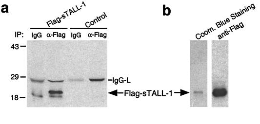
We first produced sTALL-1 in a human embryonic kidney 293 cell line. Previously, it was shown that the transmembrane TALL-1 precursor is cleaved between amino acid residues R133 and A134 to form sTALL-1 (14). To produce sTALL-1, we made a mammalian secretion expression construct in which an N-terminal Flag epitope was fused in-frame with a cDNA fragment encoding amino acids 134–285 of TALL-1. This construct was transiently or stably transfected into 293 cells. Expression of Flag-tagged sTALL-1 (Flag-sTALL-1) in the culture medium was confirmed by Western blot analysis with an anti-Flag antibody (Fig. 1). The secreted Flag-sTALL-1 also was purified with an anti-Flag antibody affinity column (Fig. 1).
Figure 1.
Expression and purification of Flag-sTALL-1. (a) Expression of Flag-sTALL-1. 293 cells were transfected with pSec-Flag-sTALL-1 or an empty control plasmid. Twenty-four hours after transfection, cell culture medium was collected and immunoprecipitated with a monoclonal anti-Flag antibody (lanes 2 and 4) or mouse IgG control (lanes 1 and 3). The immunoprecipitates were analyzed by Western blot with anti-Flag antibody. (b) Purification of Flag-sTALL-1. Conditioned medium from a 293 stable cell line expressing Flag-sTALL-1 was passed through an anti-Flag affinity column. Bound proteins were eluted with Flag peptide and analyzed by Coomassie blue staining (left lane) or Western blot with anti-Flag antibody (right lane).
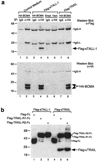
To determine whether TALL-1 binds to BCMA, we constructed a mammalian expression construct for C-terminal HA epitope-tagged BCMA (BCMA-HA). This plasmid or an empty control plasmid was transfected into 293 cells, and the transfected cell lysates were mixed with the Flag-sTALL-1 containing medium. Coimmunoprecipitation and Western blot analysis indicated that BCMA-HA interacted with Flag-sTALL-1 (Fig. 2a). As a control, BCMA-HA did not interact with proteins in control medium or Flag-tagged soluble TRAIL (Flag-sTRAIL) (Fig. 2a), another member of the TNF family (3, 5).
Figure 2.
Specific interaction between BCMA and TALL-1. (a) BCMA interacts with sTALL-1 but not sTRAIL. 293 cells were transfected with pCMV-BCMA-HA (lanes 1–4, 7, and 8) or an empty control plasmid (lanes 5 and 6). Twenty-four hours after transfection, cells were lysed and the lysates were mixed with control medium (lanes 1 and 2) or conditioned medium containing Flag-sTALL-1 (lanes 3–6) or Flag-sTRAIL (lanes 7 and 8). The mixtures were immunoprecipitated with a monoclonal anti-HA antibody (lanes 2, 4, 6, and 8) or mouse control IgG (lanes 1, 3, 5, and 7). The immunoprecipitates were analyzed by Western blot with anti-Flag antibody (Upper). The same blot was stripped and reprobed with anti-HA antibody (Lower). The higher-molecular-weight band is probably a glycosylated form of BCMA, which has been shown to be a glycoprotein (25). (b) sTALL-1 does not bind to TRAIL receptors. 293 cells were transfected with secretion expression plasmids for Flag-Fc, Flag-sTRAIL-R1-Fc, and Flag-sTRAIL-R2-Fc. Twenty-four hours after transfection, aliquots of the cell culture medium were mixed with conditioned medium containing either Flag-sTALL-1 (lanes 1–3) or Flag-sTRAIL (lanes 4–6). The mixtures were precipitated with GammaBind G Sepharose beads. Proteins bound to the beads were analyzed by Western blot with anti-Flag antibody.
To determine whether the binding of TALL-1 to BCMA is specific or whether TALL-1 nonspecifically binds to any member of the TNF receptor family, we examined whether Flag-sTALL-1 interacts with TRAIL-R1/DR4 and TRAIL-R2/DR5, two receptors for TRAIL (18, 26). To do this, conditioned medium containing Flag-sTRAIL-R1-Fc (18) or Flag-sTRAIL-R2-Fc (26) fusion protein was mixed with conditioned medium containing Flag-sTALL-1 or Flag-sTRAIL. The mixtures were precipitated with protein G-Sepharose beads, and proteins bound to the beads were analyzed by Western blot with anti-Flag antibody. This experiment indicates that Flag-sTALL-1 does not interact with TRAIL-R1 and TRAIL-R2 (Fig. 2b). As expected, Flag-sTRAIL interacted with TRAIL-R1 and TRAIL-R2 in the same experiment (Fig. 2b). Taken together, these experiments suggest that TALL-1 specifically interacts with BCMA.
BCMA Can Be Targeted to the Plasma Membrane, Where It Can Bind to sTALL-1.
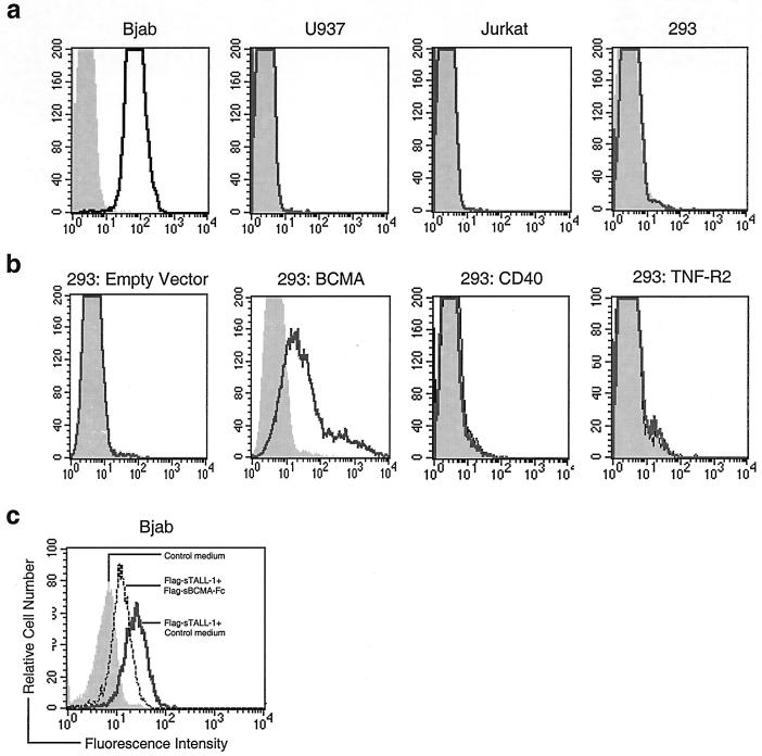
Although it has been demonstrated experimentally that BCMA can insert into canine microsomes as a glycosylated type I integral membrane protein in vitro (25), immunocytochemistry and subfractionation studies indicated that BCMA was enriched in Golgi apparatus and failed to detect BCMA on the plasma membrane (25). In addition, sequence analysis suggests that BCMA has no recognizable signal peptide at its N terminus (23–25). These observations raise the question of whether BCMA can function as a plasma membrane-bound receptor. To resolve this question, we performed flow cytometry analysis. To do this, we first tested whether the Flag-sTALL-1 protein produced by us could bind to its receptor on plasma membrane of the B lymphocyte-derived Bjab cell line, which previously has been shown to express TALL-1 receptor (14). As shown in Fig. 3a, flow cytometry analysis with a monoclonal anti-Flag antibody indicates that Flag-sTALL-1 can bind to the plasma membrane of Bjab cells, but not of the monocytic U937 cells, the T lymphoma Jurkat cells, and the embryonic kidney 293 cells (Fig. 3a). These data suggest that Bjab, but not U937, Jurkat, and 293 cells, express TALL-1 receptor. To test whether BCMA can function as a membrane receptor, 293 cells were transfected with the expression plasmid for BCMA. Twenty-four hours after transfection, cells were incubated with control medium or Flag-sTALL-1 containing medium, and flow cytometry analysis of the transfected intact cells was performed with anti-Flag antibody. As shown in Fig. 3b, Flag-sTALL-1 could bind to the plasma membrane of BCMA-transfected cells, but not of empty control plasmid, CD40, or TNF-R2-transfected cells. These data suggest that BCMA can be targeted to the plasma membrane and sTALL-1 specifically binds to the plasma membrane-bound BCMA but not other examined TNF receptor family members. This is not surprising considering that some well-established TNF receptor family members, such as human TNF-R1, TRAIL-R4 (27), and HVEM (28), also do not have a recognizable signal peptide based on structural analysis with existing software programs (data not shown). In addition, many examined TNF receptor family members, such as TNF-R1 and Fas, are found to be enriched in the Golgi apparatus and not or barely detectable on plasma membrane by immunocytochemistry and subfractionation experiments (21, 29, 30). However, flow cytometry analysis, a more sensitive method, could easily detect TNF-R1 and Fas on plasma membrane in the same cell types (29, 30).
Figure 3.
Flow cytometry analysis of sTALL-1 binding to BCMA. (a) TALL-1 receptor expression in various cell lines detected by Flag-sTALL-1. The indicated cells were incubated with control medium (shaded histograms) or Flag-sTALL-1 containing medium (solid-line histograms). Cells were washed and analyzed by flow cytometry with a monoclonal anti-Flag antibody. (b) BCMA is targeted to plasma membrane, where it can bind to sBCMA. 293 cells were transfected with the indicated expression plasmids. Twenty-four hours after transfection, cells were incubated with control medium (shaded histograms) or Flag-sTALL-1 containing medium (solid-line histograms). Flow cytometry analysis of the transfected intact cells was performed with anti-Flag antibody. (c) sBCMA blocks sTALL-1 binding to its receptor. Bjab cells were incubated with control medium (shaded histogram), Flag-sTALL-1 containing medium + control medium (solid-line histogram), or Flag-sTALL-1 containing medium + Flag-sBCMA-Fc containing medium (dashed-line histogram). The treated cells were analyzed by flow cytometry with anti-Flag antibody. Similar data have been obtained for the above flow cytometry analysis with purified Flag-sTALL-1.
Soluble BCMA Inhibits TALL-1 Binding to Bjab Cells and Inhibits sTALL-1-Triggered B Lymphocyte Proliferation.
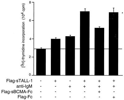
To determine whether TALL-1 signals through BCMA, we investigated whether sBCMA could block TALL-1 binding to its receptor on Bjab cells and whether sBCMA could inhibit TALL-1-triggered B lymphocyte proliferation. To do this, we produced a secreted fusion protein, Flag-sBCMA-Fc, which consisted of a N-terminal Flag tag, the extracellular domain of BCMA (amino acids 1–62), and the Fc fragment of IgG1. Flow cytometry analysis indicated that Flag-sBCMA-Fc partially blocked sTALL-1 binding to its receptor on Bjab cells (Fig. 3c). Costimulation assays indicated that Flag-sBCMA-Fc, but not Flag-Fc, significantly (P < 0.01) inhibited sTALL-1-triggered B lymphocyte proliferation (Fig. 4). These data are consistent with the conclusion that TALL-1 signals through BCMA. Because Flag-sBCMA-Fc could not completely block sTALL-1 binding to its receptor (Fig. 3c) and sTALL-1-triggered B lymphocyte proliferation (Fig. 4), this points to the possibility that an additional receptor for TALL-1 exists on B lymphocytes.
Figure 4.
Inhibition of TALL-1-triggered B cell costimulation by sBCMA. Purified B lymphocytes cultured in 96-well dishes were treated for 40 h with the following reagents: (i) TBS control buffer, (ii) Flag-sTALL-1 (100 ng/ml), (iii) anti-IgM (10 μg/ml), (iv) Flag-sTALL-1 (100 ng/ml) + anti-IgM (10 μg/ml), (v) Flag-sTALL-1 (100 ng/ml) + anti-IgM (10 μg/ml) + concentrated Flag-sBCMA-Fc (5 μl), and (vi) Flag-sTALL-1 (100 ng/ml) + anti-IgM (10 μg/ml) + concentrated Flag-Fc (5 μl). The treated cells were pulsed with [3H]thymidine for 10 h. [3H]Thymidine incorporation was measured by liquid scintillation counting.
BCMA Activates NF-κB, and This Activation Is Potentiated by sTALL-1.
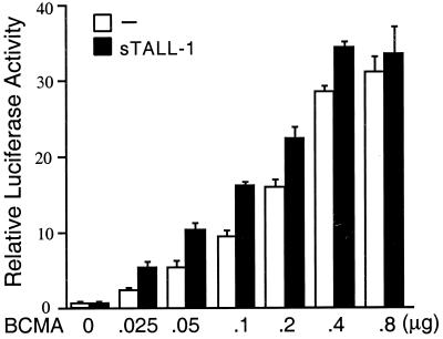
Many members of the TNF receptor family can activate NF-κB when overexpressed in mammalian cells (4, 20, 22). To determine whether BCMA can also activate NF-κB, 293 cells were transfected with a mammalian expression plasmid for BCMA. Luciferase reporter genes assays indicated that transfection of BCMA activated NF-κB in a dose-dependent manner in 293 cells (Fig. 5). Transfection of BCMA did not activate interferon response factor in reporter gene assays (data not shown), suggesting that BCMA-mediated NF-κB activation is not due to nonspecific activation of transcription and/or translation. Moreover, sTALL-1 stimulation potentiated BCMA-mediated NF-κB activation very well, especially when BCMA was expressed at low levels (Fig. 5). When BCMA was expressed at high levels, sTALL-1 stimulation could only weakly potentiate BCMA-mediated NF-κB activation. Two phenomena may account for this. First, it has been widely accepted that activation of TNF receptor family members is triggered by ligand-mediated oligomerization of the receptors. When the receptors are expressed at high levels, they may achieve maximum self-oligomerization even without their ligands. Second, at high expression levels of BCMA, downstream NF-κB activation signaling may be saturated. Taken together, these data suggest BCMA can activate NF-κB and TALL-1 can signal through BCMA.
Figure 5.
BCMA activates NF-κB, and the activation is potentiated by sTALL-1. 293 cells were transfected with 0.5 μg of NF-κB luciferase reporter plasmid and increased amounts of an expression plasmid for BCMA. Fourteen hours after transfection, cells were treated with 100 ng/ml Flag-sTALL-1 (solid bars) or left untreated (open bars) for 7 h and luciferase reporter assays were performed.
TRAF5, TRAF6, NIK, and IKKs Are Involved in BCMA-Mediated NF-κB Activation.
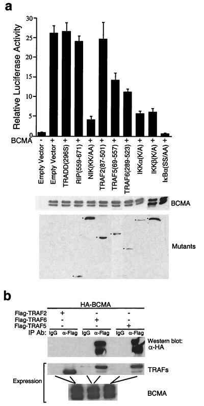
Previously, it has been shown that intracellular signaling proteins TRADD, TRAF2, TRAF5, TRAF6, RIP, NIK, IKKα, and IKKβ are involved in NF-κB activation pathways mediated by several TNF receptor family members (3, 4, 20, 22). We examined whether these proteins are also involved in NF-κB activation by BCMA. As shown in Fig. 6A, dominant negative mutants of TRAF5, TRAF6, NIK, IKKα, and IKKβ, but not of TRADD, TRAF2, and RIP, inhibited BCMA-mediated NF-κB activation. Consistent with these observations, we also found that BCMA physically interacted with TRAF5 and TRAF6, but not TRAF2, TRADD, and RIP in coimmunoprecipitation experiments (Fig. 6b). These data suggest that TRAF5, TRAF6, NIK, and IKKs, but not TRAF2, TRADD, and RIP, are involved in BCMA-mediated NF-κB activation pathways.
Figure 6.
TRAF5, TRAF6, NIK, and IKKs are involved in BCMA-mediated NF-κB activation. (a) Effects of various dominant negative mutants on BCMA-mediated NF-κB activation. 293 cells were transfected with 0.5 μg of NF-κB reporter plasmid, 0.5 μg of BCMA expression plasmid, 1 μg of control plasmid, or expression plasmids for the indicated mutants. crmA plasmid (0.5 μg) also was added to the RIP () transfections for inhibiting apoptosis. Twenty hours after transfection, luciferase assays were performed and data were treated as described in Materials and Methods. Expression of the HA-tagged BCMA and the Flag- or Myc-tagged mutants was examined by Western blot analysis with antibodies against the HA, Flag, or Myc epitopes, respectively. TRADD(296S) and IκBα(SS/AA) are not tagged and could not be detected in these experiments. (b) BCMA interacts with TRAF5 and TRAF6 but not TRAF2. 293 cells were transfected with expression plasmids for HA-BCMA and the indicated plasmids. Transfected cell lysates were immunoprecipitated with anti-Flag antibody or control IgG. The immunoprecipitates were analyzed by Western blot with anti-HA antibody (Top). Expression of the transfected proteins was detected by Western blot analysis with anti-Flag (Middle) or anti-HA antibody (Bottom).
In conclusion, our data suggest that BCMA is a B lymphocyte-specific receptor for TALL-1 and that BCMA activates NF-κB through pathways involved in TRAF5, TRAF6, NIK, and IKKs. The constitutive expression of TALL-1 by monocytes/macrophages and the specific expression of its receptor BCMA by B lymphocytes indicate that peripheral B lymphocyte proliferation, survival, and activation are critically regulated by monocytes and related cells through secretion of sTALL-1. Manipulation of the TALL-1/BCMA-signaling system may provide novel approaches for modulation of B cell-mediated immune responses and diseases.
Acknowledgments
We thank Peter Hoffman in Dr. Peter Henson's laboratory for providing human lymphocytes and Drs. David Goeddel, Zhaodan Cao, Claudio Vincenz, Gary Johnson, David Riches, John Cambier, Philippa Marrack, John Kapler, Bryant Darnay, Terry Porter, Wen-Hui Hu, Tom Mitchell, Zhonghe Zhai, and Zeng-Quan Zhou for reagents, support, and discussions. H.-B.S. is supported by the National Jewish Medical and Research Center, the American Cancer Society, the U.S. Army Breast Cancer Program, the National Natural Science Foundation of China (Grant 39925016), and the Special Funds for Major State Basic Research of China (Grant G19990539).
Abbreviations
- TNF
tumor necrosis factor
- BCMA
B cell maturation protein
- sTALL-1
soluble TALL-1
- HA
hemagglutinin
- CMV
cytomegalovirus
- IKK
IκB kinase
- NIK
NF-κB-inducing kinase
- TRAF
TNF receptor-associated factor
Footnotes
This paper was submitted directly (Track II) to the PNAS office.
Article published online before print: Proc. Natl. Acad. Sci. USA, 10.1073/pnas.160213497.
Article and publication date are at www.pnas.org/cgi/doi/10.1073/pnas.160213497
References
- 1.Smith C A, Farrah T, Goodwin R G. Cell. 1994;76:959–963. doi: 10.1016/0092-8674(94)90372-7. [DOI] [PubMed] [Google Scholar]
- 2.Grewal I S, Flavell R A. In: Immune Tolerance. Banchereau J, Dodet B, Schwartz R, Trannoy E, editors. Paris: Elsevier; 1996. pp. 125–134. [Google Scholar]
- 3.Nataga S. Cell. 1997;88:355–365. [Google Scholar]
- 4.Baker S J, Reddy E P. Oncogene. 1998;17:3261–3270. doi: 10.1038/sj.onc.1202568. [DOI] [PubMed] [Google Scholar]
- 5.Griffith T S, Lynch D H. Curr Opin Immunol. 1998;10:559–563. doi: 10.1016/s0952-7915(98)80224-0. [DOI] [PubMed] [Google Scholar]
- 6.Ashkenazi A, Dixit V M. Curr Opin Cell Biol. 1999;11:255–260. doi: 10.1016/s0955-0674(99)80034-9. [DOI] [PubMed] [Google Scholar]
- 7.Anderson D M, Maraskovsky E, Billingsley W L, Dougall W C, Tomestsko M E, Roux E R, Teepe M C, DuBose R F, Cosman D, Galibert L. Nature (London) 1997;390:175–180. doi: 10.1038/36593. [DOI] [PubMed] [Google Scholar]
- 8.Wong B R, Rho J, Arron J, Robinson E, Orlinick J, Chao M, Kalachikov S, Cayani E, Bartlett F, Frankel W N, et al. J Biol Chem. 1997;272:25190–25195. doi: 10.1074/jbc.272.40.25190. [DOI] [PubMed] [Google Scholar]
- 9.Chichepotiche Y, Bourdon P R, Xu H, Hsu Y M, Scott H, Hession C, Garcia I, Browning J L. J Biol Chem. 1997;272:32401–32410. doi: 10.1074/jbc.272.51.32401. [DOI] [PubMed] [Google Scholar]
- 10.Hahne M, Kataoka T, Schroter M, Hofmann K, Irmler M, Bodmer J, Schneider P, Bornand T, Holler N, French L E, et al. J Exp Med. 1998;188:1185–1190. doi: 10.1084/jem.188.6.1185. [DOI] [PMC free article] [PubMed] [Google Scholar]
- 11.Mauri D N, Ebner R, Montgomery R I, Kochel K D, Cheung T C, Yu G, Ruben S, Murphy M, Eisenberg R J, Cohen G H, et al. Immunity. 1998;8:21–30. doi: 10.1016/s1074-7613(00)80455-0. [DOI] [PubMed] [Google Scholar]
- 12.Kwon B, Yu K Y, Ni J, Yu G L, Jang I K, Kim Y J, Xing L, Liu D, Wang S X, Kwon B S. J Biol Chem. 1999;274:6056–6061. doi: 10.1074/jbc.274.10.6056. [DOI] [PubMed] [Google Scholar]
- 13.Shu H B, Hu W H, Johnson H. J Leukocyte Biol. 1999;65:680–683. [PubMed] [Google Scholar]
- 14.Schneider P, MacKay F, Steiner V, Hofmann K, Bodmer J L, Holler N, Ambrose C, Lawton P, Bixler S, Orbea H, et al. J Exp Med. 1999;189:1747–1756. doi: 10.1084/jem.189.11.1747. [DOI] [PMC free article] [PubMed] [Google Scholar]
- 15.Moore P A, Belvedere O, Orr A, Pieri K, LaFleur D W, Feng P, Soppet D, Charters M, Gentz R, Parmelee D, et al. Science. 1999;285:260–263. doi: 10.1126/science.285.5425.260. [DOI] [PubMed] [Google Scholar]
- 16.Mukhopadhyay A, Ni J, Zhai Y, Yu G L, Aggarwal B B. J Biol Chem. 1999;274:15978–15981. doi: 10.1074/jbc.274.23.15978. [DOI] [PubMed] [Google Scholar]
- 17.Pan G, O'Rourke K, Chinnaiyan A M, Gentz R, Ebner R, Ni J, Dixit V. Science. 1997;276:111–113. doi: 10.1126/science.276.5309.111. [DOI] [PubMed] [Google Scholar]
- 18.Macky F, Woodcock S A, Lawton P, Ambrose C, Baetscher M, Schneider P, Tschopp J, Browning J L. J Exp Med. 1999;190:1697–1710. doi: 10.1084/jem.190.11.1697. [DOI] [PMC free article] [PubMed] [Google Scholar]
- 19.Shu H B, Halpins D R, Goeddel D V. Immunity. 1997;6:751–763. doi: 10.1016/s1074-7613(00)80450-1. [DOI] [PubMed] [Google Scholar]
- 20.Hu W H, Johnson H, Shu H B. J Biol Chem. 1999;274:30603–30610. doi: 10.1074/jbc.274.43.30603. [DOI] [PubMed] [Google Scholar]
- 21.Cottin V, Van Linden A, Riches D W. J Biol Chem. 1999;274:32975–32987. doi: 10.1074/jbc.274.46.32975. [DOI] [PubMed] [Google Scholar]
- 22.Rothe M, Sarma V, Dixit V M, Goeddel D V. Science. 1995;269:1424–1427. doi: 10.1126/science.7544915. [DOI] [PubMed] [Google Scholar]
- 23.Laabi Y, Gras M P, Carbonne F, Brouet J C, Berger R, Larsen C J, Tsapis A. EMBO J. 1992;11:3897–3904. doi: 10.1002/j.1460-2075.1992.tb05482.x. [DOI] [PMC free article] [PubMed] [Google Scholar]
- 24.Laabi Y, Gras M P, Brouet J C, Berger R, Larsen C J, Tsapis A. Nucleic Acids Res. 1994;22:1147–1154. doi: 10.1093/nar/22.7.1147. [DOI] [PMC free article] [PubMed] [Google Scholar]
- 25.Gras M P, Laabi Y, Linares-Cruz G, Blondel M O, Rigaut J P, Brouet J C, Leca G, Haguenauer-Tsapis A, Tsapis A. Int Immunol. 1995;7:1093–1106. doi: 10.1093/intimm/7.7.1093. [DOI] [PubMed] [Google Scholar]
- 26.Pan G, Ni J, Wei Y F, Yu G I, Gentz R, Dixit V M. Science. 1997;277:815–817. doi: 10.1126/science.277.5327.815. [DOI] [PubMed] [Google Scholar]
- 27.Degli-Esposti M A, Dougall W C, Smolak P J, Waugh J Y, Smith C A, Goodwin R G. Immunity. 1997;7:813–820. doi: 10.1016/s1074-7613(00)80399-4. [DOI] [PubMed] [Google Scholar]
- 28.Montgomery R I, Warner M S, Lum B J. Cell. 1996;87:427–436. doi: 10.1016/s0092-8674(00)81363-x. [DOI] [PubMed] [Google Scholar]
- 29.Bennett M, Macdonald K, Chan S W, Luzio J P, Simari R, Weissberg P. Science. 1998;282:290–293. doi: 10.1126/science.282.5387.290. [DOI] [PubMed] [Google Scholar]
- 30.Jones S J, Ledgerwood E C, Prins J B, Galbraith J, Johnson D R, Pober J S, Bradley J R. J Immunol. 1999;162:1042–1048. [PubMed] [Google Scholar]


