

Abstract
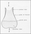
The etiology of femoral fractures in calves during assisted parturition was examined from the perspective of biomechanical force analysis. Femurs were collected from newborn calf cadavers. Their physical dimensions of length, cortical thickness, and diameter were measured from radiographs, and the data were recorded. The bones were then frozen until tested. The thawed bones were compressed axially in a materials testing machine until they broke, whereupon the breaking strength of the bones at the failure site was recorded. Fracture locations were documented radiographically, and the cortical thickness and bone diameter at the fracture site were determined. Fracture configurations and locations were similar to those found in clinical cases associated with forced extraction. The breaking strength of all femurs fell within the magnitude of forces calculated to be created when mechanical devices are used to assist delivery during dystocia. An etiology based on wedging of the femur in the maternal pelvis and resulting compression during forced extraction is suggested to account for the occurrence of supracondylar fractures of the femur of calves delivered in anterior presentation using mechanical devices in a manner commonly performed by veterinarians and owners. It is recommended that care should be exercised to correct or rule out the possibility of premature engagement of the calf's stifle into the birth canal, and thereby reduce the frequency of this type of fracture occurrence in the field.
Full text
PDF


Images in this article
Selected References
These references are in PubMed. This may not be the complete list of references from this article.
- Ferguson J. G., Dehghani S., Petrali E. H. Fractures of the femur in newborn calves. Can Vet J. 1990 Apr;31(4):289–291. [PMC free article] [PubMed] [Google Scholar]
- Hamilton G. F., Turner A. S., Ferguson J. G., Pharr J. W. Slipped capital femoral epiphysis in calves. J Am Vet Med Assoc. 1978 Jun 1;172(11):1318–1322. [PubMed] [Google Scholar]
- Hart M. B., Wu J. J., Chao E. Y., Kelly P. J. External skeletal fixation of canine tibial osteotomies. Compression compared with no compression. J Bone Joint Surg Am. 1985 Apr;67(4):598–605. [PubMed] [Google Scholar]
- Hindson J. C. Quantification of obstetric traction. Vet Rec. 1978 Apr 15;102(15):327–332. doi: 10.1136/vr.102.15.327. [DOI] [PubMed] [Google Scholar]
- Light T. R., Ogden D. A., Ogden J. A. The anatomy of metaphyseal torus fractures. Clin Orthop Relat Res. 1984 Sep;(188):103–111. [PubMed] [Google Scholar]
- Mickelsen W. D. Correction of stifle lock in bovine dystocia. Vet Med Small Anim Clin. 1976 Aug;71(8):1047–1048. [PubMed] [Google Scholar]
- Pope M. H., Outwater J. O. Mechanical properties of bone as a function of position and orientation. J Biomech. 1974 Jan;7(1):61–66. doi: 10.1016/0021-9290(74)90070-0. [DOI] [PubMed] [Google Scholar]
- Reilly D. T., Burstein A. H. Review article. The mechanical properties of cortical bone. J Bone Joint Surg Am. 1974 Jul;56(5):1001–1022. [PubMed] [Google Scholar]
- Rohlmann A., Mössner U., Bergmann G., Kölbel R. Finite-element-analysis and experimental investigation of stresses in a femur. J Biomed Eng. 1982 Jul;4(3):241–246. doi: 10.1016/0141-5425(82)90009-7. [DOI] [PubMed] [Google Scholar]
- Valliappan S., Svensson N. L., Wood R. D. Three dimensional stress analysis of the human femur. Comput Biol Med. 1977 Oct;7(4):253–264. doi: 10.1016/0010-4825(77)90031-2. [DOI] [PubMed] [Google Scholar]