Abstract
We report that females of some substrains of inbred mouse strain 129 are resistant to systemic plague due to conditionally virulent Δpgm strains of Yersinia pestis; however, fully virulent Y. pestis is not attenuated in these mice. Therefore, these mice offer a powerful system in which to map in parallel host resistance traits and opposing bacterial virulence properties for plague.
Plague in humans is caused by the gram-negative bacterium Yersinia pestis (6). The disease is 50 to 100% fatal if untreated and potentially could be caused on an epidemic scale by a malicious act. Accordingly, there is a need to understand the pathogenesis of Y. pestis and to enhance resistance to plague in people. A major virulence property of the plague-causing bacteria is the production of a set of six proteins called Yops that act within host cells to negate and modulate signaling pathways that orchestrate crucial host innate defense mechanisms (1). We had been studying YopM and had recently found that YopM counteracts innate host defenses, specifically causing a global loss of natural killer (NK) cells that could play a crucial defense role early in infection (4). In pursuit of that finding, we have been evaluating virulence of YopM− Y. pestis in several strains of mice with defects in potential innate defense mechanisms that might be directly targeted by YopM. We fortuitously found a substrain (129S2/P2) of mouse strain 129 that is resistant to systemic plague due to Yersinia pestis KIM5.
The 129S2/P2 mouse strain was of interest because it lacked expression of grancalcin (Gca−/−) (10), a protein that may modulate actin bundling in bone marrow-derived cells, such as polymorphonuclear neutrophils (PMNs) and macrophages. Accordingly, we tested whether YopM is necessary for virulence in Gca−/− mice greater than 5 weeks old infected intravenously with Y. pestis KIM5 or a YopM− mutant as described previously (4). (The Y. pestis KIM5 strain is highly attenuated when given by a peripheral route of infection due to deletion of the chromosomal pgm locus but is essentially fully virulent when it is given by an intravenous route [1, 11].) On the basis of data from two such experiments, the 50% lethal doses (LD50s) (9) for both the parent Y. pestis KIM5 and the YopM− mutant were between 5 × 10⁵ and 5 × 10⁶ CFU. This is in contrast to an LD50 of <50 CFU for Y. pestis KIM5 versus 5 × 10⁶ CFU for the YopM− mutant in C57BL/6 mice (4).
The Gca knockout had been made in a genetic background (129S2/P2) that we had not previously used in our studies. Accordingly, we tested whether it was the Gca−/− mutation or this background that was responsible for the plague resistance. The closest available match to the background strain was 129S2/Sv.Hsd (Harlan, Indianapolis, IN). As was the case for the Gca−/− mice, the LD50 for Y. pestis KIM5 in these mice was high, 2 × 10⁶ CFU, and for the YopM mutant bacteria, it was 6 × 10⁵ CFU (on the basis of the results of one experiment with female mice). We tested a second 129 mouse strain that has received extensive genetic characterization, 129P3/J (Jackson Laboratory, Bar Harbor, ME), with single doses of 104 CFU of the parent Y. pestis KIM5, and all four infected female mice survived, although they showed signs of illness (hunched posture and matted fur) (two experiments). A single similar test was made using a different Δpgm Y. pestis strain, CO99-3015.S5 (obtained from CDC, Ft. Collins, CO; a derivative of Y. pestis CO92), with the same outcome of all four mice becoming sick but surviving an intravenous challenge of 104 CFU. In a repetition of the experiment with a slightly higher dose of Y. pestis Δpgm strain CO99-3015.S5, all four mice died. These results suggested that the plague resistance likely was conferred by the 129 mouse genetic background but that different 129 substrains differed in the robustness of their resistance. This finding was interesting because there is no previous report of plague-resistant mice other than IL-10−/− mice (7), and we have not seen evidence or reported polymorphisms that would indicate that these 129 mice lack interleukin 10 (IL-10).
Dynamics of the infection.
We infected groups of three female 129S2/P2 (Gca−/−) mice intravenously with 5 × 10⁴ CFU of Y. pestis KIM5 or Y. pestis KIM5-3002 (YopM−) and analyzed the number of viable bacteria (as CFU) in liver and spleen, leukocyte populations in spleen by flow cytometry, and histopathology in liver, all as previously described (4), on day 4 postinfection (two experiments). The numbers of viable bacteria for both bacterial strains in both liver and spleen were 10- to 100-fold lower than those typically seen for infection of the susceptible C57BL/6 mouse strain (4, 7), as though the net growth of the Y. pestis strains were being limited or the bacteria were even being cleared. A robust acute inflammatory response dominated by infiltrating PMNs was present in livers of mice infected with both the parent Y. pestis KIM5 (Fig. 1, left panel) and the YopM− mutant (not illustrated) on day 4 postinfection. This histopathology was strikingly different from that in susceptible C57BL/6 mice, in which the initial inflammatory reaction dissipated and was replaced by a spreading, cell-poor, coagulative necrosis by day 4 postinfection (4) (Fig. 1, rightmost panel). When female C57BL/6 mice had been infected with the YopM− strain, the inflammatory reaction had persisted and evolved into a pregranuloma structure by day 4 (illustrated in reference 4). In the resistant female 129 mice, the continued inflammation correlated with clearance of both Y. pestis strains (data not shown).
FIG. 1.
Robust acute inflammation was under way late in infection of resistant female 129 mice with Y. pestis KIM5. Sections from formalin-fixed livers of infected female 129S2/P2 (Gca−/−) mice (left panel), noninfected female 129S2/P2 (Gca−/−) mice (middle panel), or infected female C57BL/6 mice (right panel) were stained with hematoxylin and eosin, and representative fields for day 4 postinfection are shown. The bacterial dose given to the 129S2/P2 (Gca−/−) mice was 7.7 × 10³ CFU; the dose for the susceptible female C57BL/6 mice was 102 CFU.
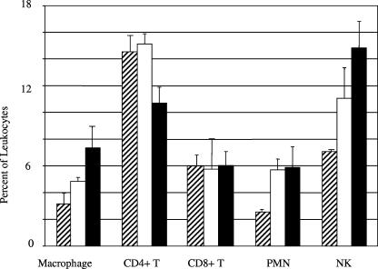
A total of 5 × 10⁶ spleen cells from the infected mice were analyzed using fluorochrome-labeled antibodies against various cell surface markers on macrophages, CD4+ T cells, CD8+ T cells, PMNs, or NK cells (Fig. 2). There was an increase in macrophages, PMNs, and NK cells due to infection with both the parent and YopM− mutant Y. pestis strains compared to uninfected mice in both experiments. This was in contrast to the previous findings with C57BL/6 mice where there was a depletion of NK cells due to YopM (4). The increases in these three cell populations were more than twofold. There was not a significant difference for CD4+ and CD8+ T cells. These findings are consistent with the acute inflammation we observed in response to infection with both strains.
FIG. 2.
Infection of female 129 mice caused an influx of macrophages, PMNs, and NK cells into the spleen. Leukocyte populations in the spleen were analyzed by flow cytometry for the percentages of macrophages, CD4+ T cells, CD8+ T cells, PMNs, and NK cells in mice infected with Y. pestis KIM5 (open bars) or the YopM− strain (closed bars) on day four postinfection. The data represent the average results ± standard deviations (error bars) from three infected mice; data for noninfected mice are provided for reference (hatched bars). The values for macrophages, PMNs, and NK cells for mice infected with both Y. pestis KIM5 and the YopM− strain differed significantly from those for noninfected mice by the unpaired t test (n = 3), with two-tailed P values of 0.011, 0.0026, and 0.034 for the three mice infected with Y. pestis KIM5 and 0.017, 0.018, and 0.0038 for the three mice infected with the YopM− strain. The same statistical test found no significant difference between the values for Y. pestis KIM5 and the YopM− mutant for macrophages, PMNs, NK cells, or CD8+ T cells. The difference in values for CD4+ T cells in mice infected with the parent and mutant strains was statistically significant but was not always observed.
Male 129 mice were more susceptible to lethal disease than female mice.
Initially we were using mice of mixed sexes for our experiments, because we had not previously found a sex-related difference in plague susceptibility (e.g., in C57BL/6 mice [4, 7]). However, in this study, we observed large variability that traced to sex, with males being clearly more susceptible than females. In one experiment involving groups of four mice infected with the parent Y. pestis KIM5, doses as low as 70 CFU were lethal to all of the males (LD50 of <70 CFU), whereas for female mice, the LD50 was >4 × 10⁵ CFU. Likewise, when groups of four 129P3/J mice were infected with a dose of 104 CFU of Y. pestis KIM5 or 104 CFU of Y. pestis Δpgm strain CO99-3015.S5, all male mice died, whereas the females survived (two experiments). A less dramatic phenomenon of sex-related differences in susceptibility to plague has been noted previously for BALB/c mice infected with Y. pestis KIM5 (5).
The scatter in the data from experiments using mixed sexes had masked a small but consistently greater lethality of the parent Y. pestis KIM5 compared to the YopM− mutant in female 129S2/P2 (Gca−/−) mice in our original LD50 assays (this was also evidenced by slower clearance of the parent strain than of the YopM− mutant from organs of infected female mice [data not shown]). In contrast, the virulence of the YopM− mutant was considerably reduced compared to that of the parent strain in the male 129S2/P2 (Gca−/−) mice. When males of these mice were given 104 CFU of Y. pestis KIM5, all died; whereas seven out of eight survived infection by the same number of the YopM− mutant (two experiments). When the surviving males were analyzed as described above for histopathology and cellular response to infection, their responses resembled those of female mice (Fig. 1 and 2) infected with a nonlethal dose of either Y. pestis strain.
Female 129 substrain mice are susceptible to fully virulent (Pgm+) Y. pestis CO99-3015.
In contrast to Y. pestis Δpgm strain KIM5 or CO99-3015.S5, the fully virulent Y. pestis CO99-3015.S0 was highly virulent for female and male 129P3/J mice. Groups of four female or four male 129P3/J mice were infected by subcutaneous injection of 0.1 ml of phosphate-buffered saline (PBS) containing the organisms (three experiments; doses tested were 103, 1.6 × 10², and 2 CFU), by intravenous injection of organisms in 0.1 ml of PBS (1 experiment; dose of 104 CFU), or by intranasal delivery of organisms in 20 μl of PBS (one experiment; dose of 4 × 10³ CFU), and female and male mice were equally susceptible. This finding indicates that the resistance of 129 mice may be overcome by a property encoded in the pgm locus. So far, this 102-kb locus has been characterized for a siderophore-based iron acquisition system that is essential for infection from a peripheral route (6), several genes required for production of biofilm and survival in the flea vector (2, 6), and an operon required for replication in infected macrophages exposed to the activating cytokine gamma interferon (8). The latter operon encodes several metabolic enzymes that by an unknown mechanism permit the bacteria to limit production of the bactericidal product nitric oxide by stimulated macrophages in vitro (8). However, none of these genes is essential for lethality from an intravenous route of infection.
Discussion and conclusion.
In summary, we have found three 129 substrains of mice for which females are strongly plague resistant, causing the parent Y. pestis KIM5 to approach the avirulence of a YopM− mutant, which in other mouse strains is attenuated by 4 to 5 orders of magnitude. These mice respond to infection of both Y. pestis strains by a robust maintenance of NK cells, the hallmark of the response to the YopM− strain by susceptible female C57BL/6 mice (4), suggesting that the enhanced innate defense(s) in these mice may be one(s) targeted by YopM. We speculate that the innate response in these mice bypasses YopM's effect, as though removing the target for YopM (e.g., by making it no longer susceptible to YopM) or counteracting YopM's effects through an antagonistic pathway. Our findings point to the 102-kb pgm locus as being crucial for the high level of virulence of Y. pestis in female 129 substrain mice, and it is possible that one or more properties encoded by this locus specifically target the same host defenses as YopM does.
Other strains of mice tested so far (six inbred strains and Swiss Webster outbred mice) have been found to be highly susceptible to plague, although there are small differences in the LD50 and mean time to death (3, 12). Accordingly, the 129 substrains identified in our study represent a new tool for plague research. In particular, the 129P3/J mice may provide a good system in which to identify genes specific for resistance to plague, because this strain is being extensively characterized genetically. It is conceivable that resistance to systemic plague in 129 substrain mice is due to a single gene that can be mapped and identified by classical backcross analysis. These mice offer an outstanding opportunity to dissect in parallel both Y. pestis virulence properties and host defenses to plague and make an important step toward learning how to confer resistance to humans.
Acknowledgments
This study was supported by PHS grant AI41668.
We thank Jürgen Roes and Anthony W. Segal (University College London) for their kind gift of breeding pairs of Gca−/− mice. We gratefully acknowledge Clarissa Cowan, Alexander V. Philipovskiy, Michael E. Gray, and Annette M. Uittenbogaard for their assistance with animal handling. Assistance from Donald A. Cohen with analysis of flow cytometry data is gratefully acknowledged.
All animal experiments were conducted under a protocol that was approved by the Institutional Animal Care and Use committee of the University of Kentucky and followed institutional and federal guidelines.
Editor: V. J. DiRita
Footnotes
Published ahead of print on 5 September 2006.
REFERENCES
- 1.Cornelis, G. R. 2002. The Yersinia Ysc-Yop ‘type III’ weaponry. Nat. Rev. Mol. Cell Biol. 3:742-752. [DOI] [PubMed] [Google Scholar]
- 2.Jarrett, C. O., E. Deak, K. E. Ischerwood, P. C. Oyston, E. R. Fischer, A. R. Whitney, S. D. Kobayashi, F. R. DeLeo, and B. J. Hinnebusch. 2004. Transmission of Yersinia pestis from an infectious biofilm in the flea vector. J. Infect. Dis. 190:783-792. [DOI] [PubMed] [Google Scholar]
- 3.Jones, S. M., F. Day, A. J. Stagg, and E. D. Williamson. 2001. Protection conferred by a fully recombinant subunit vaccine against Yersinia pestis in male and female mice of four inbred strains. Vaccine 19:358-366. [DOI] [PubMed] [Google Scholar]
- 4.Kerschen, E. J., D. A. Cohen, A. M. Kaplan, and S. C. Straley. 2004. The plague virulence protein YopM targets the innate immune response by causing a global depletion of NK cells. Infect. Immun. 72:4589-4602. [DOI] [PMC free article] [PubMed] [Google Scholar]
- 5.Mecsas, J., G. Franklin, W. A. Kuziel, R. R. Brubaker, S. Falkow, and D. E. Moiser. 2004. CCR5 mutation and plague protection. Nature 427:606. [DOI] [PubMed] [Google Scholar]
- 6.Perry, R. D., and J. D. Fetherston. 1997. Yersinia pestis—etiologic agent of plague. Clin. Microbiol. Rev. 10:35-66. [DOI] [PMC free article] [PubMed] [Google Scholar]
- 7.Philipovskiy, A. V., C. Cowan, C. R. Wulff-Strobel, S. H. Burnett, E. J. Kerschen, D. A. Cohen, A. M. Kaplan, D. A. Cohen, and S. C. Straley. 2004. Antibody against V antigen prevents Yop-dependent growth of Yersinia pestis. Infect. Immun. 73:1532-1542. [DOI] [PMC free article] [PubMed] [Google Scholar]
- 8.Pujol, C., J. P. Grabenstein, R. D. Perry, and J. B. Bliska. 2005. Replication of Yersinia pestis in interferon gamma-activated macrophages requires ripA, a gene encoded in the pigmentation locus. Proc. Natl. Acad. Sci. USA 102:12909-12914. [DOI] [PMC free article] [PubMed] [Google Scholar]
- 9.Reed, L. J., and H. Muench. 1938. A simple method of estimating fifty percent endpoints. Am. J. Hyg. 27:493-497. [Google Scholar]
- 10.Roes, J., B. K. Choi, D. Power, P. Xu, and A. W. Segal. 2003. Granulocyte function in grancalcin-deficient mice. Mol. Cell. Biol. 23:826-830. [DOI] [PMC free article] [PubMed] [Google Scholar]
- 11.Une, T., and R. R. Brubaker. 1984. In vivo comparison of avirulent Vwa− and Pgm− or Pstr phenotypes of yersiniae. Infect. Immun. 43:895-900. [DOI] [PMC free article] [PubMed] [Google Scholar]
- 12.Wake, A. 1980. Genetically controlled natural resistance of mice to plague infection and its relationship to genetically controlled cell-mediated immune resistance, p. 179-184. In E. Skamene and P. A. L. Landy (ed.), Genetic control of natural resistance to infection and malignancy, 1st ed. Academic Press, New York, N.Y.


