Abstract
Caf1, a chaperone-usher protein from Yersinia pestis, is a major protective antigen in the development of subunit vaccines against plague. However, recombinant Caf1 forms polymers of indeterminate size. We report the conversion of Caf1 from a polymer to a monomer by circular permutation of the gene. Biophysical evaluation confirmed that the engineered Caf1 was a folded monomer. We compared the immunogenicity of the engineered monomer with polymeric Caf1 in antigen presentation assays to CD4 T-cell hybridomas in vitro, as well as in the induction of antibody responses and protection against subcutaneous challenge with Y. pestis in vivo. In C57BL/6 mice, for which the major H-2b-restricted immunodominant CD4 T-cell epitopes were intact in the engineered monomer, immunogenicity and protective efficacy were preserved, although antibody titers were decreased 10-fold. Disruption of an H-2d-restricted immunodominant CD4 T-cell epitope during circular permutation resulted in a compromised T-cell response, a low postvaccination antibody titer, and a lack of protection of BALB/c mice. The use of circular permutation in vaccine design has not been reported previously.
Yersinia pestis is thought to be the etiological agent of epidemic diseases referred to as the Black Death over many centuries (26). In recent years, infections with Y. pestis have been reported on all continents except Australia (3). The discovery of multiple antibiotic-resistant strains of Y. pestis in Madagascar (13) and the possible use of Y. pestis as a biological weapon has led to the classification of plague as a re-emerging disease by the World Health Organization.
The virulence factor Caf1, encoded by the pFra plasmid of Y. pestis, is induced to form a protein capsule during growth above 33°C, and strains lacking Caf1 show increased susceptibility to phagocytosis by macrophages in vitro (11). However, infection of experimental animals has not demonstrated a clear role for Caf1 in virulence in vivo (9, 26).
Current vaccines for Y. pestis consist of either killed whole cells resulting from heat or formaldehyde treatment or live attenuated strains. Killed whole cells require multiple doses, induce frequent side effects, and incur large manufacturing expenses under stringent microbiological containment conditions (37). Live attenuated strains are also highly problematic and are not licensed for use in humans (30). Protein subunit vaccines can overcome many of these problems, and recombinant forms, produced in a nonpathogenic strain, lead to a large reduction in costs. Vaccination with recombinant Caf1 (rCaf1) (20, 35) alone or in combination with other Y. pestis proteins (37) confers protection against plague in mice (35). Sera from humans recovering from plague contain significant titers of anti-Caf1 antibodies (27), and the efficacy of vaccination with Caf1 in humans is being evaluated (38).
The design of new immunogens for effective, protective immunity is one of the “14 Grand Challenges in Global Health” (28), and the extracellular chaperone-usher (CU) polymers (36) are a large group of promising bacterial vaccine candidates. The CU system secretes polymeric surface proteins from many gram-negative bacteria, including human pathogens responsible for plague, meningitis, and urinary tract and gastrointestinal infections (31). CU polymers are composed of small immunoglobulin-like subunits linked by a “donor strand complementation” mechanism, in which a β-strand is donated by the next molecule in the chain to create polymers of stable monomers joined by noncovalent links (32). Naturally occurring CU proteins generate defined structures (8, 25), whereas recombinant CU proteins form either aggregated structures (20) or vaccine-chaperone dimers (18) or are unstable (14). Future development of protein therapeutics is being driven toward easily assayed final products that are not defined simply by the manufacturing process (5), and one important step is the design of new regulatory-friendly protein therapeutics. A homogenous quaternary structure is a major objective, and prospective vaccines based upon isolated CU subunits do not yet meet these criteria (40). Immunization of mice with Caf1 monomerized by heating in sodium dodecyl sulfate confers limited protection in the mouse model (20), and it is not clear whether denatured Caf1 remains monomeric after injection.
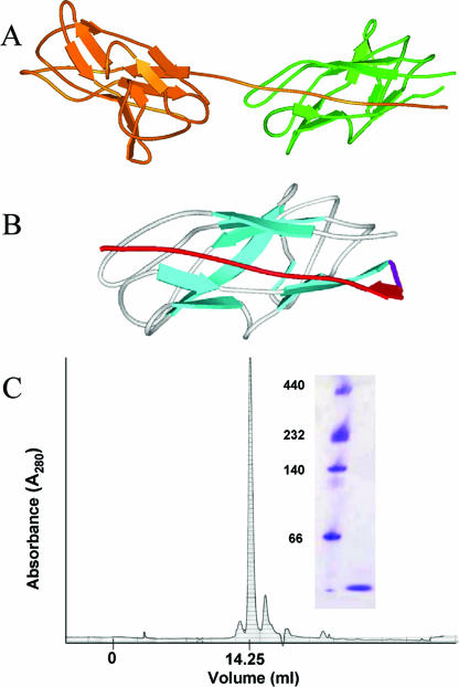
In the present study we evaluate the antigenicity of a stable monomeric form of Caf1 created by a protein engineering technique known as circular permutation. Proteins with closely spaced N and C termini can be treated as circular molecules such that the existing termini can be joined with a short linker sequence and new termini introduced in almost any surface loop along the sequence. This has been used to investigate the role of amino acid sequence upon protein folding and to introduce new functions into existing proteins by adding additional amino acids at the new termini. The situation in polymeric Caf1 is different since the N terminus of one monomer is far from its own C terminus but very close to that of its neighbor (Fig. 1A). Hence, by moving the N-terminal strand to the C terminus we can try to mimic polymeric structure in a monomeric format. Although many circularly permuted proteins have been described (22), this is the first immunological analysis of one that is a candidate vaccine antigen.
FIG. 1.
Design and creation of a folded monomeric Caf1 protein. (A) Ribbon diagram of Caf1 dimer (taken from PDB file 1P5U [40]) illustrating the donor strand complementation critical for correct folding of Caf1 polymer. (B) Energy minimized model of cpCaf1 structure based on 1P5U showing the engineered flexible linker (magenta) and repositioned N-terminal strand (red). (C) Schematic representation of rCaf1 and its mutant derivatives. Amino acid residue numbering of the mature protein is used in the numbering scheme, and as in panel B the engineered flexible linker is shown in magenta and repositioned N-terminal strand in red. (D) Superose 12 size exclusion chromatography of cpCaf1 (50 mM phosphate buffer [pH 7.4]) produced a single peak corresponding to the expected monomer size. CpCaF1 (16.1 kDa) elutes at a volume of 14.25 ml compared to carbonic anhydrase (29 kDa, 13.75 ml) and bovine serum albumin (66 kDa; 12.25 ml). Native polymeric Caf1 elutes in the void volume 6 ml (21). The inset shows Coomassie blue-stained native polyacrylamide gel electrophoresis. Lane 1, molecular mass markers (in kilodaltons); lane 2, purified cpCaf1.
MATERIALS AND METHODS
Expression, purification, and cloning of cpCaf1.
All engineered versions of Caf1 were produced as Tol fusion proteins (1), and sequences for cloning into pTol vectors were generated by PCR using the plasmid pAH34L (21) encoding Caf1 antigen (accession number X61996) as a template. The mature Caf1 sequence (residues 1 to 149) was truncated at the N and C termini (Fig. 1C) to create ΔN-Caf1(19-149) and ΔC-Caf1(1-122) deletion mutants, respectively, by using specific primer pairs in a PCR. For circularly permuted Caf1 (cpCaf1), the circular permutation method (Fig. 1B and C) attaches a copy of the donor strand (residues 1 to 15; shown in red) to the C terminus of the target protein (cyan) using a short linker (purple), while also removing the equivalent sequence from the N terminus. Thus, the primary sequence of circularly permuted Caf1 (cpCaf1) begins with the dipeptide glycine-serine (GS) (from the fusion protein used to enhance expression levels (1), followed by the wild-type Caf1 sequence P16 through S146. This is joined by a flexible linker (TGSGNG), to the stabilizing strand removed from the original N terminus (1ADLTASTTATATLVE15). The resulting protein which therefore consists of a shuffled wild-type sequence plus linker has a predicted molecular mass of 16,181 Da, a pI of 4.61, and an ɛ280 value of 5,960 M−1 cm−1.
Circular permutation using PCR was performed essentially as previously described (22). Briefly, two copies of the gene coding for Caf1 antigen (lacking signal sequence) were amplified by PCR: one with the sequence coding for a flexible N- to C-terminal linker (residues TGSGNG) at the 5′ end and another with the same sequence at the 3′ end. Overlap PCR created a tandem copy joined by the linker connecting one N and one C terminus (16) which was cloned into plasmid pGEM-T (Promega, United Kingdom). This was used as the PCR template in a third PCR using two further primers that amplified the “cpCaf1” region from P16 of the 5′ DNA copy of Caf1 to G15 of the 3′ copy of Caf1 (via the linker).
The resulting ΔN-Caf1, ΔC-Caf1, and cpCaf1 were cloned into pTolT in the pTOL vector system (1) to produce a Tol-Caf1 fusion protein separable by thrombin cleavage.
For protein expression and purification, TUNER cells (Novagen, Nottingham, United Kingdom) were transformed with pTolT-cpCaf1. Cells were prepared and harvested, and protein isolation was performed essentially as described previously (1). Briefly, cells were grown at 37°C to an optical density at 600 nm of ∼0.6, induced with 1 mM IPTG (isopropyl-β-d-thiogalactopyranoside), and grown for ∼18 h at 18°C. The cells were then sonicated, soluble cpCaf1 was purified by using nitrilotriacetic acid agarose affinity (QIAGEN, United Kingdom), and thrombin cleavage was performed as described by Anderluh et al. (1).
Size exclusion chromatography was performed at a constant flow rate of 0.5 ml/min using a Superose 12 column (GE Healthcare) and cpCaf1 in 50 mM phosphate buffer (pH 7.4), and native polyacrylamide gel electrophoresis (PAGE) was performed using an 8 to 25% gel. Matrix-assisted laser desorption ionization-time of flight (MALDI-TOF) analysis of cpCaf1 was performed using an Applied Biosystems Voyager DE STR.
Structural analysis.
The circular dichroism (CD) of Caf1 samples was analyzed by using a JASCO-810 spectropolarimeter (JASCO UK, Ltd., Essex, United Kingdom). For far-UV CD, samples were diluted to ∼0.5 mg/ml in 50 mM phosphate buffer (pH 7.4), and a 0.1-mm-path-length cuvette was used. For far-UV CD thermal analysis, protein samples were diluted to ∼0.1 mg/ml and analyzed in a 1-mm capped cuvette at 210 nm over a gradient of increasing temperature (1°C/min). The CD spectra changed very little between 5 and 30°C; thus, 30°C was used in the normalization procedure as fully folded, and 95°C was normalized as fully unfolded. Exact amino acid concentrations were calculated by using molar extinction coefficients.
Differential scanning calorimetry was performed by using a Micro-Cal VP-DSC (cell volume of 0.52 ml; MicroCal Europe, Milton Keynes, United Kingdom). Samples were scanned with a scan rate of 1°C/min, and a filtering period of 16 s with protein at 0.25 to 0.45 mg/ml and an appropriate buffer in the reference cell. All solutions were gently degassed prior to use. The data were corrected for the buffer baseline (buffer scans performed under identical conditions) and analyzed by using standard MicroCal ORIGIN V.7 software.
The intrinsic fluorescence of 20 μg of each protein diluted in 400 μl of 50 mM phosphate buffer (pH 7.4) was measured with an excitation wavelength of 280 nm, and emission data were collected from 295 to 450 nm in a Cary Eclipse fluorometer (Varian, Victoria, Australia) with excitation and emission bandwidths of 5 nm. An appropriate buffer spectrum was subtracted.
Immunogenicity of cpCaf1.
Cells were grown in culture medium (RPMI 1640 medium containing 3 mM l-glutamine, 50 μM 2-mercaptoethanol, 10% [vol/vol] fetal bovine serum, and 30 μg of gentamicin [Sigma, United Kingdom]/ml). Macrophages were grown from femoral bone marrow cells of BALB/c (H-2d) and BALB.B (H-2b) mice in the presence of macrophage colony-stimulating factor as described previously (12) and activated by treatment overnight with 1 ng of gamma interferon (R&D Systems, Abingdon, United Kingdom)/ml before use as antigen-presenting cells in T-cell assays. The T-cell hybridomas specific for four epitopes of Caf1 (7-21/Ad, 48-61/Ab, 123-136/Ad, and 134-147/Ab) have been described elsewhere (23).
Macrophages (4 × 10⁴/well) in flat-bottom 96-well microtiter plates were fixed in 1% paraformaldehyde for 4 min and washed either before or after treatment with Caf1 antigen, cpCaf1, or the relevant synthetic peptides for 5 h. T-cell hybridoma cells (4 × 10⁴/well) were added, and the plates were incubated for 24 h before culture supernatants were collected. The response of T-cell hybridomas was measured as the interleukin-2 content of supernatants measured in a bioassay as the proliferation of CTLL-2 cells (3 × 10⁴/well) for 24 h, including 38.4 kBq of [3H]thymidine (specific activity, 74 GBq/mmol; Amersham International, Plc, Buckinghamshire, United Kingdom). The radioactivity was quantitated by using a direct beta counter (Matrix 9600; Packard Instrument Company, Meridan, CT). The results were plotted as the mean counts per minute of triplicate wells ± the standard deviation, which was considered to be proportional to the T-cell response. Experiments were repeated at least twice, and essentially the same results were obtained. Linear regression analysis was performed on the plots of T-cell hybridoma responses to determine whether the slope of the antigen titrations deviated significantly from zero, using Prism version 4.0 (GraphPad Software, Inc., San Diego, CA).
Groups of six BALB/c or C57BL/6 mice were immunized intramuscularly with 10 μg of rCaf1 adsorbed to the adjuvant Alhydrogel on days 1 and 21, blood samples were obtained on day 28 after primary immunization, and anti-Caf1 antibodies were measured by enzyme-linked immunosorbent assay (ELISA) using wells coated with rCaf1 and endpoint titers defined as the serum dilution, yielding an absorbance of 0.1 above background, as described previously (39).
Mice were subcutaneously challenged with 58 CFU per mouse of Y. pestis strain GB (one median lethal dose is ∼1 CFU/mouse for BALB/c mice and <5 CFU/mouse for C57BL/6 mice) on day 37 after primary immunization. Mice were observed for 14 days postchallenge to determine their protected status.
Binding of the protective anti-rCaf1 monoclonal antibody F104AG1 (2) to rCaf1 and cpCaf1 was determined by ELISA. Duplicate wells of microtiter 96-well immunoassay plates (Immulon 1; Dynex Technologies, Southampton, United Kingdom) were coated with a range of doses of rCaf1, cpCaf1, or recombinant V antigen of Y. pestis as a negative control in 15 mM Na2CO3, 35 mM NaHCO3, and 3 mM NaN3 (pH 9.3) and incubated at 4°C for 18 h. Plates were washed in 200 mM Tris-HCl-190 mM NaCl-0.05% Tween 20 (pH 7.4), blocked in 5% bovine serum albumin-0.9% NaCl for 1 h at room temperature, and washed again before incubation for 1 h with 0.64 μg of F104AG1 monoclonal antibody/ml (determined by titration) diluted in 0.5% bovine serum albumin-0.9% NaCl. Plates were washed again and incubated for 1 h with 1:1,000 goat anti-mouse immunoglobulin G (IgG)-peroxidase conjugate (Sigma Chemical Co.) diluted in 0.5% bovine serum albumin-0.9% NaCl; the reaction was then developed with the liquid substrate system 2,2′-azino-bis(3-ethylbenzthiazoline-5-sulfonic acid) (Sigma Chemical Co.), and the absorbance was measured at 405 nm.
RESULTS
Creation of monomeric Caf1 by truncation and circular permutation.
Using the published crystal structure of Caf1 (Fig. 1A), we designed a monomeric circularly permutated form of Caf1, termed cpCaf1, in which the N-terminal donor strand was relocated after a flexible linker to the C terminus, substituting for the strand donated by the neighboring monomer (Fig. 1B and C). Size exclusion chromatography and native gel electrophoresis demonstrated that cpCaf1 was a monomer of the expected size (Fig. 1D and inset).
Solution structure of cpCaf1.
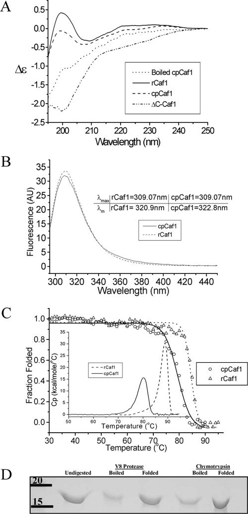
The structures of monomeric cpCaf1 and polymeric rCaf1 (20) were compared by biophysical analyses. Far-UV CD spectroscopy measures the protein secondary structure, and the immunoglobulin-type fold of rCaf1 gives rise to a distinctive CD spectrum with 210-nm negative and 198-nm positive maxima (Fig. 2A). rCaf1 heated at >95°C for >5 min displays a strongly negative spectrum typical of denatured proteins. The CD spectrum of cpCaf1 closely resembles the distinctive spectrum of folded polymeric rCaf1 (20) but demonstrates a smaller intensity. This may indicate that some of the Caf1 CD signal results from stabilization in the polymeric state. That the two proteins share a similar fold was further supported by comparison of their fluorescence emission spectra, which are essentially identical (Fig. 2B).
FIG. 2.
Structural similarity of cpCaf1 and rCaf1. (A) Far-UV CD spectra of ∼0.5 mg of rCaf1, C-terminal deletion mutant (ΔC-Caf1), native cpCaf1, and heat-denatured cpCaf1 (>95°C for >5 min, path length of 0.2 nm)/ml. (B) Intrinsic fluorescence of cpCaf1 and rCaf1 (λex [excitation wavelength] = 280 nm). The λmax (peak wavelength) and λm (barycentric mean emission wavelength of 295 to 450 nm) values are shown. (C) Normalized CD intensity at 210 nm (30→95°C at 1°C/min) of ∼0.1 mg of cpCaf1 and rCaf1/ml. The midpoints (Tm) were 83°C for rCaf1 and 76.2°C for cpCaf1. The inset shows differential scanning calorimetry of 0.25 to 0.45 mg/ml at 1°C/min for cpCaf1 (solid line) and rCaf1 (dashed line). (D) Coomassie blue-stained sodium dodecyl sulfate-polyacrylamide gel electrophoresis of an overnight digestion of heat-denatured (>95°C for >5 min) cpCaf1 and native cpCaf1 (in phosphate-buffered saline [pH 7.4]) with V8 protease and chymotrypsin. A total of 10 μg of cpCaf1 was incubated with either 0.5 μg of V8 protease or 0.03 μg of chymotrypsin.
Analysis of the Caf1 structure (40) indicated that simple truncation of the Caf1 protein might also prevent polymerization. We created C-terminal (ΔC-Caf1 1-122) and N-terminal (ΔN-Caf1 19-149) deletion mutants of Caf1. Of the two truncated proteins, only ΔC-Caf1 could be purified successfully. It was natively monomeric but showed the CD spectrum of a fully unfolded protein (Fig. 2A).
Most stably folded proteins are cooperative structures that thermally unfold over a small temperature range, whereas poorly folded polypeptides show more gradual temperature effects. To test our hypothesis that the fold and stability of cpCaf1 resemble those of Caf1, the thermal stability was measured by CD and differential scanning calorimetry. The results show (Fig. 2C) that both proteins show highly cooperative unfolding. Measured by CD, monomeric cpCaf1 unfolds with a transition temperature (Tm) of 82°C compared to rCaf1 at 86°C. Similarly, differential scanning calorimetry showed the Tm (79.8°C versus 88.4°C) and integrated calorimetric enthalpy of unfolding (ΔHcal) (81.4 versus 121 kcal mol−1) of cpCaf1 to be less than for rCaf1. Detailed interpretation of these data was prevented by the irreversibility of the thermal unfolding, but it shows that cpCaf1 has adopted a cooperatively folded single-domain structure similar to rCaf1 but with a reduced stability. This may be due to the removal of stabilizing intermolecular contacts compared to the polymer or to effects of the linker design (see below). However, the monomeric cpCaf1 is clearly a close structural mimic of wild-type rCaf1 and is, even with a slightly reduced unfolding temperature, sufficiently stable for use in vivo. Unfolded proteins are very sensitive to the action of proteolytic enzymes (proteases), thus affecting their stability and antigen processing behavior but, as expected, the stably folded cpCaf1 was, like rCaf1, resistant to protease digestion (Fig. 2D). These data are similar to those obtained from other monomerized CU proteins, such as the FimH-FimC hybrid protein (4) and the very similar scCaf1 recently reported by Zavaliov et al. (41), neither of which have been subjected to immunological investigation.
Immunogenicity of cpCaf1.
We mapped the major immunodominant major histocompatibility complex (MHC) class II-restricted epitopes of rCaf1 in two inbred strains of mice (23). Two H-2d-restricted epitopes were recognized by CD4 T cells from BALB/c mice and two H-2b-restricted epitopes by C57BL/6 mice. One H-2d-restricted epitope (Caf17-20) was disrupted between residues 15 and 16 by insertion of the linker during circular permutation This allowed investigation of the impact of this epitope on the immunogenicity of cpCaf1 in mice of the H-2d haplotype compared to H-2b mice for which both immunodominant epitopes are intact in cpCaf1.
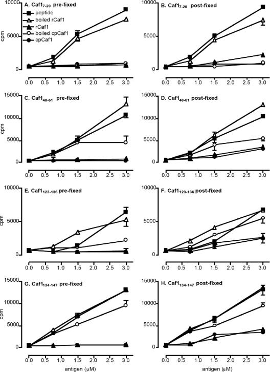
We used antigen presentation to T-cell hybridomas in vitro as a sensitive technique to study the role of cpCaf1 structure in antigenicity (23, 24). We measured the responses of Caf1-specific T-cell hybridomas to cpCaf1 and rCaf1 presented by bone marrow macrophages. A T-cell hybridoma specific for Caf17-20 recognized rCaf1 but not cpCaf1 (Fig. 3A), confirming disruption of the epitope in cpCaf1. T-cell hybridoma responses to the remaining H-2d-restricted epitope (123-136), as well as both H-2b-restricted epitopes (48-61 and 134-147), confirmed their integrity in cpCaf1 (Fig. 3D, F, and H). Fixing of antigen-presenting cells prevents the uptake and processing of folded proteins for presentation to T cells, affecting responses to denatured proteins or to synthetic peptides to a much lesser degree (15, 24, 29). We fixed macrophages either before (prefixed, Fig. 3C, E, and G) or after (postfixed, Fig. 3D, F, and H), adding rCaf1 or cpCaf1, to determine whether uptake and processing were required. Postfixed macrophages presented epitopes 48-61, 123-136, and 134-147 from intact rCaf1 and intact cpCaf1 to a similar degree, and responses to denatured rCaf1 and cpCaf1 were greater than those seen to the intact antigens (Fig. 3D, F, and H). In contrast, prefixed macrophages failed to present intact cpCaf1 or rCaf1 but presented all three epitopes when cpCaf1 or rCaf1 were heat denatured immediately prior to the assay, as expected of unfolded protein antigens (Fig. 3C, E, and G). In fact, responses to heat-denatured rCaf1 were of the same magnitude as the responses to synthetic peptides representing each epitope. These results show that intact cpCaf1, like rCaf1, behaves as a folded protein in antigen presentation assays.
FIG. 3.
T-cell hybridoma responses to cpCaf1 in vitro. Response of F1-specific T-cell hybridomas to rCaf1, cpCaf1, and synthetic peptides presented by bone marrow macrophages measured as the amount of interleukin-2 released and detected in bioassay using tritiated thymidine incorporation in CTLL-2 cells. Macrophages were fixed in paraformaldehyde either before (A, C, E, and G) or after (B, D, F, and H) incubation with a molar dose range of antigens that were either intact or heat denatured by boiling. The relevant synthetic peptide for each of four F1 epitopes were used as positive controls, and cells incubated in the absence of antigen (“0” on the x axis) were the negative control. Linear regression analysis was applied to the antigen titrations shown in the figure to determine whether the slope of the titrations deviated significantly from zero. The slope of the titrations of responses to cpCaf1 were significantly different from zero in panels D, F, and H, with P values of ≤0.0025, but not in panels C, E, and G. The slopes were also significantly different from zero for responses to boiled cpCaf1 in panels C to H, with P values of ≤0.01.
Immunization with cpCaf1 induced anti-rCaf1 IgG antibodies (Table 1) . The mean titers of cpCaf1-immunized C57BL/6 mice were ∼10-fold lower than those immunized with rCaf1, and the titers were more than 100-fold lower in cpCaf1-immunized mice than in rCaf1-immunized BALB/c mice (Table 1).
TABLE 1.
Antibody responses and survival after challenge of mice in vivoa
| Group | Mouse | Challenge (no. of survivors/ total no.) | Mean ± SEM
|
|
|---|---|---|---|---|
| MTTD (days) | Antibody titer | |||
| cpCaf1 | BALB/c | 1/6 | 9.6 ± 0.60 | 1:83 ± 30 |
| C57BL/6 | 6/6 | >14 | 1:1,967 ± 463 | |
| rCaf1 | BALB/c | 6/6 | >14 | 1:15,333 ± 3,291 |
| C57BL/6 | 6/6 | >14 | 1:13,333 ± 3,955 | |
| ΔC-Caf1 | BALB/c | 0/6 | 4.5 ± 1.3 | ND |
| C57BL/6 | 1/6 | 5.3 ± 0.61 | 1:75 ± 25 | |
| Adjuvant only | BALB/c | 0/6 | 3.8 ± 0.54 | ND |
| C57BL/6 | 0/6 | 5.3 ± 0.33 | ND | |
Groups of six BALB/c or C57BL/6 mice were immunized intramuscularly with 10 μg of rCaf1, cpCaf1, or ΔC-Caf1 antigen adsorbed to Alhydrogel adjuvant on days 1 and 21 and bled on day 28. Anti-Caf11 antibody was measured by ELISA, and the results are expressed as the mean IgG endpoint titers. The same mice were challenged with 58 CFU per mouse of Y. pestis strain GB (one median lethal dose is ∼1 CFU/mouse for BALB/c mice and <5 CFU/mouse in C57BL/6 mice) on day 37 after primary immunization. Mice were observed for 14 days postchallenge to determine their protected status. The number of animals surviving at 14 days is reported. MTTD, mean time to death. ND, not detectable.
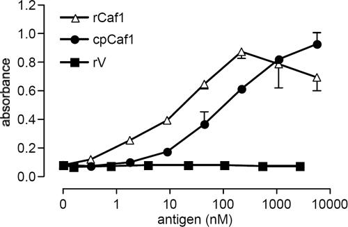
Protection of BALB/c and C57BL/6 mice immunized with rCaf1 or cpCaf1 in adjuvant was determined by challenge with a lethal dose of Y. pestis, and their survival was compared to nonimmunized and ΔC-Caf1-immunized mice. C57BL/6 mice were fully protected by cpCaf1 against lethal Y. pestis challenge (Table 1). In contrast, the majority of BALB/c mice immunized with the cpCaf1 protein did not survive challenge. Finally, we showed that a previously characterized protective anti-Caf1 monoclonal antibody (2) bound cpCaf1 efficiently, although ∼5-fold less well than it bound rCaf1, by ELISA (Fig. 4). The specificity of the antibody was confirmed by the lack of binding to recombinant V antigen of Y. pestis.
FIG. 4.
Recognition of rCaf1 and cpCaf1 by a protective monoclonal anti-Caf1 antibody. Plates were coated with the dose range shown of rCaf1, cpCaf1, or V antigen of Y. pestis as a negative control; incubated with 0.64 μg of the protective monoclonal antibody F1-04-A-F1/ml; and developed by ELISA as described in Materials and Methods. The results are plotted as the mean absorbance at 405 nm for duplicate wells.
DISCUSSION
We have used circular permutation to engineer a folded monomeric form of the bacterial chaperone-usher protein Caf1 from Yersinia pestis. We compared the immunogenicity of the engineered monomer with polymeric Caf1 in antigen presentation assays to CD4 T-cell hybridomas in vitro as well as in the induction of antibody responses and protection against challenge with Y. pestis in vivo.
The structures of rCaf1 and cpCaf1 were compared by biophysical analysis, and cpCaf1 was shown to be monomeric by size exclusion chromatography and native polyacrylamide gel electrophoresis, providing clear evidence for the monomeric state of cpCaf1 and contrasting with the evidence for aggregation obtained for rCaf1 (20). Far-UV CD measurements showed that the secondary structures of cpCaf1 and rCaf1 were very similar. The intrinsic fluorescence spectra also suggested that the aromatic residues of cpCaf1 are in an environment very similar to that of the equivalent residues in rCaf1; this is particularly significant since two of the four tyrosines in the Caf1 sequence interact with the amino-terminal “donor strand” known to be essential for the stability of the polymeric Caf1 structure (40). Both rCaf1 and cpCaf1 showed cooperative behavior by rapidly unfolding over a small temperature range, which is indicative of a single stable fold. The results of differential scanning calorimetry also confirmed the similarity of rCaf1 and cpCaf1. However, the calorimetric enthalpy of the unfolding transition was less in cpCaf1, indicating a reduced number of stabilizing interactions. The loss of stabilizing intermolecular contacts (Fig. 1A and B) is the most obvious explanation of the reduction in thermal stability compared to rCaf1, but linker design (Fig. 1B and C) could also play a role. The linker, designed to allow the translated strand flexibility to fold in the IgG domain, is longer than in a recently reported cp protein, scCaf1 (41), and linker size has been linked to changes in stability (17). Intact cpCaf1 and rCaf1 were both resistant to proteolysis unless thermally denatured, a finding consistent with the retention of native structure by cpCaf1.
We investigated the immunogenicity of cpCaf1 in vitro using T-cell hybridomas specific for four previously defined MHC class II-restricted CD4 T-cell epitopes within Caf1 (23). The primary sequences of rCaf1 and cpCaf1 are almost the same except that in cpCaf1 the amino-terminal amino acids 1 to 15 were relocated to the carboxy terminus separated by the 6-amino-acid linker. The Caf1 epitope 7-20 was thus disrupted in cpCaf1, a finding consistent with the absence of response to cpCaf1 by a T-cell hybridoma specific for the epitope 7-20. T-cell hybridomas specific for the remaining three epitopes responded to cpCaf1 presented by macrophages, confirming that these epitopes were intact in cpCaf1. An unusual feature of antigen presentation of rCaf1 is that T-cell responses to all of the major epitopes are low compared to the synthetic peptide controls, in contrast to our reported responses to other microbial antigens (24). This low response pattern of rCaf1 is retained by cpCaf1 and is consistent with the property of resistance to proteolysis of rCaf1 and that of cpCaf1, which we show in Fig. 2D. The presentation of MHC class II-restricted T-cell epitopes usually requires the uptake and processing of intact proteins, whereas denaturation by heat or chemical substitution allows presentation of the same epitopes by fixed antigen-presenting cells resulting from binding cell surface MHC molecules in the absence of antigen uptake or processing (7, 33, 34). Macrophages fixed before antigen exposure were unable to present any of the three epitopes from rCaf1 and cpCaf1, whereas prior heat denaturation permitted presentation of the three epitopes with an efficiency similar to that of the synthetic peptides. Collectively, both the pattern of antigen presentation and resistance to proteolysis show that cpCaf1 behaves similarly to intact folded rCaf1.
The immunogenicity of cpCaf1 in vivo was investigated by measuring antibodies that bound rCaf1 in serum from mice immunized with either cpCaf1 or rCaf1. Immunization with cpCaf1 and rCaf1 induced similar antibody titers that were within an order of magnitude in C57BL/6 mice, but titers were 100-fold less in cpCaf1-immunized BALB/c mice. In protection experiments, all C57BL/6 mice immunized with either cpCaf1 or rCaf1 were protected against a lethal challenge with Y. pestis. However, BALB/c mice immunized with cpCaf1 were much more susceptible since only one mouse survived compared to the survival of all BALB/c immunized with rCaf1. Serum antibody induced by cpCaf1 was measured against native rCaf1 in ELISA, showing that antibody responses were largely cross-reactive between cpCaf1 and rCaf1. However, the slightly lower antibody titers in cpCaf1-immunized mice may reflect a limitation on the degree of cross-reactivity, or it might be a result of polymeric rCaf1 being more immunogenic than the monomeric form. In support of the former view, we showed that a previously characterized protective anti-Caf1 monoclonal antibody (2) bound cpCaf1, demonstrating that an important protective antibody epitope of Caf1 is preserved in cpCaf1. However, binding was ∼5-fold weaker than to rCaf1, suggesting the antibody was not fully cross-reactive.
The lower anti-Caf1 antibody titer and lack of protection in BALB/c mice may result from reduced T-cell help after immunization with cpCaf1, which lacks one of the two immunodominant H-2d-restricted T-cell epitopes identified in this haplotype (23). In contrast, higher antibody titers and protection in cpCaf1-immunized C57BL/6 mice were consistent with the presence of both immunodominant H-2b-restricted T-cell epitopes in cpCaf1. It has previously been reported that a threshold number of immunodominant helper T-cell epitopes are required to stimulate IgG responses to conformational antibody epitopes from the same antigen (10). Antibody responses to conformational epitopes are thought to correlate with the efficacy of protection, as shown in a number of experimental infection models (15). Hence, it is unlikely that the presence of only one of the two major helper T-cell epitopes of Caf1 can provide the threshold level of T-cell help to induce protective antibody responses against Y. pestis challenge in BALB/c mice. An alternative explanation, that cpCaf1 lacks the complete repertoire of conformational Caf1 antibody epitopes required for protection of BALB/c mice, is unlikely given that C57BL/6 mice were fully protected by cpCaf1. Furthermore, the carboxy-terminal truncate ΔC-Caf1 is also unfolded, as well as lacking one epitope from each of the MHC haplotypes, which is consistent with its inability to protect either strain of mice. Since monomeric truncates lose structure and immunogenicity, whereas rCaf1 is immunogenic and polymeric, the circular permutation approach is currently the only method to make immunogenic monomers. Although our studies on cpCaf1 have shown that folded monomeric versions of CU proteins can be protective, it has also revealed the importance of maintaining the original linear epitopes of the wild type. This could be achieved in cpCaf1 by simply extending the protein to include a complete N terminus; however, such a loose peptide would be prone to protease digestion and not meet our design criteria of wild-type-like stability. We are currently investigating other designs which may overcome this limitation and provide a more immunogenic protein.
In conclusion, previous work on circularly permuted proteins was restricted to the study of enzymes (6, 19), whereas our objective was to create circularly permuted Caf1 as the model for a new family of antibacterial vaccines. We generated a circularly permuted monomeric form of Caf1 of Y. pestis that retains the structural and immunogenic characteristics of polymeric Caf1, demonstrating the successful application of protein engineering by circular permutation to vaccine design. Stable monomers of polymeric microbial proteins could provide candidate subunit vaccines against a wide range of microbial infections.
Acknowledgments
We thank Helen Ridley for excellent technical assistance, Harry Gilbert and Danny Altmann for helpful discussions, the U.S. Navy for the gift of anti-Caf1 monoclonal antibody F104AG1, and Joe Gray for MALDI-TOF mass spectrometry analysis. We thank the Defence Science and Technology Laboratory for financial support and the Wellcome Trust for equipment grants 56232, 40422, and 55979.
These studies were carried out in strict accordance with the Animals (Scientific Procedures) Act 1986.
Editor: J. B. Bliska
Footnotes
Published ahead of print on 18 September 2006.
REFERENCES
- 1.Anderluh, G., I. Gokce, and J. H. Lakey. 2003. Expression of proteins using the third domain of the Escherichia coli periplasmic-protein TolA as a fusion partner. Protein Expr. Purif. 28:173-181. [DOI] [PubMed] [Google Scholar]
- 2.Anderson, G. W., Jr., P. L. Worsham, C. R. Bolt, G. P. Andrews, S. L. Welkos, A. M. Friedlander, and J. P. Burans. 1997. Protection of mice from fatal bubonic and pneumonic plague by passive immunization with monoclonal antibodies against the F1 protein of Yersinia pestis. Am. J. Trop. Med. Hyg. 56:471-473. [DOI] [PubMed] [Google Scholar]
- 3.Anisimov, A. P., L. E. Lindler, and G. B. Pier. 2004. Intraspecific diversity of Yersinia pestis. Clin. Microbiol. Rev. 17:434-464. [DOI] [PMC free article] [PubMed] [Google Scholar]
- 4.Barnhart, M. M., F. G. Sauer, J. S. Pinkner, and S. J. Hultgren. 2003. Chaperone-subunit-usher interactions required for donor strand exchange during bacterial pilus assembly. J. Bacteriol. 185:2723-2730. [DOI] [PMC free article] [PubMed] [Google Scholar]
- 5.Brandau, D. T., L. S. Jones, C. M. Wiethoff, J. Rexroad, and C. R. Middaugh. 2003. Thermal stability of vaccines. J. Pharm. Sci. 92:218-231. [DOI] [PubMed] [Google Scholar]
- 6.Buchwalder, A., H. Szadkowski, and K. Kirschner. 1992. A fully active variant of dihydrofolate-reductase with a circularly permuted sequence. Biochemistry 31:1621-1630. [DOI] [PubMed] [Google Scholar]
- 7.Carrasco-Marin, E., J. E. Paz-Miguel, P. Lopez-Mato, C. Alvarez-Dominguez, and F. Leyva-Cobian. 1998. Oxidation of defined antigens allows protein unfolding and increases both proteolytic processing and exposes peptide epitopes which are recognized by specific T cells. Immunology 95:314-321. [DOI] [PMC free article] [PubMed] [Google Scholar]
- 8.Choudhury, D., A. Thompson, V. Stojanoff, S. Langermann, J. Pinkner, S. J. Hultgren, and S. D. Knight. 1999. X-ray structure of the FimC-FimH chaperone-adhesin complex from uropathogenic Escherichia coli. Science 285:1061-1066. [DOI] [PubMed] [Google Scholar]
- 9.Davis, K. J., D. L. Fritz, M. L. Pitt, S. L. Welkos, P. L. Worsham, and A. M. Friedlander. 1996. Pathology of experimental pneumonic plague produced by fraction 1-positive and fraction 1-negative Yersinia pestis in African green monkeys (Cercopithecus aethiops). Arch. Pathol. Lab. Med. 120:156-163. [PubMed] [Google Scholar]
- 10.DeFranco, A. L. 1999. B-lymphocyte activation, p. 225-261. In W. E. Paul (ed.), Fundamental immunology, 4th ed. Lippincott-Raven, Philadelphia, Pa.
- 11.Du, Y., R. Rosqvist, and A. Forsberg. 2002. Role of fraction 1 antigen of Yersinia pestis in inhibition of phagocytosis. Infect. Immun. 70:1453-1460. [DOI] [PMC free article] [PubMed] [Google Scholar]
- 12.Fischer, H. G., B. Opel, K. Reske, and A. B. Reske-Kunz. 1988. Granulocyte-macrophage colony-stimulating factor-cultured bone marrow-derived macrophages reveal accessory cell function and synthesis of MHC class II determinants in the absence of external stimuli. Eur. J. Immunol. 18:1151-1158. [DOI] [PubMed] [Google Scholar]
- 13.Guiyoule, A., G. Gerbaud, C. Buchrieser, M. Galimand, L. Rahalison, S. Chanteau, P. Courvalin, and E. Carniel. 2001. Transferable plasmid-mediated resistance to streptomycin in a clinical isolate of Yersinia pestis. Emerg. Infect. Dis. 7:43-48. [DOI] [PMC free article] [PubMed] [Google Scholar]
- 14.Hultgren, S. J., F. Lindberg, G. Magnusson, J. Kihlberg, J. M. Tennent, and S. Normark. 1989. The PapG adhesin of uropathogenic Escherichia coli contains separate regions for receptor binding and for the incorporation into the pilus. Proc. Natl. Acad. Sci. USA 86:4357-4361. [DOI] [PMC free article] [PubMed] [Google Scholar]
- 15.Ionescu-Matiu, I., R. C. Kennedy, J. T. Sparrow, A. R. Culwell, Y. Sanchez, J. L. Melnick, and G. R. Dreesman. 1983. Epitopes associated with a synthetic hepatitis B surface antigen peptide. J. Immunol. 130:1947-1952. [PubMed] [Google Scholar]
- 16.Iwakura, M., and T. Nakamura. 1998. Effects of the length of a glycine linker connecting the N and C termini of a circularly permuted dihydrofolate reductase. Protein Eng. 11:707-713. [DOI] [PubMed] [Google Scholar]
- 17.Iwakura, M., T. Nakamura, C. Yamane, and K. Maki. 2000. Systematic circular permutation of an entire protein reveals essential folding elements. Nat. Struct. Biol. 7:580-585. [DOI] [PubMed] [Google Scholar]
- 18.Langermann, S., R. Mollby, J. E. Burlein, S. R. Palaszynski, C. G. Auguste, A. DeFusco, R. Strouse, M. A. Schenerman, S. J. Hultgren, J. S. Pinkner, J. Winberg, L. Guldevall, M. Soderhall, K. Ishikawa, S. Normark, and S. Koenig. 2000. Vaccination with FimH adhesin protects cynomolgus monkeys from colonization and infection by uropathogenic Escherichia coli. J. Infect. Dis. 181:774-778. [DOI] [PubMed] [Google Scholar]
- 19.Luger, K., U. Hommel, and K. Kirschner. 1988. An enzyme with a circularly permuted amino-acid sequence is fully active. Biol. Chem. Hoppe-Seyler 369:868-869. [Google Scholar]
- 20.Miller, J., E. D. Williamson, J. H. Lakey, M. J. Pearce, S. M. Jones, and R. W. Titball. 1998. Macromolecular organisation of recombinant Yersinia pestis F1 antigen and the effect of structure on immunogenicity. FEMS Immunol. Med. Microbiol. 21:213-221. [DOI] [PubMed] [Google Scholar]
- 21.Morton, M., H. S. Garmory, S. D. Perkins, A. M. O'Dowd, K. F. Griffin, A. K. Turner, A. M. Bennett, and R. W. Titball. 2004. A Salmonella enterica serovar Typhi vaccine expressing Yersinia pestis F1 antigen on its surface provides protection against plague in mice. Vaccine 22:2524-2532. [DOI] [PubMed] [Google Scholar]
- 22.Mullins, L. S., K. Wesseling, J. M. Kuo, J. B. Garrett, and F. M. Raushel. 1994. Transposition of protein sequences: circular permutation of ribonuclease-T1. J. Am. Chem. Soc. 116:5529-5533. [Google Scholar]
- 23.Musson, J. A., M. Morton, N. Walker, H. M. Harper, H. V. McNeill, E. D. Williamson, and J. H. Robinson. 2006. Sequential proteolytic processing of the capsular Caf1 antigen of Yersinia pestis for MHC class II-restricted presentation to T lymphocytes. J. Biol. Chem. 281:26129-26135. [DOI] [PubMed] [Google Scholar]
- 24.Musson, J. A., N. Walker, H. Flick-Smith, E. D. Williamson, and J. H. Robinson. 2003. Differential processing of CD4 T-cell epitopes from the protective antigen of Bacillus anthracis. J. Biol. Chem. 278:52425-52431. [DOI] [PubMed] [Google Scholar]
- 25.Normark, S. 2000. Anfinsen comes out of the cage during assembly of the bacterial pilus. Proc. Natl. Acad. Sci. USA 97:7670-7672. [DOI] [PMC free article] [PubMed] [Google Scholar]
- 26.Perry, R. D., and J. D. Fetherston. 1997. Yersinia pestis: etiologic agent of plague. Clin. Microbiol. Rev. 10:35-66. [DOI] [PMC free article] [PubMed] [Google Scholar]
- 27.Poland, J. D., and D. T. Dennis. 1999. Diagnosis and clinical manifestations, p. 43-51. Plague manual: epidemiology, distribution, surveillance, and control. WHO/CDS/CSR/EDC/99.2. World Health Organization, Geneva, Switzerland.
- 28.Ritvo, P., K. Wilson, D. Willms, R. Upshur, A. Goldman, D. Kelvin, K. L. Rosenthal, A. Rinfret, R. Kaul, and M. Krahn. 2005. Vaccines in the public eye. Nat. Med. 10(Suppl. 1):S20-S24. [DOI] [PubMed] [Google Scholar]
- 29.Robinson, J. H., and A. A. Delvig. 2002. Diversity in MHC class II antigen presentation. Immunology 105:252-262. [DOI] [PMC free article] [PubMed] [Google Scholar]
- 30.Russell, P., S. M. Eley, S. E. Hibbs, R. J. Manchee, A. J. Stagg, and R. W. Titball. 1995. A comparison of plague vaccine, USP and EV76 vaccine induced protection against Yersinia pestis in a murine model. Vaccine 13:1551-1556. [DOI] [PubMed] [Google Scholar]
- 31.Sauer, F. G., M. Barnhart, D. Choudhury, S. D. Knight, G. Waksman, and S. J. Hultgren. 2000. Chaperone-assisted pilus assembly and bacterial attachment. Curr. Opin. Struct. Biol. 10:548-556. [DOI] [PubMed] [Google Scholar]
- 32.Sauer, F. G., K. Futterer, J. S. Pinkner, K. W. Dodson, S. J. Hultgren, and G. Waksman. 1999. Structural basis of chaperone function and pilus biogenesis. Science 285:1058-1061. [DOI] [PubMed] [Google Scholar]
- 33.Seixas, E., C. Cross, S. Quin, and J. Langhorne. 2001. Direct activation of dendritic cells by the malaria parasite, Plasmodium chabaudi chabaudi. Eur. J. Immunol. 31:2970-2978. [DOI] [PubMed] [Google Scholar]
- 34.Sette, A., L. Adorini, S. M. Colon, S. Buus, and H. M. Grey. 1989. Capacity of intact proteins to bind to MHC class II molecules. J. Immunol. 143:1265-1267. [PubMed] [Google Scholar]
- 35.Titball, R. W., and E. D. Williamson. 2004. Yersinia pestis (plague) vaccines. Expert Opin. Biol. Ther. 4:965-973. [DOI] [PubMed] [Google Scholar]
- 36.Vetsch, M., C. Puorger, T. Spirig, U. Grauschopf, E. U. Weber-Ban, and R. Glockshuber. 2004. Pilus chaperones represent a new type of protein-folding catalyst. Nature 431:329-333. [DOI] [PubMed] [Google Scholar]
- 37.Williamson, E. D., S. M. Eley, A. J. Stagg, M. Green, P. Russell, and R. W. Titball. 2000. A single dose sub-unit vaccine protects against pneumonic plague. Vaccine 19:566-571. [DOI] [PubMed] [Google Scholar]
- 38.Williamson, E. D., H. C. Flick-Smith, C. Lebutt, C. A. Rowland, S. M. Jones, E. L. Waters, R. J. Gwyther, J. Miller, P. J. Packer, and M. Irving. 2005. Human immune response to a plague vaccine comprising recombinant F1 and V antigens. Infect. Immun. 73:3598-3608. [DOI] [PMC free article] [PubMed] [Google Scholar]
- 39.Williamson, E. D., P. M. Vesey, K. J. Gillhespy, S. M. Eley, M. Green, and R. W. Titball. 1999. An IgG1 titre to the F1 and V antigens correlates with protection against plague in the mouse model. Clin. Exp. Immunol. 116:107-114. [DOI] [PMC free article] [PubMed] [Google Scholar]
- 40.Zavialov, A. V., J. Berglund, A. F. Pudney, L. J. Fooks, T. M. Ibrahim, S. MacIntyre, and S. D. Knight. 2003. Structure and biogenesis of the capsular F1 antigen from Yersinia pestis: preserved folding energy drives fiber formation. Cell 113:587-596. [DOI] [PubMed] [Google Scholar]
- 41.Zavialov, A. V., V. M. Tischenko, L. J. Fooks, B. O. Brandsdal, J. Aqvist, V. P. Zav'yalov, S. Macintyre, and S. D. Knight. 2005. Resolving the energy paradox of chaperone/usher-mediated fibre assembly. Biochem. J. 389:685-694. [DOI] [PMC free article] [PubMed] [Google Scholar]


