Abstract
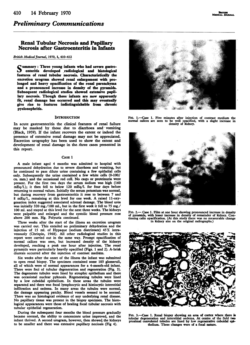
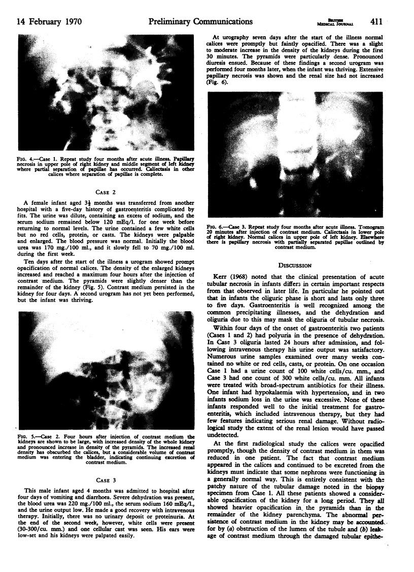
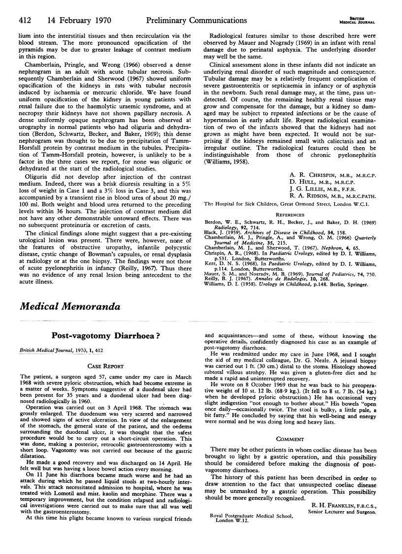
Three young infants who had severe gastroenteritis developed radiological and histological features of renal tubular necrosis. Characteristically the excretion urogram showed renal enlargement with prolonged and heavy opacification of the renal parenchyma and a pronounced increase in density of the pyramids. Subsequent radiological studies showed extensive papillary necrosis. Though these infants are now apparently fit, renal damage has occurred and this may eventually give rise to features indistinguishable from chronic pyelonephritis.
Full text
PDF


Images in this article
Selected References
These references are in PubMed. This may not be the complete list of references from this article.
- BLACK J. Renal tubular damage in infantile gastro-enteritis. Arch Dis Child. 1959 Apr;34(174):158–165. doi: 10.1136/adc.34.174.158. [DOI] [PMC free article] [PubMed] [Google Scholar]
- Berdon W. E., Schwartz R. H., Becker J., Baker D. H. Tamm-Horsfall proteinuria. Its relationship to prolonged nephrogram in infants and children and to renal failure following intravenous urography in adults with multiple myeloma. Radiology. 1969 Mar;92(4):714–722. doi: 10.1148/92.4.714. [DOI] [PubMed] [Google Scholar]
- Chamberlain M. J., Pringle A., Wrong O. M. Oliguric renal failure in the nephrotic syndrome. Q J Med. 1966 Apr;35(138):215–235. [PubMed] [Google Scholar]
- Reilly B. J. Infantile pyelonephritis. A preliminary report. Ann Radiol (Paris) 1967;10(3):268–272. [PubMed] [Google Scholar]


