Fractures caused by osteoporosis affect one in two women and one in five men over the age of 50, resulting in an estimated annual cost to the health services of around £1.8bn (€2.7bn; $3.5bn) in the United Kingdom and €30bn in all of Europe.1 2 Most patients with osteoporosis are managed in primary care, but a minority will benefit from referral to specialised centres. In recent years considerable advances have been made both in the identification of people at high risk of fracture and in therapeutic options to reduce the risk of fracture. This review focuses on these areas and also on the partnership that is required between primary and secondary care to optimise the management of patients with osteoporosis.
What is osteoporosis?
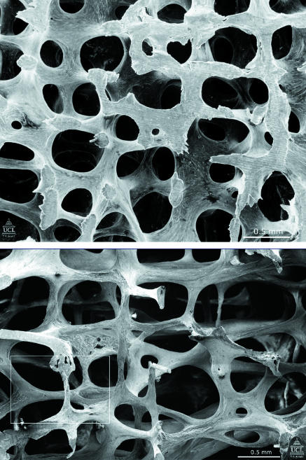
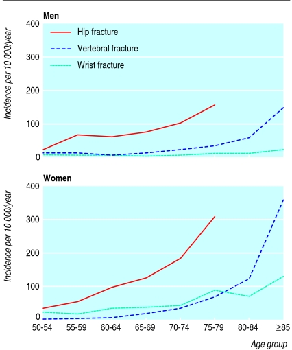
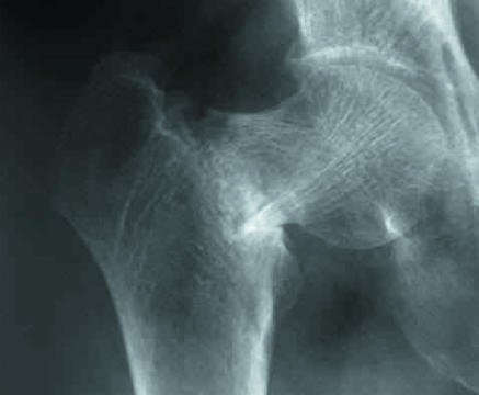
Osteoporosis results from reduced bone mass and disruption of the micro-architecture of bone (fig 1), giving decreased bone strength and increased risk of fracture, particularly of the spine, hip, wrist, humerus, and pelvis. The risk of fractures increases steeply with age (fig 2) and most of those affected are over 75.1 2 Globally, osteoporotic fractures caused an estimated 5.8 million disability adjusted life years in the year 2000w1 and are also associated with increased mortality. Hip fractures (fig 3) result in loss of independence for at least a third of people with osteoporosis, and vertebral fractures (fig 4) cause height loss, chronic pain, and difficulty with normal daily activities.
Fig 1 Scanning electron micrographs to show the structure of L3 vertebra in a 31 year old woman (top) and in a 70 year old woman (bottom). Note that many of the plate-like structures have become converted to thin rods
Fig 2 Epidemiology of osteoporotic fractures in men and women. Reprinted with permission28
Fig 3 Fracture of the femoral neck
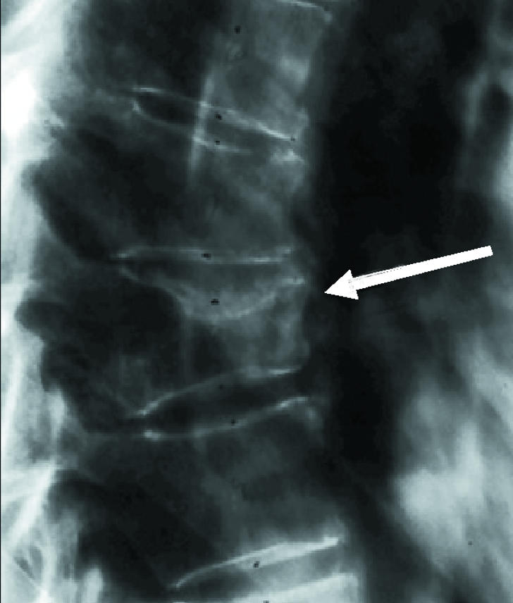
Fig 4 Vertebral fracture
What causes osteoporosis?
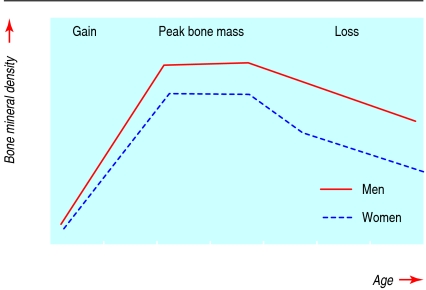
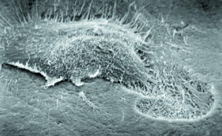
Age related bone loss starts in the fourth or fifth decade of life (fig 5). It occurs as a result of increased bone breakdown by osteoclasts (fig 6) and decreased bone formation by osteoblasts.3 The role of oestrogen deficiency in menopausal and age related bone loss in women is well documented, and bone mass in elderly men is also positively related to oestrogen levels. Vitamin D insufficiency and secondary hyperparathyroidism are common in elderly people and may contribute. Other possible factors are reduced physical activity with ageing and decreased production of insulin-like growth factors.
Fig 5 Age related changes in bone mass throughout life in women and men. Peak bone mass is attained in the third decade of life and age related bone loss probably starts around the age of 40 in both men and women. In women, bone loss accelerates around the time of the menopause
Fig 6 Scanning electron micrograph of an osteoclast resorbing bone
Genetic factors have a strong influence on peak bone mass, which is attained during the third decade of life and is an important determinant of bone mass later in life. Nutrition, particularly calcium and vitamin D intake, hormonal status, and physical activity also influence peak bone mass.
Although low bone mass has a major role in the pathogenesis of fracture, factors related to falling—risk of falling, protective response, and energy absorption— make an important contribution. In addition, aspects of bone composition and structure that may not be captured by bone mineral density measurements, such as bone size and geometry, and bone structure and material, contribute to increased bone fragility.
Methods
We searched PubMed with the terms osteoporosis plus randomised controlled trials (124 hits), systematic reviews (118), meta-analyses (218), and Cochrane database (15). We also searched using the terms risk factors plus osteoporosis and systematic reviews (34) or meta-analyses (58). We searched the “epub ahead of print” sections of the relevant specialist journals.
Ongoing research
Evaluation of new treatments, including:
A human monoclonal antibody to the receptor activator of NFkB ligand (RANKL), which is given subcutaneously once every six months
Oral calciomimetic drugs that stimulate intermittent production of parathyroid hormone
Selective oestrogen receptor modulators with mixed oestrogenic and anti-oestrogenic effects
Inhibitors of sclerostin, a protein produced by bone that is a negative regulator of bone formation, and its signalling pathway
Investigation of the causes and management of poor compliance and persistence
Assessment of the long term effects of anti-resorptive treatments on bone strength
Who is at risk of osteoporosis?
Lower peak bone mass, increased bone loss at the menopause, and greater longevity all confer a greater risk of osteoporosis in women than in men, and the disease is most commonly seen in postmenopausal women. Some of the risk factors for osteoporosis (box 1) are at least partially independent of bone mineral density,w2-w6 whereas the effect of others on fracture risk is mediated solely through reduced bone mineral density. Oral glucocorticoids, which are taken by about 1% of the population and 2.5% of those aged over 75, are a common cause of osteoporosis, and there are specific national guidelines for the prevention and management of glucocorticoid induced osteoporosis (see additional educational resources box).
Box 1 Risk factors for osteoporosis
Independent of bone mineral density
Age
Previous fragility fracture
Maternal history of hip fracture
Oral glucocorticoid therapy
Current smoking
Alcohol intake ≥3 units/day
Rheumatoid arthritis
Body mass index ≤19
Falls
Depending on bone mineral density
Untreated hypogonadism
Malabsorption
Endocrine disease
Chronic renal disease
Chronic liver disease
Chronic obstructive pulmonary disease
Immobility
Drugs (aromatase inhibitors, androgen deprivation therapy)
How does osteoporosis present?
Osteoporosis often presents as a clinically evident fracture. A low trauma fracture (following a fall from standing height or less) in someone aged over 45 should trigger the suspicion of osteoporosis. In other cases, osteoporosis may present as backache, height loss, spinal deformity, or radiological osteopenia.
Although most fractures due to osteoporosis present clinically, vertebral fractures may be asymptomatic in as many as two thirds of patients.4 It is important to detect these fractures since they carry a high risk of further fractures in the spine and elsewhere.5 Lateral x rays of the spine should be considered in patients with height loss or spinal deformity.
Who should be treated?
The World Health Organization's definition of osteoporosis is based on bone mineral density in the spine and proximal femur measured with dual energy x ray absorptiometry (DXA). Osteoporosis is classified as a bone mineral density 2.5 or more standard deviations below normal peak bone mass—that is, a T score ≤−2.5.6 Other techniques—for example, ultrasound of the calcaneus and peripheral DXA measurements—cannot be used in the same way to diagnose osteoporosis but are useful as a preliminary assessment of risk where access to axial DXA is inadequate.
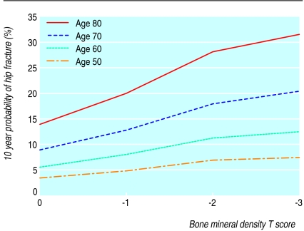
Although osteoporosis indicates a high likelihood of fracture, many fragility fractures occur in people with bone density values above the defined level.7 Fractures can be better predicted by adding clinical risk factors that contribute to fracture risk independently of bone mineral density (fig 7; box 1).8 This approach is being developed under the auspices of the WHO and will be delivered in the form of an algorithm that enables the probability of a fracture to be calculated from clinical risk factors with or without bone mineral density values. Intervention thresholds based on cost effectivenessw7 can be used to make a decision about treatment.9
Fig 7 Age affects fracture risk independently of bone mineral density. For any given bone density, the fracture probability increases with age. For example, at a T score of −2, the 10 year hip fracture probability at the age of 50 is around 5% but at the age of 80 it is around 30%29
People who have already had a fragility fracture are at greatly increased risk of sustaining a further fracture,5 and pharmacological intervention should be started promptly in such cases. Bone density measurement is not always required to confirm the diagnosis of osteoporosis, particularly in older patients, but is useful where the trauma preceding the fracture is uncertain or where other causes of fracture are suspected. Secondary causes of osteoporosis should be excluded (box 2).
Box 2 Routine investigations to exclude secondary causes of osteoporosis
Full blood count and erythrocyte sedimentation rate
Liver and renal function tests
Bone function tests (calcium, phosphate, and alkaline phosphatase)
Serum immunoglobulins and paraproteins, urinary Bence-Jones proteins
Thyroid function tests
Management of osteoporosis
Non-pharmacological measures
Falls have an important role in the pathogenesis of fragility fractures, particularly in frail and elderly people. Multiple medical and environmental factors increase the risk of falling and many of these are modifiable. Multifaceted interventions have been shown to reduce the frequency of falling but not fractures.10
Lifestyle measures to improve bone health include maintaining adequate dietary calcium intake and normal vitamin D status.w8 w9 Appropriate levels of exercise should be recommended and smoking and alcohol abuse discouraged.11 Physiotherapy and pain relief are important in managing fractures.
Pharmacological interventions
Therapeutic options for osteoporosis have increased considerably over recent years. Although most patients with osteoporosis can be managed in primary care, some patients benefit from specialist assessment: younger men and women, patients who continue to fracture despite treatment, and those who require assessment for anabolic treatments. Anabolic and intravenous treatments are generally instigated by hospital specialists; thereafter, shared care between primary and secondary organisations is appropriate.
Currently licensed treatments (table) include the bisphosphonates, raloxifene and hormone replacement therapy (which prevent bone resorption), strontium ranelate (uncertain mechanism of action), and parathyroid hormone peptides (anabolic). Without head to head comparison trials with fracture end points, the efficacy of these drugs cannot be directly compared. Some, but not all, have proved efficacy against vertebral and non-vertebral fractures, including hip fractures,w10 and this is an important factor influencing choice. Safety, tolerability, and cost are important considerations, and NICE (the National Institute for Health and Clinical Excellence) is currently assessing the cost effectiveness of different interventions in the primary and secondary prevention of osteoporotic fractures.w11
Pharmacological interventions for osteoporosis
| Intervention | Dosing regimen | Route of administration | Licensed indication |
|---|---|---|---|
| Alendronate | 70 mg once weekly, or 5 mg or 10 mg once daily | Oral | Postmenopausal osteoporosis; glucocorticoid-induced osteoporosis; osteoporosis in men |
| Etidronate | 400 mg daily for 2 weeks every 3 months | Oral | Postmenopausal osteoporosis; glucocorticoid-induced osteoporosis |
| Ibandronate | 150 mg once monthly | Oral | Postmenopausal osteoporosis |
| 3 mg once every 3 months | Intravenous injection | Postmenopausal osteoporosis | |
| Risedronate | 35 mg once weekly, or 5 mg once daily | Oral | Postmenopausal osteoporosis; glucocorticoid-induced osteoporosis |
| Raloxifene | 60 mg once daily | Oral | Postmenopausal osteoporosis |
| Strontium ranelate | 2 g once daily | Oral | Postmenopausal osteoporosis |
| Teriparatide | 20 µg once daily | Subcutaneous injection | Postmenopausal osteoporosis |
| Parathyroid hormone 1-84 | 100 µg once daily | Subcutaneous injection | Postmenopausal osteoporosis |
Bisphosphonates
Alendronate, etidronate, ibandronate, and risedronate are approved for treating postmenopausal osteoporosis. Alendronate, etidronate, and risedronate are approved for glucocorticoid induced osteoporosis, and alendronate is approved for osteoporosis in men. Because alendronate and risedronate have been shown to reduce vertebral and non-vertebral fractures, including hip fractures,12 13 14 15 w12 w13 they are considered first line options for preventing postmenopausal osteoporosis. Oral bisphosphonates must be taken fasting, with a full glass of water, and the individual must be upright and stay sitting or standing without taking food or drink for the next 30-60 minutes. Bisphosphonates are generally well tolerated but may be associated with upper gastrointestinal side effects, particularly if the dosing regimen is not adhered to.
An intravenous formulation of ibandronate is approved for postmenopausal osteoporosis. It is given as an injection over 15-30 seconds every three months. Antifracture efficacy has not been directly shown for this formulation or for the oral 150 mg once monthly regimen, but it is assumed from a bridging study based on changes in bone mineral density.16 17
Strontium ranelate
Strontium ranelate (a sachet mixed with water and taken daily) reduces vertebral and non-vertebral (including hip) fractures in postmenopausal women with osteoporosis.18 19 Adverse events are generally mild and include diarrhoea and headache. The spectrum of anti-fracture efficacy of strontium ranelate makes it an alternative front line option to alendronate or risedronate,w14 particularly in people for whom these drugs are contraindicated or are not tolerated.
Raloxifene
Raloxifene reduces the risk of vertebral fractures, but has not been shown to prevent fractures at other sites.20 w15 Side effects include hot flushes, leg cramps, and a threefold increase in the relative risk of venous thromboembolism. Raloxifene also protects against breast cancer.21 It can be regarded as a second line option in younger postmenopausal women with vertebral osteoporosis.
Parathyroid hormone peptides
Teriparatide (recombinant 1-34 parathyroid hormone), given as a subcutaneous daily injection of 20 µg, reduces vertebral and non-vertebral fractures in postmenopausal women with osteoporosis.22 w16 Preotact, the full 1-84 parathyroid hormone peptide, has recently been approved and is given in the same way in a daily dose of 100 µg. Neither of these interventions has been shown to reduce hip fractures. Because they cost more than other options, they are reserved for patients with severe osteoporosis who are unable to tolerate or seem to be unresponsive to other treatments.
Hormone replacement therapy
Because the risk-benefit balance of hormone replacement therapy is generally unfavourable in older postmenopausal women, it is regarded as a second line treatment option.23 It is an appropriate option in younger postmenopausal women at high risk of fracture, particularly those with vasomotor symptoms.
Calcium and vitamin D
The available evidence does not support a role for calcium and vitamin D alone in preventing osteoporotic fractures, except in institutionalised elderly people.24 25 w17 Calcium and vitamin D supplements should be prescribed with other treatments for osteoporosis since the evidence base for their efficacy in preventing fractures is derived from studies in which calcium and vitamin D were routinely administered.
Monitoring of treatment
Whether treatment response should be monitored and, if so, whether bone density measurements or biochemical markers should be used, is unclear. Compliance and persistence with osteoporosis treatments need to be improved26; possible approaches include better patient education and the use of intermittent dosing regimens, such as once weekly or once monthly oral bisphosphonate therapy and three monthly intravenous ibandronate. Even longer dosing intervals are expected in the near future, with the likely approval of once yearly intravenous zoledronic acid.27
Summary points
Fragility fractures caused by osteoporosis affect one in two women and one in five men over the age of 50
Vertebral fractures in particular remain under-recognised and under-treated and are associated with poor health outcomes
A range of pharmacological treatments is effective in reducing the risk of fracture
Poor patient compliance and persistence with prescribed treatments for osteoporosis are common but may be improved by better patient education
A minority of patients will benefit from assessment in secondary care for starting anabolic treatments or interventional techniques, particularly when other treatments have failed
Patient's story
My name is Jean Marsh and I am 73 years old. I had “sailed” through the menopause by the age of 45 and led an active life. My health was excellent until I was 58 years old, when one morning I noticed a dull ache between my shoulder blades. This got progressively worse and I saw my GP a few days later. He couldn't identify the problem and gave me painkillers. Later that evening I was in terrible pain. The next day I asked for an x ray and this showed collapse of the T7 vertebra.
My GP gave me a leaflet from the National Osteoporosis Society, which I joined immediately. Of the free booklets that I received, How to Cope was wonderful and I remember crying with joy that someone understood what I was going through.
The pain lasted for about 10 weeks, lessening a little as the weeks went by. I was terrified of falling, so going out was difficult. I couldn't lie down in bed and had to sleep propped up by lots of pillows. Clothes were difficult because of my new shape.
I was prescribed etidronate followed by HRT. My bone density increased during this time, and my fear of falling gradually disappeared. I now lead a very busy, normal life. I do have a weakness in my spine, which aches when I do too much gardening, but other than that I am now fine.
Tips for non-specialists
Patients at risk of osteoporosis can be identified by using clinical risk factors
Treatment should be started as rapidly as possible in patients presenting with a fragility fracture
The patient's preference is an important consideration in choosing treatment options
Compliance and persistence with treatments for osteoporosis are poor but can be improved by better patient education
Audit should focus on high risk groups, such as patients taking glucocorticoids and patients with previous fragility fracture
Additional educational resources
Sambrook P, Cooper C. Osteoporosis. Lancet 2006;367:2010-8.
Department of Health and Human Services. Bone health and osteoporosis: a report of the surgeon general. Rockville: US Department of Health and Human Services, Office of the Surgeon General, 2004.
Guidelines Working Group for the Bone and Tooth Society, National Osteoporosis Society, and Royal College of Physicians. Glucocorticoid-induced osteoporosis: guidelines for prevention and treatment. London: Royal College of Physicians, 2002. (www.rcplondon.ac.uk/pubs/books/glucocorticoid/index.asp)
Bone Research Society website picture gallery (www.brsoc.org.uk/gallery/default.htm)
Information resources for patients
National Osteoporosis Society (www.nos.org.uk)—UK charity that raises awareness of osteoporosis and provides patient support and education. The website contains links to the BMJ, several professional and patient societies, and the Department of Health and a number of educational resources for patients and professionals
International Osteoporosis Foundation (www.iofbonehealth.org)—a non-profit organisation that advocates the early prevention, diagnosis, and treatment of osteoporosis and related bone diseases and provides educational material for patients and healthcare professionals. A comprehensive teaching package on vertebral fractures is provided on this website
Supplementary Material
We thank T R Arnett, University College London, for the scanning electron micrographs (figures 1 and 6).
Competing interests: KESP has been reimbursed by the Alliance for Better Bone Health for attending a scientific conference. JEC has received payment for consultancy work and speaking engagements from Procter & Gamble, Eli Lilly, GSK/Roche, Amgen, Pfizer, Servier, Shire, Novartis and Nycomed and has been reimbursed for attending scientific conferences by Procter & Gamble, Eli Lilly, and Servier. She receives funding for a grant from Procter & Gamble and has acted as an expert witness in several cases of glucocorticoid-induced osteoporosis and in an alendronate patent dispute.
References
- 1.Dolan P, Torgerson DJ. The cost of treating osteoporotic fractures in the United Kingdom female population. Osteoporos Int 2000;11:551-2. [DOI] [PubMed] [Google Scholar]
- 2.Cummings SR, Melton III LJ. Epidemiology and outcomes of osteoporotic fractures. Lancet 2002;359:1761-7. [DOI] [PubMed] [Google Scholar]
- 3.Compston JE. Sex steroids and bone. Physiol Rev 2001;81:419-47. [DOI] [PubMed] [Google Scholar]
- 4.Cooper C, Atkinson EJ, O'Fallon WM, Melton LJ. Incidence of clinically diagnosed vertebral fractures: a population-based study in Rochester, Minnesota, 1985-1989. J Bone Miner Res 1992;7:221-7. [DOI] [PubMed] [Google Scholar]
- 5.Klotzbuecher CM, Ross PD, Landsman PB, Abbott III TA, Berger M. Patients with prior fractures have an increased risk of future fractures: a summary of the literature and statistical synthesis. J Bone Miner Res 2000;15:721-39. [DOI] [PubMed] [Google Scholar]
- 6.WHO Study Group. Assessment of fracture risk and its application to postmenopausal osteoporosis. World Health Organ Tech Ser 1984;(843). [PubMed]
- 7.Siris ES, Chen Y-T, Abbott TA, Barrett-Connor E, Miller PD, Wehren LE, et al. Bone mineral density thresholds for pharmacological interventions to prevent fractures. Arch Intern Med 2004;164:1108-12. [DOI] [PubMed] [Google Scholar]
- 8.Kanis JA. Diagnosis of osteoporosis and assessment of fracture risk. Lancet 2002;359:1929-36. [DOI] [PubMed] [Google Scholar]
- 9.Kanis JA, Johnell O, Oden A, de Laet C, Oglesby A, Jönsson B. Intervention thresholds for osteoporosis. Bone 2002;31:26-31. [DOI] [PubMed] [Google Scholar]
- 10.Close J, Ellis M, Hooper R, Glucksman E, Jackson S, Swift C. Prevention of falls in the elderly trial (PROFET): a randomised controlled trial. Lancet 1999;353:93-7. [DOI] [PubMed] [Google Scholar]
- 11.Hind K, Burrows M. Weight-bearing exercise and bone mineral accrual in children and adolescents: a review of controlled trials. Bone 2006. Sep 4(online publication). [DOI] [PubMed]
- 12.Black D, Cummings S, Karpf D, Cauley JA, Thompson DE, Nevitt MC, et al. Randomised trial of effect of alendronate on risk of fracture in women with existing vertebral fractures. Lancet 1996;348:1535-41. [DOI] [PubMed] [Google Scholar]
- 13.Harris ST, Watts NB, Genant HK, McKeever CD, Hangartner T, Keller M, et al. Effect of risedronate treatment on vertebral and non vertebral fractures in women with postmenopausal osteoporosis. A randomized controlled trial. JAMA 1999;282:1344-52. [DOI] [PubMed] [Google Scholar]
- 14.Reginster JY, Minne H, Sorensen O, Hooper M, Roux C, Brandi ML, et al. Randomized trial of the effects of risedronate on vertebral fractures in women with established postmenopausal osteoporosis. Osteoporos Int 2000;11:83-91. [DOI] [PubMed] [Google Scholar]
- 15.McClung MR, Geusens P, Miller PD, Zippel H, Benson WG, Roux C, et al. Effect of risedronate on the risk of hip fracture in elderly women. Hip Intervention Program Study Group. N Engl J Med 2001;344:333-40. [DOI] [PubMed] [Google Scholar]
- 16.Delmas PD, Recker RR, Chesnut CH 3rd, Skag A, Stakkestad JA, Emkey R, et al. Daily and intermittent oral ibandronate normalize bone turnover and provide significant reduction in vertebral fracture risk: results from the BONE study. Osteoporos Int 2004;15:792-8. [DOI] [PubMed] [Google Scholar]
- 17.Delmas PD, Adami S, Strugala C, Stakkestad JA, Reginster JY, Felsenberg D, et al. Intravenous ibandronate injections in postmenopausal women with osteoporosis: one-year results from the dosing intravenous administration study. Arthritis Rheum 2006;54:1838-46. [DOI] [PubMed] [Google Scholar]
- 18.Meunier PJ, Roux C, Seeman E, Ortolani S, Badurski J, Spector TD, et al. The effects of strontium ranelate on the risk of vertebral fracture in women with postmenopausal osteoporosis. N Engl J Med 2004;350:459-68. [DOI] [PubMed] [Google Scholar]
- 19.Reginster JY, Seeman E, De Vernejoul MC, Adami S, Compston J, Phenekos C, et al. Strontium ranelate reduces the risk of nonvertebral fractures in postmenopausal women with osteoporosis: Treatment of Peripheral Osteoporosis (TROPOS) study. J Clin Endocrinol Metab 2005;90:2816-22. [DOI] [PubMed] [Google Scholar]
- 20.Ettinger B, Black DM, Mitlak BH, Knickerbocker RK, Nickelsen T, Genant HK, et al. Reduction of vertebral fracture risk in postmenopausal women with osteoporosis treated with raloxifene: results from a 3-year randomized clinical trial. JAMA 1999;282:637-45. [DOI] [PubMed] [Google Scholar]
- 21.Cummings SR, Eckert S, Krueger KA, Grady D, Powles TJ, Cauley JA, et al. The effect of raloxifene on risk of breast cancer in postmenopausal women. JAMA 1999;281:2189-97. [DOI] [PubMed] [Google Scholar]
- 22.Neer RM, Arnaud CD, Zanchetta JR, Prince R, Gaich GA, Reginster JY. Effect of parathyroid hormone on vertebral bone mass and fracture incidence among postmenopausal women with osteoporosis. N Engl J Med 2001;344:1434-41. [DOI] [PubMed] [Google Scholar]
- 23.Compston J. How to manage osteoporosis after the menopause. Best Pract Res Clin Rheumatol 2005;19:1007-19. [DOI] [PubMed] [Google Scholar]
- 24.Bischoff-Ferrari HA, Willett WC, Wong JB, Giovannucci E, Dietrich T, Dawson-Hughes B. Fracture prevention with vitamin D supplementation: a meta-analysis of randomized controlled trials. JAMA 2005;293:2257-64. [DOI] [PubMed] [Google Scholar]
- 25.Boonen S, Bischoff-Ferrari HA, Cooper C, Lips P, Ljunggren O, Meunier PJ, et al. Addressing the musculoskeletal components of fracture risk with calcium and vitamin D: a review of the evidence. Calcif Tissue Int 2006;78:257-70. [DOI] [PubMed] [Google Scholar]
- 26.Compston JE, Seeman E. Compliance with osteoporosis therapy is the weakest link. Lancet 2006;368:973-4. [DOI] [PubMed] [Google Scholar]
- 27.Reid IR, Brown JP, Burckhardt P, Horowitz Z, Richardson P, Treschel U, et al. Intravenous zoledronic acid in postmenopausal women with low bone mineral density. N Engl J Med 2002;346:653-61. [DOI] [PubMed] [Google Scholar]
- 28.Dennison E, Cole Z, Cooper C. Diagnosis and epidemiology of osteoporosis. Curr Opinion Rheumatol 2005;17:456-61. [DOI] [PubMed] [Google Scholar]
- 29.Kanis JA, Johnell O, Oden A, Dawson A, De Laet C, Jonsson B. et al. Ten year probabilities of osteoporotic fractures according to BMD and diagnostic thresholds. Osteoporosis Int 2001;12:989-95 [DOI] [PubMed] [Google Scholar]
Associated Data
This section collects any data citations, data availability statements, or supplementary materials included in this article.


