Abstract
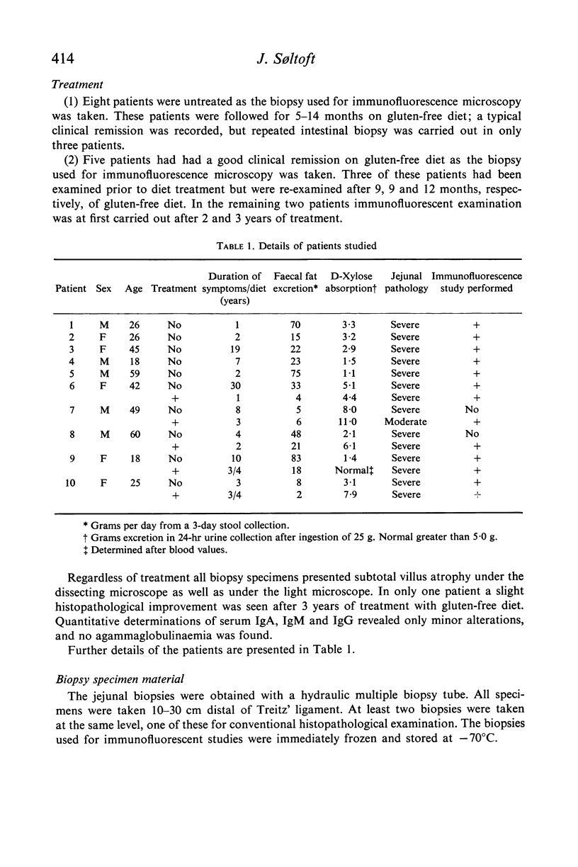
Immunofluorescent microscopy studies of jejunal biopsies from ten patients with gluten-induced enteropathy are reported. Eight biopsies were examined before and five after onset of gluten-free diet.
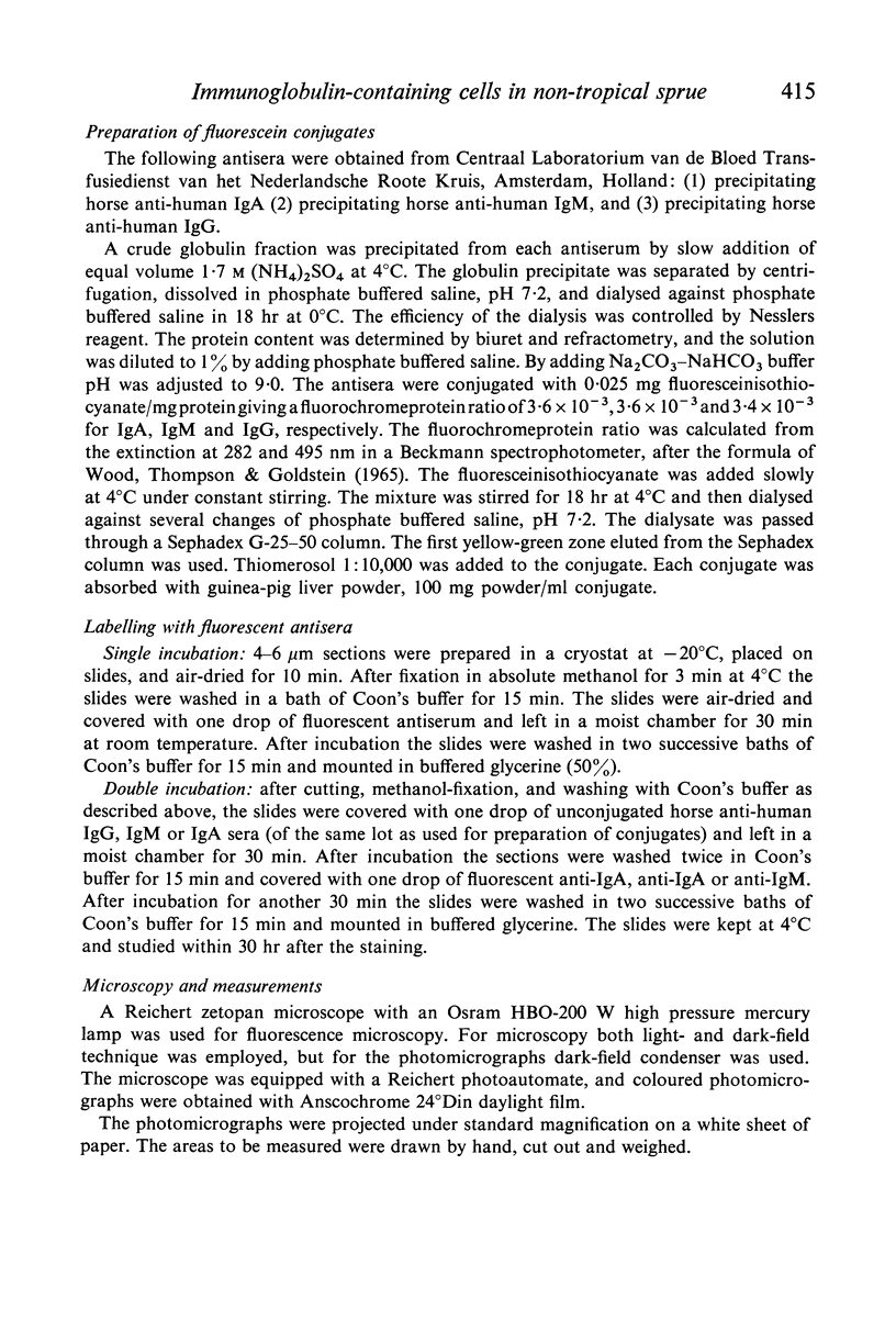
Fluorescent antisera specifically reacting with IgA, IgG and IgM were applied, and accordingly the cells were differentiated and quantitated.
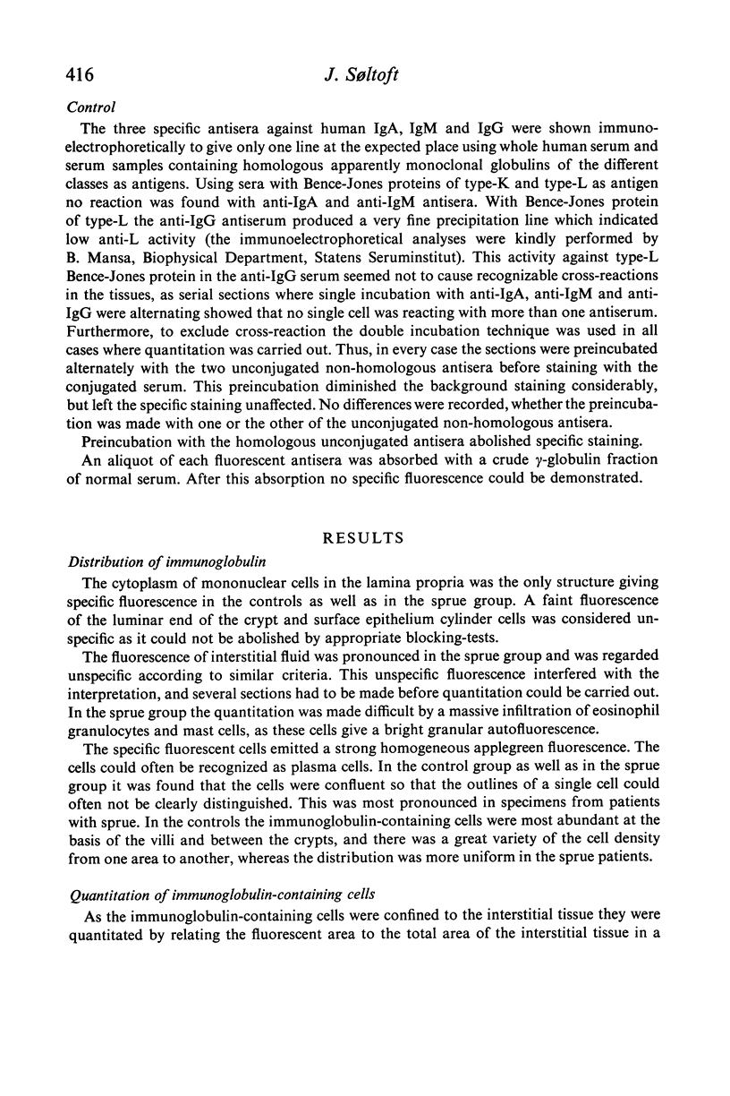
The immunofluorescence microscopy revealed an increased amount of immunoglobulin-containing cells in both treated and untreated cases of sprue. The distribution of the immunoglobulin-containing cells within the three immunoglobulin classes was significantly altered as the proportion of IgM- and IgG-containing cells was elevated.
Full text
PDF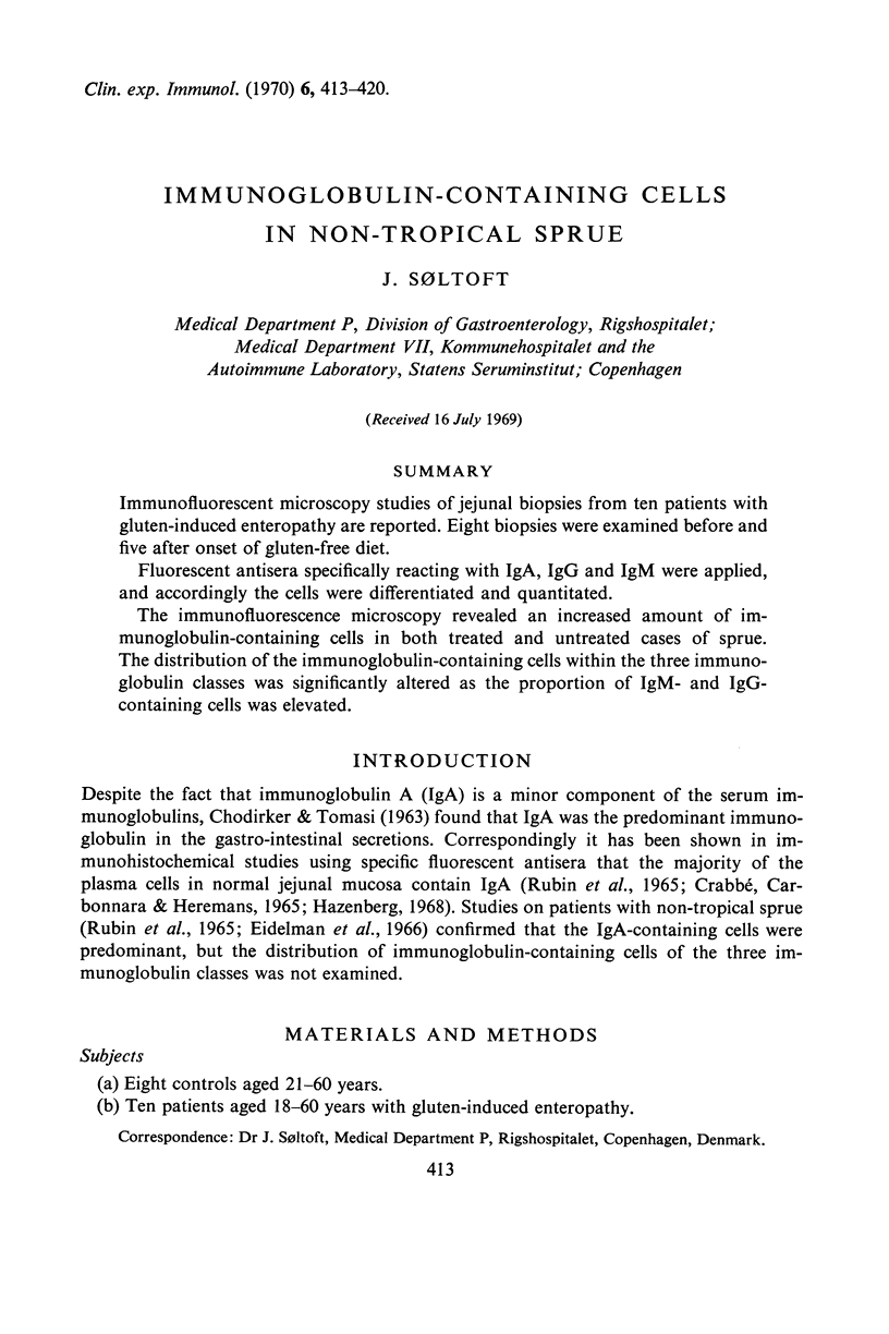
Selected References
These references are in PubMed. This may not be the complete list of references from this article.
- CHODIRKER W. B., TOMASI T. B., Jr GAMMA-GLOBULINS: QUANTITATIVE RELATIONSHIPS IN HUMAN SERUM AND NONVASCULAR FLUIDS. Science. 1963 Nov 22;142(3595):1080–1081. doi: 10.1126/science.142.3595.1080. [DOI] [PubMed] [Google Scholar]
- CRABBE P. A., CARBONARA A. O., HEREMANS J. F. THE NORMAL HUMAN INTESTINAL MUCOSA AS A MAJOR SOURCE OF PLASMA CELLS CONTAINING GAMMA-A-IMMUNOGLOBULIN. Lab Invest. 1965 Mar;14:235–248. [PubMed] [Google Scholar]
- RUBIN W., FAUCI A. S., MARVIN S. F., SLEISENGER M. H., JEFRIES G. H. IMMUNOFLUORESCENT STUDIES IN ADULT CELIAC DISEASE. J Clin Invest. 1965 Mar;44:475–485. doi: 10.1172/JCI105161. [DOI] [PMC free article] [PubMed] [Google Scholar]
- Söltoft J. Immunoglobulin-containing cells in normal jejunal mucosa and in ulcerative colitis and regional enteritis. Scand J Gastroenterol. 1969;4(4):353–360. doi: 10.3109/00365526909180616. [DOI] [PubMed] [Google Scholar]
- Wood B. T., Thompson S. H., Goldstein G. Fluorescent antibody staining. 3. Preparation of fluorescein-isothiocyanate-labeled antibodies. J Immunol. 1965 Aug;95(2):225–229. [PubMed] [Google Scholar]


