

Abstract
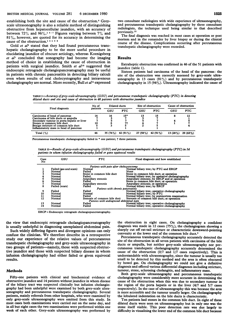
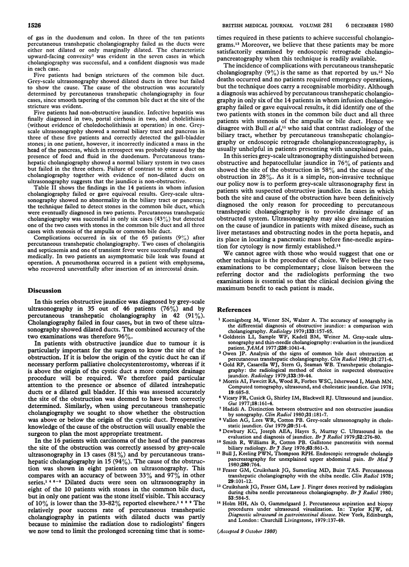
Fifty-one patients with suspected obstructive jaundice and 14 without jaundice in whom disease of the biliary tract was suspected but infusion cholangiography had been unhelpful were examined by grey-scale ultrasonography and percutaneous transhepatic cholangiography and the findings analysed retrospectively. Grey-scale ultrasonography distinguished between obstructive and hepatocellular jaundice in 35 out of 46 patients (76%) and indicated the site of the obstruction in 27 (58%) and the cause of the obstruction in 13 (28%). Percutaneous transhepatic cholangiography distinguished between obstructive and hepatocellular jaundice in 42 of the patients (91%) and indicated the site of the obstruction in 42 (91%) and the cause in 29 (63%). In the 14 patients without jaundice percutaneous transhepatic cholangiography showed bile-duct stones in one an ampullary stenosis in three. It is concluded that grey-scale ultrasonography and percutaneous transhepatic cholangiography are complementary examinations and that ultrasonography should always be undertaken first as it is a non-invasive procedure that may provide the surgeon with all the diagnostic information he requires. Percutaneous transhepatic cholangiography should be performed when grey-scale ultrasonography has shown dilated bile ducts but failed to provide adequate diagnostic information. Cholangiography is also required when preoperative percutaneous drainage of the bile duct is contemplated. In those patients in whom grey-scale ultrasonography shows non-dilated ducts endoscopic retrograde cholangiopancreatography is probably the contract examination of choice.
Full text
PDF
Selected References
These references are in PubMed. This may not be the complete list of references from this article.
- Bull J., Keeling P. W., Thompson R. P. Endoscopic retrograde cholangiopancreatography for unexplained upper abdominal pain. Br Med J. 1980 Mar 15;280(6216):764–764. doi: 10.1136/bmj.280.6216.764. [DOI] [PMC free article] [PubMed] [Google Scholar]
- Cruikshank J. G., Fraser G. M., Law J. Finger doses received by raediologists during Chiba needle percutaneous cholangiography. Br J Radiol. 1980 Jun;53(630):584–585. doi: 10.1259/0007-1285-53-630-584. [DOI] [PubMed] [Google Scholar]
- Dewbury K. C., Joseph A. E., Hayes S., Murray C. Ultrasound in the evaluation and diagnosis of jaundice. Br J Radiol. 1979 Apr;52(616):276–280. doi: 10.1259/0007-1285-52-616-276. [DOI] [PubMed] [Google Scholar]
- Fraser G. M., Cruikshank J. G., Sumerling M. D., Buist T. A. Percutaneous transhepatic cholangiography with the Chiba Needle. Clin Radiol. 1978 Jan;29(1):101–112. doi: 10.1016/s0009-9260(78)80175-5. [DOI] [PubMed] [Google Scholar]
- Gold R. P., Casarella W. J., Stern G., Seaman W. B. Transhepatic cholangiography: the radiological method of choice in suspected obstructive jaundice. Radiology. 1979 Oct;133(1):39–44. doi: 10.1148/133.1.39. [DOI] [PubMed] [Google Scholar]
- Goldstein L. I., Sample W. F., Kadell B. M., Weiner M. Gray-scale ultrasonography and thin-needle cholangiography. Evaluation in the jaundiced patient. JAMA. 1977 Sep 5;238(10):1041–1044. [PubMed] [Google Scholar]
- Hadidi A. Distinction between obstructive and non-obstructive jaundice by sonography. Clin Radiol. 1980 Mar;31(2):181–187. doi: 10.1016/s0009-9260(80)80151-6. [DOI] [PubMed] [Google Scholar]
- Koenigsberg M., Wiener S. N., Walzer A. The accuracy of sonography in the differential diagnosis of obstructive jaundice: a comparison with cholangiography. Radiology. 1979 Oct;133(1):157–165. doi: 10.1148/133.1.157. [DOI] [PubMed] [Google Scholar]
- Morris A., Fawcitt R. A., Wood R., Forbes W. S., Isherwood I., Marsh M. N. Computed tomography, ultrasound, and cholestatic jaundice. Gut. 1978 Aug;19(8):685–688. doi: 10.1136/gut.19.8.685. [DOI] [PMC free article] [PubMed] [Google Scholar]
- Owen J. P. Analysis of the signs of common bile duct obstruction at percutaneous transhepatic cholangiography. Clin Radiol. 1980 May;31(3):271–276. doi: 10.1016/s0009-9260(80)80216-9. [DOI] [PubMed] [Google Scholar]
- Smith R., Williams R., Cotton P. B. Gallstone pancreatitis with normal biliary radiology. Br J Surg. 1976 Nov;63(11):861–863. doi: 10.1002/bjs.1800631106. [DOI] [PubMed] [Google Scholar]
- Vallon A. G., Lees W. R., Cotton P. B. Grey-scale ultrasonography in cholestatic jaundice. Gut. 1979 Jan;20(1):51–54. doi: 10.1136/gut.20.1.51. [DOI] [PMC free article] [PubMed] [Google Scholar]
- Vicary F. R., Cusick G., Shirley I. M., Blackwell R. J. Ultrasound and jaundice. Gut. 1977 Feb;18(2):161–164. doi: 10.1136/gut.18.2.161. [DOI] [PMC free article] [PubMed] [Google Scholar]