Abstract
Diketo acids such as L-731,988 are potent inhibitors of HIV-1 integrase that inhibit integration and viral replication in cells. These compounds exhibit the unique ability to inhibit the strand transfer activity of integrase in the absence of an effect on 3′ end processing. To understand the reasons for this distinct inhibitory profile, we developed a scintillation proximity assay that permits analysis of radiolabeled inhibitor binding and integrase function. High-affinity binding of L-731,988 is shown to require the assembly of a specific complex on the HIV-1 long terminal repeat. The interaction of L-731,988 with the complex and the efficacy of L-731,988 in strand transfer can be abrogated by the interaction with target substrates, suggesting competition between the inhibitor and the target DNA. The L-731,988 binding site and that of the target substrate are thus distinct from that of the donor substrate and are defined by a conformation of integrase that is only adopted after assembly with the viral end. These results elucidate the basis for diketo acid inhibition of strand transfer and have implications for integrase-directed HIV-1 drug discovery efforts.
The emergence of HIV-1 strains resistant to the current generation of reverse transcriptase and protease inhibitors highlights the necessity of developing new antivirals with novel mechanisms of action. Along with reverse transcriptase and protease, integrase is one of three enzymes encoded in the HIV-1 genome. Integrase is essential for viral replication (1, 2), making it an attractive but unexploited target for antiretroviral therapy.
Integrase catalyzes two reactions that are required for the insertion of the reverse-transcribed viral genome into the host DNA (3–5). In the first reaction, endonucleolytic cleavage, the terminal two 3′ nucleotides are removed from the U3 and U5 regions at each end of the HIV-1 DNA. After 3′ end processing, integrase catalyzes strand transfer between the recessed viral DNA ends and the cellular DNA. Both of these reactions can be recapitulated in vitro, using recombinant integrase and synthetic oligonucleotide substrates (3, 5, 6). The sequence-specific oligonucleotide representing the U3 or U5 end of HIV-1 is referred to as the viral end or donor substrate, and the nonspecific oligonucleotide that mimics the cellular DNA is termed the target substrate. In vitro, integrase also catalyzes a disintegration reaction. In this reaction integrase excises the viral DNA end and joins adjacent target sequences from a branched oligonucleotide. Because this substrate mimics an integration intermediate, disintegration is referred to as the reverse of integration (7).
Although unique with respect to substrate(s), 3′ processing, strand transfer, and disintegration have similar requirements. Each activity is mediated by a multimeric complex, requiring at least one intact integrase active site and the conserved amino acids Asp64, Asp116, and Glu152 (8–10). A specific U3 or U5 viral DNA sequence is required to assemble a stable, enzymatically active complex (11–14). Either manganese or magnesium is essential for assembly of the stable complex and as a catalytic cofactor (13–16).
We recently reported the identification of a series of diketo acids (DKAs) that prevent HIV-1 replication by inhibiting strand transfer and integration (17). Unlike previously described inhibitors, the DKAs inhibit strand transfer at concentrations that are significantly below the concentration required to inhibit either 3′ processing or disintegration. Inasmuch as 3′ processing of the viral DNA ends is observed in HIV-1 infected cells at concentrations of the inhibitor that block integration and suppress viral replication (17), the antiviral activity of the DKAs is due exclusively to their effect on strand transfer. Resistance to the DKAs maps to residues within the integrase active site, validating integrase itself as the target for inhibition (17). The DKAs represent a unique class of antiviral agents, the ability of which to differentiate between strand transfer and 3′ processing suggests that they may be exploited to dissect the integration reaction in greater detail.
To study interactions with integrase and determine the mechanism of action, a novel scintillation proximity assay (SPA) was developed, using a representative DKA, L-731,988. L-731,988 is shown to selectively bind the integrase donor substrate complex and inhibit strand transfer by competing with the target DNA. DKAs thus recognize a conformation of the integrase active site that is defined only subsequent to assembly on a specific viral DNA end. Because binding of the DKA is incompatible with the target substrate, these results suggest that the target site itself is configured upon assembly of the stable complex.
Materials and Methods
Materials.
Oligonucleotides were from Midland Certified Reagent (Midland, TX). The N-terminal epitope-tagged enzymes were made by ligating annealed oligonucleotides encoding the FLAG peptide amino acid sequence (5′-pTATGGACTACAAGGACGACGATGACAAGGG)/(5′-pTACCCTTGTCATCGTCCTTGTAGTCCA-3′) to NdeI-digested pET3c NL4–3 integrase expression plasmids (18). Proteins were purified as described (18). [3H]L-731,988 was labeled by palladium-catalyzed gas tritiation of 4-[1-(4-fluorobenzyl)-4-iodopyrrole-2-yl]-2,4-diketobutanoic acid (described in ref. 19), purified by HPLC, and stored in 2:1 EtOH:H2O containing l-ascorbic acid. Dilutions were made in 100% DMSO, and all reactions were performed in 10% DMSO.
SPA with Immobilized Integrase.
FLAG-integrase was immobilized on streptavidin-poly(vinyl toluene) SPA beads (Amersham Pharmacia) as follows. Beads were resuspended to10 mg/ml in buffer A (30 mM Hepes (pH 7.8)/30 mM MnCl2/120 μg/ml BSA/5 mM 2-mercaptoethanol) with 150 mM NaCl. Anti-FLAG biotinylated antibody Bio M2 (Sigma) was added to 250 nM and incubated at room temperature for 30 min. The beads were washed, and FLAG-integrase (final concentration 28 μg/ml) in buffer B (50 mM Tris/1 M NaCl/2 mM 2-mercaptoethanol/20% glycerol) was added to beads resuspended at 2 mg/ml. After 1–2 h at room temperature, the beads were washed and dispensed at 0.4 mg/ml in buffer A + 50 mM NaCl into a microtiter plate with test DNA and [3H]L-731,988 as appropriate. The plates were incubated overnight in the dark at room temperature, and scintillation was measured on a Hewlett Packard Top Count (3 min per well). In saturation binding assays, the DNA concentration was 25 nM. [3H]L-731,988 was titrated or used at 35 nM. The DNAs used were U5 (5′-TAGTCAGTGTGGAAAATCTCTAGCAGT/complement), U3 (5′-CGTTGGGAGTGAATTAGCCCTTCCAGT/complement) (20), NS Δ3′/NS (5′-CGCAGAAGTTAACACTTTCGGATATTTCAG/(5′-GACTGAAATATCCGAAAGTGTTAACTTCTGCG), and Dumbbell (5′-TGCTAGTTCTAGCAGGCCCTTGGGCCGGCGCTTGCGCC).
SPA with Immobilized DNA.
The 2′,3′-dideoxy U5 was made by adding 2′,3′-dideoxyATP (Roche Molecular Biochemicals) to 5′-GTGTGGAAAATCTCTAGC, using terminal transferase (Roche Molecular Biochemicals), and gel purifying the resulting 19-mer. The 3′ biotinylated U5 complement (5′-ACTGCTAGAGATTTTCCACAC) was annealed to the dideoxy strand. The 2′,3′-dideoxy U5 DNA (final concentration, 365 nM) was added to 10 mg/ml SPA beads and incubated as described above. NL4–3 integrase (final concentration, 500 nM) was added to 2-mg/ml beads with immobilized DNA. After incubation for 1–2 h, the complexes were dispensed into a microtiter plate containing buffer A with 50 mM NaCl, [3H]L-731,988 and target DNA (5–1,000 nM). Target DNAs used for competition were (5′-TATGGACTACAAGGACGACGATGACAAGGG-3′/TACCCTTGTCATCGTCCTTGTAGTCCA-3′ and 5′-CGCAGAAGTTAACACTTTCGGATATTTCAG-3′/complement). Single-stranded 20-mers used for competition were G-rich (5′-GGGGCCGCGGGGCGGGGGGG-3′) and T-rich (5′-TTTATTTATTATTATTTATT). Incubation and quantification were as described.
Integrase Assay.
The microtiter plate assay for strand transfer activity was performed as described (16,18). Assays for strand transfer and 3′ processing used FLAG-integrase assembled on SPA beads as described above. Beads were incubated for 60 min at 37°C in buffer A with 50 mM NaCl, 50 nM 32P-labeled U5 viral ends, and L-731,988 or 10% DMSO. The beads were pelleted (5 min at 1,400 × g) and resuspended in loading buffer (80% formamide in 90 mM Tris/64.6 mM boric acid/2.5 mM EDTA, pH 8.3), and the reactions were analyzed by 20% PAGE. Analysis of the supernatants revealed that strand transfer products remain associated with the beads. Bands were quantified by using a Molecular Dynamics Storm 840 phosphorimager. Disintegration assays were performed by using 50 nM 32P-labeled dumbbell substrate.
Exonuclease Protection Assay.
The 32P-labeled 2′,3′-dideoxy oligonucleotide was bound to beads as described above. After incubation with integrase, the beads were diluted 1:4 into buffer A with 10% DMSO and target DNA (1.4–1,000 nM). After overnight incubation at room temperature, beads were washed twice each in buffer A with 250 mM NaCl and 50 mM NaCl, and then incubated for 5 min at room temperature in 5 units/μl exonuclease III (Promega). The reactions were analyzed by 20% PAGE and autoradiography.
Results
FLAG-SPA Design and Activity of Epitope-Tagged Integrase Attached to Beads.
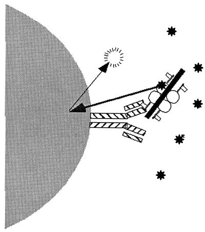
To study the interaction between the DKA inhibitors and integrase, we developed an assay to permit the evaluation of both integrase activity and inhibitor binding. NL4–3 integrase was modified with a FLAG-epitope tag at the amino terminus (FLAG-integrase) and immobilized onto streptavidin-coated SPA beads by means of biotinylated anti-FLAG antibodies (Fig. 1). The immobilized integrase was used in SPAs with a representative radiolabeled DKA, [3H]-labeled L-731,988. In scintillation proximity, enhanced scintillation is observed only when the radiolabel is in proximity to the SPA beads; thus an increase in signal indicates direct interaction between the inhibitor and the immobilized enzyme (Fig. 1) (21, 22). Specific binding of L-731,988 is defined as the signal that requires integrase and can be competed by the presence of structurally related inhibitors (data not shown and as seen in Figs. 3 and 5).
Figure 1.
Schematic of the FLAG-SPA. Biotinylated anti-FLAG antibody (striped bars) is bound to SPA beads. FLAG-integrase (circles with flags) associates with the SPA beads via interaction with the anti-FLAG antibody; the immobilized enzyme binds [3H]L-731,988 (✷) and/or DNA (solid bar). [3H]L-731,988 stimulates scintillation specifically when brought into close proximity to the SPA beads (arrows) by association with integrase.
Figure 3.
Stimulation of binding of L-731,988 to FLAG-integrase by HIV-1 donor DNA. (A) FLAG-integrase associated with SPA beads was incubated with labeled L-731,988 titrated in the absence (●) or presence (■) of 100 nM U5 DNA. (B) FLAG-integrase associated with SPA beads was incubated with labeled L-731,988 (35 nM) and unlabeled L-731,988 titrated in the absence (●) or presence (■) of 100 nM U5 DNA. FLAG-Asp116Ala associated with SPA beads was incubated with labeled L-731,988 and a titration of unlabeled L-731,988 in the presence of 100 nM U5 DNA (×). (C) FLAG-integrase associated with SPA beads was incubated with L-731,988 with a titration of U5 viral ends (■, n = 7), a nonspecific DNA containing a 2-bp 3′ overhang (×, n = 4), or a disintegration dumbbell (●, n = 2). Averaged results of multiple experiments are shown and the standard error is indicated.
Figure 5.
Direct competition of target DNA for the L-731,988 binding site. (A) Competition of target DNA for L-731,988. L-731,988 was incubated with NL4–3 integrase assembled on SPA beads coated with 2′,3′-dideoxy donor DNA. The ability of two nonspecific target DNA sequences to compete for inhibitor binding was determined. A representative experiment done with duplicate samples is shown with bars indicating standard error. ●, ▴, target DNAs 1 and 2 as described; ■, unlabeled L-731,988; ○ and ×, single-stranded G-rich and T-rich 20-mer. (B) Exonuclease protection of donor DNA in the presence of target DNA. NL4–3 integrase was assembled on SPA beads coated with 32P-labeled, 2′,3′- dideoxy donor DNA. The assembled beads were incubated overnight with target DNA, washed, and treated with exonuclease III. Lane 1, no integrase; lanes 2–9, beads with assembled integrase incubated with a titration of target DNA; lane 2, 1,000 nM; lane 3, 333 nM; lane 4, 111 nM; lane 5, 37 nM; lane 6, 12.3 nM; lane 7, 4.1 nM; lane 8, 1.4 nM; lane 9, 0.45 nM; lane 10, no target DNA added; lane 11, no exonuclease III treatment.
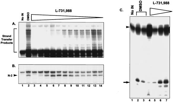
To assess the enzymatic competence of the immobilized enzyme, the activity of the immobilized integrase and inhibition by L-731,988 were evaluated by using radiolabeled substrates. Neither the 3′ processing, strand transfer, or disintegration activities of integrase nor the inhibition by DKAs were affected by the introduction of the epitope tag or by immobilization (Fig. 2). The IC50s determined for L-731,988 in all three reactions were comparable to the IC50s determined under standard solution conditions (17), thus validating the system for studies of DKA binding. As observed in solution assays, L-731,988 preferentially inhibited strand transfer; the IC50 determined for strand transfer was between 86 and 192 nM, whereas the IC50s for 3′ processing or disintegration were more than 100-fold higher (Fig. 2A versus 2B and 2C).
Figure 2.
Activity of immobilized FLAG-integrase and inhibition by L-731,988. FLAG-integrase was assembled on the SPA beads and incubated with 32P-labeled U5 viral ends in the absence or presence of L-731,988. The beads were pelleted and resuspended in gel loading buffer. Lane 1, no FLAG-integrase; lane 2, SPA beads with FLAG-integrase; lanes 3–14, FLAG-integrase with a titration of L-731,988; lane 3, 100 μM; lane 4, 33 μM; lane 5, 11 μM; lane 6, 3.7 μM; lane 7, 1.23 μM; lane 8, 0.41 μM; lane 9, 0.14 μM; lane 10, 0.045 μM; lane 11, 0.015 μM; lane 12, 0.005 μM; lane 13, 0.0016 μM; lane 14, 0.0005 μM. (A) Inhibition of strand transfer (1-h exposure). (B) Inhibition of 3′ processing. N-2 processing product is indicated (arrowhead) (15-min exposure). (C) Inhibition of disintegration. The cleaved (arrow) and uncleaved (arrowhead) disintegration products are indicated. Lane 1, dumbbell probe; lane 2, FLAG-integrase in solution; lane 3, FLAG-integrase associated with SPA beads; lanes 4–7, FLAG-integrase associated with SPA beads with a titration of L-731,988; lane 4, 100 μM; lane 5, 10 μM; lane 6, 1 μM; lane 7, 0.1 μM.
High-Affinity Binding of L-731,988 Requires Assembly of a Catalytically Active Complex.
In initial experiments, the SPA was used to evaluate the interaction between L-731,988 and integrase in the absence of viral DNA ends. Although 100 nM L-731,988 is sufficient to inhibit strand transfer, a specific interaction between the immobilized enzyme and L-731,988 was not detected (Fig. 3A), even when micromolar concentrations of the inhibitor were used. The weak signal observed under these conditions was not diminished with an excess of unlabeled inhibitor; therefore all binding observed with integrase alone was judged to be nonspecific (Fig. 3B). The apparently weak interaction between the inhibitor and the isolated enzyme was also seen in equilibrium dialysis studies where binding was observed under high salt conditions only when much higher concentrations (20–50 μM) of L-731,988 were used (data not shown).
Given the potency of L-731,988 in strand transfer reactions mediated by the assembled integrase donor complex (approximately 100 nM), binding between L-731,988 and integrase might be enhanced in the context of the donor substrate. U5 viral DNA substrates were therefore included in the SPA. In the presence of a U5 oligonucleotide at 100 nM, L-731,988 bound with a Kd of 75 ± 10 nM (Fig. 3A). The Kd measured under these conditions approximates the IC50 for inhibition of strand transfer, suggesting a functional correlation between binding and inhibition. Binding of the labeled DKA was abolished when unlabeled inhibitors were included in the assay (Fig. 3B) and by mutations in integrase associated with DKA resistance (data not shown) (17). Therefore, in contrast to the interaction observed with integrase alone, the SPA signal observed in the presence of the U5 substrate reflects a specific interaction between integrase and L-731,988.
Mutations in integrase that engender resistance to L-731,988 are proximal to the catalytic resides Asp64 and Glu152. To determine whether an intact active site is required for L-731,988 binding, epitope-tagged integrase mutants with inactivating substitutions in either of two catalytic residues were evaluated for the ability to bind inhibitor in the presence of U5 viral ends. Neither the Asp116Ala (Fig. 3B) nor the Asp64Ala (data not shown) mutant was competent to bind L-731,988. As published, the Asp116Ala and Asp64Ala mutants were active in complementation assays using the assembly-defective mutants His12Ala and Lys264Asp (9, 23–26). Complexes assembled with these complementation partners were susceptible to inhibition by L-731,988 (data not shown); thus binding to the catalytically active subunit(s) in the complex is sufficient for DKA inhibition.
High-Affinity Binding of L-731,988 Is Specific for Complexes Assembled on Viral DNA Ends.
The discordance in the affinities for L-731,988 measured in the presence or absence of the viral substrate suggests specific recognition of the assembled strand transfer complex. Assembly of an enzymatically active complex requires specific interactions between integrase and the HIV-1 DNA end. To determine whether the high-affinity interaction observed for L-731,988 reflects assembly of a strand transfer complex, binding of the inhibitor was compared in the presence of U5 DNA and nonspecific sequence oligonucleotides (Fig. 3C). As anticipated from the previous result, the U5 substrate enhanced binding of L-731,988 to the immobilized integrase, and the magnitude of this effect was strictly dependent on the concentration of U5 (Fig. 3C). Increasing the concentration of the U5 viral DNA enhanced L-731,988 binding with maximum binding seen at a concentration of U5 DNA between 10 and 100 nM. Interestingly, a dose-dependent decrease in L-731,988 binding was observed at DNA concentrations greater than 100 nM.
In contrast to the results obtained with U5 DNA, the interaction between L-731,988 and integrase was not enhanced in the presence of nonspecific DNA (NSΔ3′), an unrelated double-stranded blunt-ended DNA of similar length with a 2-nucleotide 5′ overhang that mimics the processed viral end (Fig. 3C). As observed with the U5 sequence, U3 viral ends were able to promote the L-731,988 interaction; however, a 3-fold higher concentration of U3 DNA was required to achieve this effect (data not shown). This difference likely reflects the lower efficiency with which the U3 substrate is used in catalysis (20).
The previous results suggest binding of L-731,988 requires assembly with HIV-1-specific donor substrates. Although viral specific sequences are also required for disintegration (7, 20), when a U5 dumbbell disintegration substrate was evaluated for the ability to promote inhibitor binding (Fig. 3C), binding of L-731,988 was not observed at any concentration of substrate. This result was surprising; however, the lack of DKA binding with the disintegration substrate is consistent with poor inhibition in the disintegration assay (Fig. 2).
The DKA Binding Site Overlaps with the Target DNA Binding Site in Integrase.
Disintegration substrates, such as the dumbbell used in Figs. 2 and 3, mimic integration intermediates and thus should occupy both the donor and target DNA binding sites in the active complex (27–29). L-731,988 was not bound in the presence of disintegration substrates, suggesting that the target DNA may occupy a site required for DKA binding. The observation that a decrease in inhibitor binding was seen when higher concentrations of donor DNA were included in the SPA would be consistent with this hypothesis, because at higher concentrations the DNA may occupy both the donor and target binding sites.
To test whether the target substrate competes with L-731,988, we examined the effect of target DNA concentration and target DNA preincubation on the apparent potency of L-731,988 in strand transfer (Fig. 4 A and B, respectively). Reactions were performed in staged enzymatic assays using complexes assembled on immobilized U5 donor substrates (15, 16, 18). The effect of target DNA concentration was assessed by measuring inhibition by L-731,988 at seven concentrations of target substrate. The effect of target DNA preincubation was investigated by assembling a complex loaded with the target substrate under noncatalytic conditions as follows. Integrase-donor substrate complexes were assembled in manganese, the complexes were washed, and the buffer was exchanged with buffer containing no divalent metal. Target DNA was introduced to the immobilized complexes in the absence of divalent cation, and the complexes were incubated for 0–30 min with target DNA before the addition of L-731,988. Strand transfer was initiated with 2.5 mM MnCl2, and IC50s were determined for each preincubation condition.
Figure 4.
Competition between L-731,988 and target DNA substrates in strand transfer. (A) Effect of target concentration on inhibition. In a staged microtiter plate assay (18), integrase was assembled on donor DNA and washed. After assembly, the reactions were incubated with L-731,988 (0–0.4 μM) and a titration of target DNA ranging from 0 to 125 nM. The effect of increasing amounts of target DNA on the IC50 for the inhibition of strand transfer by L-731,988 is shown. (B) The effect of target preincubation on inhibition. Integrase was assembled on donor DNA in the presence of MnCl2. Unbound integrase and MnCl2 were removed, and target DNA was added in the absence of divalent cation. Target DNA was incubated for 0–30 min. At the specified time, L-731,988 was titrated into the reactions, and strand transfer was initiated with MnCl2. The IC50 for the inhibition of strand transfer activity by L-731,988 as a function of time of preincubation with the target is shown.
Inhibition by L-731,988 depended on the concentration of target in the strand reaction and the time of preincubation with the target. Consistent with the loss of inhibitor binding seen at high concentrations of donor DNA (Fig. 3), higher target substrate concentrations reduced L-731,988 efficacy (Fig. 4A). Prior addition of target DNA to the complex also diminished the ability of L-731,988 to inhibit strand transfer. After 30 min, the inhibitory potency of L-731,988 decreased by more than 100-fold as compared with the concurrent addition of the inhibitor and the target substrate (Fig. 4B).
Binding of the target substrate abrogates DKA inhibition, suggesting competition for overlapping sites on the strand transfer complex. The effect of the target substrate on inhibitor binding itself was therefore assessed directly in the SPA. To eliminate potential complications resulting from strand transfer, integrase complexes were assembled onto a catalytically incompetent 2′,3′-dideoxy U5 donor DNA, and binding of L-731,988 was measured in the presence of target DNA. Consistent with competition between the inhibitor and target, the addition of either of two different target substrates to these complexes resulted in a dose-dependent decrease in the amount of labeled L-731,988 bound to the complex similar to that observed when unlabeled L-731,988 was used as the competitor (Fig. 5A). Even at the highest concentration of target DNA included in the study, the donor DNA remained resistant to exonuclease digestion (Fig. 5B), indicating the continued presence of integrase and discounting possible effects on complex stability. Single-stranded DNA, which is not a substrate for strand transfer, did not reduce binding of L-731,988 (Fig. 5A). Binding of the target DNA and binding of the DKA are thus mutually exclusive, and the ability of the DNA to compete with the inhibitor is associated with functional relevance as a substrate.
Discussion
L-731,988 was used as the prototype DKA to investigate the mechanism of action of this novel class of HIV-1 antiviral agents that selectively block the strand transfer activity of integrase (17). We have demonstrated that L-731,988 recognizes integrase exclusively in the context of catalytically active protein assembled on the viral DNA end. L-731,988 binds within the integrase active site and inhibits strand transfer by competing with the target DNA substrate. The inhibitor and, by implication, the target DNA require a configuration of the enzyme that is defined only subsequent to assembly of the specific complex; therefore, the binding site(s) for the DKAs and the target DNA are structurally distinct from that of the donor substrate. These studies thus elucidate the mechanistic basis for DKA inhibition. The results provide insights into the strand transfer reaction and have implications for drug discovery efforts.
The high-affinity interaction between integrase and L-731,988 exhibits many of the same absolute requirements associated with the specific catalytic activities of integrase. High-affinity binding (i) requires assembly on specific viral DNA donor substrates, with the U5 end preferred over U3, (ii) is competed with by DNAs that are competent to serve as target substrates for strand transfer or occupy the target site in the context of the disintegration reaction (7, 27–29), and (iii) requires an intact integrase catalytic center in the context of an active multimer. The ability to function as a donor substrate in strand transfer and 3′ processing correlates with the ability to promote inhibitor binding because the U5 end is more efficient than U3 in both contexts (20). Function as a target DNA substrate in strand transfer also correlates with the ability to compete with L-731,988, because single-stranded DNAs that do not function as target substrates do not compete. In addition, target DNAs that perform better as substrates in strand transfer reaction because of differences in length or sequence are also better competitors in L-731,988 SPA binding assays (A.S.E., unpublished observations). The SPA results are also consistent with studies of enzymatic function showing that target DNA binding decreases the effectiveness of inhibition by L-731,988 in the strand transfer reaction.
Together the data suggest that the target DNA and the inhibitor occupy the same or overlapping sites on integrase in the complex. L-731,988 binds with approximately 1,000-fold lower affinity to the uncomplexed enzyme as compared with the strand transfer complex (10–20 μM vs. 100 nM for the complex); therefore the inhibitor/target binding site(s) is defined upon assembly of the strand transfer and is structurally distinct from the binding site for viral DNA. These results account for the DKA's unique ability to selectively inhibit strand transfer.
Although distinct from that of the viral donor substrate, the binding site for the DKA inhibitors is within the active site. Binding in the active site is suggested by the inability of L-731,988 to bind the active site mutants Asp64Ala and Asp116Ala. The proximity of the DKA binding site to the active site is supported by earlier studies in which DKA resistance was mapped to mutations at Thr66 and either Met154 or Ser153, close to the catalytic amino acids Asp64 and Glu152, respectively (17).
Crystallographic studies using the integrase catalytic core domain have recently shown that the inhibitor 5CITEP 1-(5-chloroindol-3-yl)-3-hydroxy-3-(2H-tetrazol-5-yl)-pro-penone binds centrally in the active site between Asp64, Asp116, and Glu152 (30). 5ClTEP may be a structural homolog of the DKAs in that the diketo linker of 5CITEP connects an isosteric acid replacement (the tetrazol) to a hydrophobic ring system. Interestingly, the keto tetrazol portion of 5CITEP makes contacts in the integrase active site that are identical or close to those predicted for the DKAs on the basis of resistance (17). In the 5CITEP structure, the nitrogen atoms of the tetrazole ring are hydrogen-bonded toThr66, Asn155, Lys159, and Lys156, and Glu152 is within hydrogen-bonding distance of the enol hydroxyl.
Given the results presented herein with the DKAs, it is therefore somewhat surprising that 5CITEP binds in the absence of donor substrate and that substantial conformational changes in the protein are not observed in the 5CITEP complex (30). Although it is not known whether 5CITEP inhibition is mechanistically analogous to that of the DKA, it is possible that the 5CITEP structure represents the less interesting weak micromolar binding mode observed for the DKAs in the absence of substrate. Whether this interaction is predictive of the high-affinity DKA inhibited complex is therefore unclear. In the 5CITEP structure, the authors did note a change in the orientation of the Glu152 side chain. Integrase crystal structures can be strikingly different in the positioning of the active site residues, particularly Glu152. The conformational flexibility exhibited by Glu152 is consistent with the notion that the active site itself is flexible and may not adopt a well-defined conformation until integrase has assembled on the viral end (31–33). Recognition of the specifically engaged active integrase structure by L-731,988 suggests that this class of compounds may provide a tool for probing the structure of the active conformation. The ability of the DKAs to discern the enzymatically relevant complex can also be exploited to address other outstanding issues in the field of integrase biochemistry, including the stoichiometry of the minimal catalytically active integrase/DNA complex and the number of active sites required.
Our findings have provided additional insights into the biochemical reaction and have important implications for drug discovery efforts. Integrase inhibitors that disrupt earlier steps in integration (e.g., assembly) have thus far not proved to be effective inhibitors of preintegration complexes or of viral replication (34–36). The DKAs are the first inhibitors of integrase whose antiviral activity clearly results from inhibition of integration (17). The marked difference in potency for inhibition of strand transfer compared with 3′ processing and disintegration exhibited by these compounds and the demonstration that the DKAs (i) require a defined strand transfer complex and (ii) are specifically competitive with the target substrate in strand transfer indicate that the nature of the assay used can dramatically influence the ability to find biologically relevant compounds; assays that rely exclusively on 3′ processing or disintegration would fail to uncover compounds with this unique and validated antiviral mechanism.
Abbreviations
- DKA
diketo acid
- SPA
scintillation proximity assay
- 5CITEP
1-(5-chloroindol-3-yl)-3-hydroxy-3-(2H-tetrazol-5-yl)-pro-penone
Footnotes
This paper was submitted directly (Track II) to the PNAS office.
Article published online before print: Proc. Natl. Acad. Sci. USA, 10.1073/pnas.200139397.
Article and publication date are at www.pnas.org/cgi/doi/10.1073/pnas.200139397
References
- 1.LaFemina R L, Schneider C L, Robbins H L, Callahan P L, LeGrow K, Roth E, Schleif W A, Emini E A. J Virol. 1992;66:7414–7419. doi: 10.1128/jvi.66.12.7414-7419.1992. [DOI] [PMC free article] [PubMed] [Google Scholar]
- 2.Wiskerchen M, Muesing M A. J Virol. 1995;69:376–386. doi: 10.1128/jvi.69.1.376-386.1995. [DOI] [PMC free article] [PubMed] [Google Scholar]
- 3.Sherman P A, Fyfe J A. Proc Natl Acad Sci USA. 1990;87:5119–5123. doi: 10.1073/pnas.87.13.5119. [DOI] [PMC free article] [PubMed] [Google Scholar]
- 4.LaFemina R L, Callahan P L, Cordingley M G. J Virol. 1991;65:5624–5630. doi: 10.1128/jvi.65.10.5624-5630.1991. [DOI] [PMC free article] [PubMed] [Google Scholar]
- 5.Bushman F D, Craigie R. Proc Natl Acad Sci USA. 1991;88:1339–1343. doi: 10.1073/pnas.88.4.1339. [DOI] [PMC free article] [PubMed] [Google Scholar]
- 6.Katz R A, Merkel G, Kulkosky J, Leis J, Skalka A M. Cell. 1990;63:87–95. doi: 10.1016/0092-8674(90)90290-u. [DOI] [PubMed] [Google Scholar]
- 7.Chow S A, Vincent K A, Ellison V, Brown P O. Science. 1992;255:723–726. doi: 10.1126/science.1738845. [DOI] [PubMed] [Google Scholar]
- 8.Drelich M, Wilhelm R, Mous J. Virology. 1992;188:459–468. doi: 10.1016/0042-6822(92)90499-f. [DOI] [PubMed] [Google Scholar]
- 9.Engelman A, Craigie R. J Virol. 1992;66:6361–6369. doi: 10.1128/jvi.66.11.6361-6369.1992. [DOI] [PMC free article] [PubMed] [Google Scholar]
- 10.Kulkosky J, Jones K S, Katz R A, Mack J P, Skalka A M. Mol Cell Biol. 1992;12:2331–2338. doi: 10.1128/mcb.12.5.2331. [DOI] [PMC free article] [PubMed] [Google Scholar]
- 11.Shibagaki Y, Holmes M L, Appa R S, Chow S A. Virology. 1997;230:1–10. doi: 10.1006/viro.1997.8466. [DOI] [PubMed] [Google Scholar]
- 12.Ellison V, Gerton J, Vincent K A, Brown P O. J Biol Chem. 1995;270:3320–3326. doi: 10.1074/jbc.270.7.3320. [DOI] [PubMed] [Google Scholar]
- 13.Ellison V, Brown P O. Proc Natl Acad Sci USA. 1994;91:7316–7320. doi: 10.1073/pnas.91.15.7316. [DOI] [PMC free article] [PubMed] [Google Scholar]
- 14.Vink C, Lutzke R A, Plasterk R H. Nucleic Acids Res. 1994;22:4103–4110. doi: 10.1093/nar/22.20.4103. [DOI] [PMC free article] [PubMed] [Google Scholar]
- 15.Wolfe A L, Felock P J, Hastings J C, Blau C U, Hazuda D J. J Virol. 1996;70:1424–1432. doi: 10.1128/jvi.70.3.1424-1432.1996. [DOI] [PMC free article] [PubMed] [Google Scholar]
- 16.Hazuda D J, Felock P J, Hastings J C, Pramanik B, Wolfe A L. J Virol. 1997;71:7005–7011. doi: 10.1128/jvi.71.9.7005-7011.1997. [DOI] [PMC free article] [PubMed] [Google Scholar]
- 17.Hazuda D J, Felock P, Witmer M, Wolfe A, Stillmock K, Grobler J, Espeseth A, Gabryelski L, Schleif W, Blau C, et al. Science. 2000;287:646–650. doi: 10.1126/science.287.5453.646. [DOI] [PubMed] [Google Scholar]
- 18.Hazuda D J, Hastings J C, Wolfe A L, Emini E A. Nucleic Acids Res. 1994;22:1121–1122. doi: 10.1093/nar/22.6.1121. [DOI] [PMC free article] [PubMed] [Google Scholar]
- 19.Selnick, H., Hazuda, D., Egbertson, M., Guare, J., Wai, J., Young, S., Clark, D. & Medina, J. (1999) U.S. Patent WO 9962513; (1999) Chem. Abstr. 132, 22866.
- 20.van Gent D C, Elgersma Y, Bolk M W, Vink C, Plasterk R H. Nucleic Acids Res. 1991;19:3821–3827. doi: 10.1093/nar/19.14.3821. [DOI] [PMC free article] [PubMed] [Google Scholar]
- 21.Bosworth N, Towers P. Nature (London) 1989;341:167–168. doi: 10.1038/341167a0. [DOI] [PubMed] [Google Scholar]
- 22.Cook N D, Jessop R A, Robinson P S, Richards A D, Kay J. Adv Exp Med Biol. 1991;306:525–528. doi: 10.1007/978-1-4684-6012-4_70. [DOI] [PubMed] [Google Scholar]
- 23.Vincent K A, Ellison V, Chow S A, Brown P O. J Virol. 1993;67:425–437. doi: 10.1128/jvi.67.1.425-437.1993. [DOI] [PMC free article] [PubMed] [Google Scholar]
- 24.Lutzke R A, Vink C, Plasterk R H. Nucleic Acids Res. 1994;22:4125–4131. doi: 10.1093/nar/22.20.4125. [DOI] [PMC free article] [PubMed] [Google Scholar]
- 25.Engelman A, Bushman F D, Craigie R. EMBO J. 1993;12:3269–3275. doi: 10.1002/j.1460-2075.1993.tb05996.x. [DOI] [PMC free article] [PubMed] [Google Scholar]
- 26.van Gent D C, Vink C, Groeneger A A, Plasterk R H. EMBO J. 1993;12:3261–3267. doi: 10.1002/j.1460-2075.1993.tb05995.x. [DOI] [PMC free article] [PubMed] [Google Scholar]
- 27.Chow S A, Brown P O. J Virol. 1994;68:3896–3907. doi: 10.1128/jvi.68.6.3896-3907.1994. [DOI] [PMC free article] [PubMed] [Google Scholar]
- 28.Gerton J L, Herschlag D, Brown P O. J Biol Chem. 1999;274:33480–33487. doi: 10.1074/jbc.274.47.33480. [DOI] [PubMed] [Google Scholar]
- 29.Gerton J L, Brown P O. J Biol Chem. 1997;272:25809–25815. doi: 10.1074/jbc.272.41.25809. [DOI] [PubMed] [Google Scholar]
- 30.Goldgur Y, Craigie R, Cohen G H, Fujiwara T, Yoshinaga T, Fujishita T, Sugimoto, Endo T, Murai H, Davies D R. Proc Natl Acad Sci USA. 1999;96:13040–13043. doi: 10.1073/pnas.96.23.13040. [DOI] [PMC free article] [PubMed] [Google Scholar]
- 31.Wlodawer A. In: Advances in Virus Research. Maramorosch K, Murphy F A, Shatkin A J, editors. Vol. 52. New York: Academic; 1999. pp. 335–350. [Google Scholar]
- 32.Chen Z, Yan Y, Munshi S, Li Y, Zugay-Murphy J, Xu B, Witmer M, Felock P, Wolfe A, Sardana V, et al. J Mol Biol. 2000;296:521–533. doi: 10.1006/jmbi.1999.3451. [DOI] [PubMed] [Google Scholar]
- 33.Lubkowski J, Dauter Z, Yang F, Alexandratos J, Merkel G, Skalka A M, Wlodawer A. Biochemistry. 1999;38:13512–13522. doi: 10.1021/bi991362q. [DOI] [PubMed] [Google Scholar]
- 34.Pommier Y, Neamati N. In: Advances in Virus Research. Maramorosch K, Murphy F, Shatkin A, editors. Vol. 52. New York: Academic; 1999. p. 427. [DOI] [PubMed] [Google Scholar]
- 35.Farnet C M, Wang B, Lipford J R, Bushman F D. Proc Natl Acad Sci USA. 1996;93:9742–9747. doi: 10.1073/pnas.93.18.9742. [DOI] [PMC free article] [PubMed] [Google Scholar]
- 36.Hazuda D, Felock P J, Hastings J C, Pramanik B, Wolfe A L. Drug Des Discov. 1997;15:17–24. [PubMed] [Google Scholar]


