Abstract
AIMS—To compare the levels of conus medullaris in preterm and term neonates; to show the time of ascent to normal; and to evaluate the babies with low conus medullaris levels for tethered cord syndrome. METHODS—Levels were assessed using ultrasonography in 41 preterm and 64 term neonates. RESULTS—In the preterm group the conus medullaris level in one infant (2.4%) was below L4. In three infants (7.2%) it was between L2 and L3 and in 37 infants (90.4%) it was above L2. In the term group it was below L4 in one baby (1.6%), between L2 and L3 in four (6.3%), and above L2 in 57 babies (92.1%). The difference in the conus medullaris levels between term and preterm neonates and genders was not significant. Two patients, one with a conus medullaris level at L4-L5, and the other at L2-L3, had Down's syndrome. CONCLUSION—The ascent of conus medullaris seems to occur early in life. It is important to follow up patients with conus medullaris levels at or below the 4th lumbar vertebra for the development of tethered cord syndrome. Keywords: conus medullaris; spinal cord; tethered cord; ultrasonography
Full Text
The Full Text of this article is available as a PDF (115.5 KB).
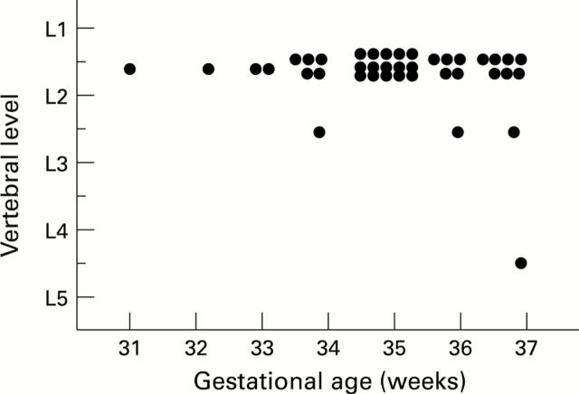
Figure 1 .
Vertebral level of conus medullaris in preterm babies.
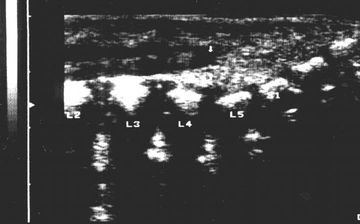
Figure 2 .
Ultrasonographic appearance of conus medullaris.
Selected References
These references are in PubMed. This may not be the complete list of references from this article.
- Barson A. J. The vertebral level of termination of the spinal cord during normal and abnormal development. J Anat. 1970 May;106(Pt 3):489–497. [PMC free article] [PubMed] [Google Scholar]
- DiPietro M. A. The conus medullaris: normal US findings throughout childhood. Radiology. 1993 Jul;188(1):149–153. doi: 10.1148/radiology.188.1.8511289. [DOI] [PubMed] [Google Scholar]
- Ghazi S. R., Gholami S. Allometric growth of the spinal cord in relation to the vertebral column during prenatal and postnatal life in the sheep (Ovis aries). J Anat. 1994 Oct;185(Pt 2):427–431. [PMC free article] [PubMed] [Google Scholar]
- Gusnard D. A., Naidich T. P., Yousefzadeh D. K., Haughton V. M. Ultrasonic anatomy of the normal neonatal and infant spine: correlation with cryomicrotome sections and CT. Neuroradiology. 1986;28(5-6):493–511. doi: 10.1007/BF00344103. [DOI] [PubMed] [Google Scholar]
- Hill C. A., Gibson P. J. Ultrasound determination of the normal location of the conus medullaris in neonates. AJNR Am J Neuroradiol. 1995 Mar;16(3):469–472. [PMC free article] [PubMed] [Google Scholar]
- O'Connor J. F., Cranley W. R., McCarten K. M., Radkowski M. A. Radiographic manifestations of congenital anomalies of the spine. Radiol Clin North Am. 1991 Mar;29(2):407–429. [PubMed] [Google Scholar]
- Vettivel S. Vertebral level of the termination of the spinal cord in human fetuses. J Anat. 1991 Dec;179:149–161. [PMC free article] [PubMed] [Google Scholar]
- Wolf S., Schneble F., Tröger J. The conus medullaris: time of ascendence to normal level. Pediatr Radiol. 1992;22(8):590–592. doi: 10.1007/BF02015359. [DOI] [PubMed] [Google Scholar]
- Yamada S., Zinke D. E., Sanders D. Pathophysiology of "tethered cord syndrome". J Neurosurg. 1981 Apr;54(4):494–503. doi: 10.3171/jns.1981.54.4.0494. [DOI] [PubMed] [Google Scholar]


