Abstract
Background: Measurements of the subarachnoid space during routine cranial sonography may provide an indirect method of monitoring brain growth in preterm infants.
Methods: The width of the subarachnoid space was measured on coronal views during head sonography. Initial scans (within five days of birth) were compared with follow up scans.
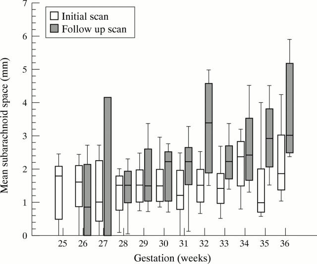
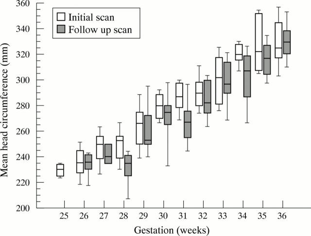
Results: A total of 361 scans were performed on 201 preterm infants. The mean width of the subarachnoid space was < 3.5 mm for 95% of initial scans. It was slightly larger in neonates born closer to term, the equivalent of an increase of 0.02 mm/gestational week (95% confidence interval 0 to 0.10 mm) for initial scans. When the scans of all infants, born at 24–36 gestational weeks who were 36 weeks corrected gestational age were compared, the mean (SD) subarachnoid space was 60% larger for follow up scans than for intial scans: 3.2 (1.38) v 1.95 (1.35) mm (p = 0.002) or the equivalent of a mean increase of 0.20 mm/week (95% confidence interval 0.15 to 0.30 mm) for follow up scans. At 36 weeks corrected gestational age, mean head circumference was not different between those having initial or follow up scans (33.0 (2.0) v 32.2 (1.9) cm; p = 0.31).
Conclusions: The mean subarachnoid space is normally < 3.5 mm in preterm infants. The difference between initial and follow up scans suggests reduced brain growth in extrauterine preterm babies.
Full Text
The Full Text of this article is available as a PDF (85.6 KB).
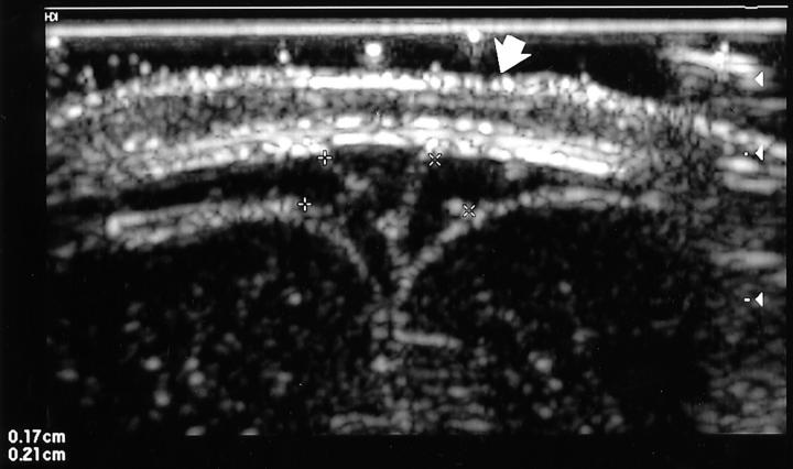
Figure 1 .
Coronal scan of an infant at 33 weeks of gestational age. The subarachnoid space is measured in perpendicular fashion, with electronic calipers, from the edge of the triangular sagittal sinus to the surface of the cortex. The right subarachnoid space is 0.17 cm, and the left is 0.21 cm, resulting in a mean measurement of 1.9 mm. Note that the skin line of the scalp is evident (arrow), indicating that there is no effective pressure from the transducer.
Figure 2 .
Box plot of subarachnoid space measurements at each gestational week of age. Bars show interquartile range, the middle line indicates the median, and whiskers indicate the 10th and 90th centiles. For initial scans, n = 201 (range 6–29 for each week), for follow up scans, n = 160 (range 3–22 for each week).
Figure 3 .
Box plot of head circumference measurements at each gestational week of age. Bars show interquartile range, the middle line indicates the median, and whiskers indicate the 10th and 90th centiles. For initial scans, n = 196 (range 6–25 for each week), and for follow up scans, n = 156 (range 4–25 for each week).
Selected References
These references are in PubMed. This may not be the complete list of references from this article.
- Ajayi-Obe M., Saeed N., Cowan F. M., Rutherford M. A., Edwards A. D. Reduced development of cerebral cortex in extremely preterm infants. Lancet. 2000 Sep 30;356(9236):1162–1163. doi: 10.1016/s0140-6736(00)02761-6. [DOI] [PubMed] [Google Scholar]
- Beeby P. J., Bhutap T., Taylor L. K. New South Wales population-based birthweight percentile charts. J Paediatr Child Health. 1996 Dec;32(6):512–518. doi: 10.1111/j.1440-1754.1996.tb00965.x. [DOI] [PubMed] [Google Scholar]
- Crawford M. Placental delivery of arachidonic and docosahexaenoic acids: implications for the lipid nutrition of preterm infants. Am J Clin Nutr. 2000 Jan;71(1 Suppl):275S–284S. doi: 10.1093/ajcn/71.1.275S. [DOI] [PubMed] [Google Scholar]
- Dobbing J., Sands J. Head circumference, biparietal diameter and brain growth in fetal and postnatal life. Early Hum Dev. 1978 Apr;2(1):81–87. doi: 10.1016/0378-3782(78)90054-3. [DOI] [PubMed] [Google Scholar]
- Frankel D. A., Fessell D. P., Wolfson W. P. High resolution sonographic determination of the normal dimensions of the intracranial extraaxial compartment in the newborn infant. J Ultrasound Med. 1998 Jul;17(7):411–418. doi: 10.7863/jum.1998.17.7.411. [DOI] [PubMed] [Google Scholar]
- Howard E., Benjamins J. A. DNA, ganglioside and sulfatide in brains of rats given corticosterone in infancy, with an estimate of cell loss during development. Brain Res. 1975 Jul 4;92(1):73–87. doi: 10.1016/0006-8993(75)90528-4. [DOI] [PubMed] [Google Scholar]
- Inder T. E., Huppi P. S., Warfield S., Kikinis R., Zientara G. P., Barnes P. D., Jolesz F., Volpe J. J. Periventricular white matter injury in the premature infant is followed by reduced cerebral cortical gray matter volume at term. Ann Neurol. 1999 Nov;46(5):755–760. doi: 10.1002/1531-8249(199911)46:5<755::aid-ana11>3.0.co;2-0. [DOI] [PubMed] [Google Scholar]
- Libicher M., Tröger J. US measurement of the subarachnoid space in infants: normal values. Radiology. 1992 Sep;184(3):749–751. doi: 10.1148/radiology.184.3.1509061. [DOI] [PubMed] [Google Scholar]
- O'Shea T. M., Kothadia J. M., Klinepeter K. L., Goldstein D. J., Jackson B. G., Weaver R. G., 3rd, Dillard R. G. Randomized placebo-controlled trial of a 42-day tapering course of dexamethasone to reduce the duration of ventilator dependency in very low birth weight infants: outcome of study participants at 1-year adjusted age. Pediatrics. 1999 Jul;104(1 Pt 1):15–21. doi: 10.1542/peds.104.1.15. [DOI] [PubMed] [Google Scholar]
- Simon N. P., Brady N. R., Stafford R. L. Catch-up head growth and motor performance in very-low-birthweight infants. Clin Pediatr (Phila) 1993 Jul;32(7):405–411. doi: 10.1177/000992289303200704. [DOI] [PubMed] [Google Scholar]
- Thomson G. D., Teele R. L. High-frequency linear array transducers for neonatal cerebral sonography. AJR Am J Roentgenol. 2001 Apr;176(4):995–1001. doi: 10.2214/ajr.176.4.1760995. [DOI] [PubMed] [Google Scholar]
- Whitelaw A., Thoresen M. Antenatal steroids and the developing brain. Arch Dis Child Fetal Neonatal Ed. 2000 Sep;83(2):F154–F157. doi: 10.1136/fn.83.2.F154. [DOI] [PMC free article] [PubMed] [Google Scholar]


