

Full Text
The Full Text of this article is available as a PDF (107.8 KB).
Figure 1 .

Radiograph taken several hours after birth suggesting respiratory distress syndrome I–II. No other abnormalities can be seen.
Figure 2 .

Radiograph taken on admission to the neonatal intensive care unit, showing a cyst located retrocardially.
Figure 3 .
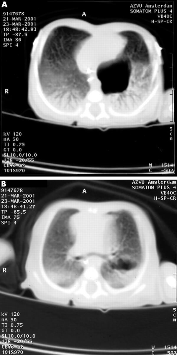
Thoracic computed tomography showing the cystic malformation located retrocardially with its origin in a proximal bronchus without connection to the lung parenchyma. Other parts of the lung are normal.