

Full Text
The Full Text of this article is available as a PDF (110.9 KB).
Figure 1 .
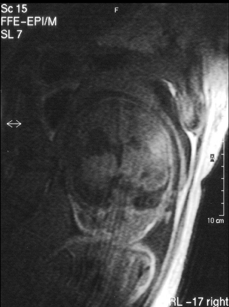
Antenatal magnetic resonance imaging, T1 weighted, coronal view, showing dilatation of the left lateral ventricle with communication to the subarachnoid space and intracerebral right sided fresh haemorrhage.
Figure 2 .

Antenatal magnetic resonance imaging, T2 weighted, coronal view showing same features as in fig 1 with clear demarcation of the fresh right sided intracerebral haemorrhage.