Abstract
Background: Magnetic resonance (MR) imaging of the neonate has been restricted by the need to transport the sick baby to the large magnetic resonance scanners and often the need for sedation or anaesthesia in order to obtain good quality images. Ultrasound is the reference standard for neonatal imaging.
Objective: To establish a dedicated neonatal MR system and compare the clinical usefulness of MR imaging with ultrasound imaging.
Design: Prospective double blind trial.
Setting: Neonatal intensive care unit, Sheffield.
Main outcome measures: Imaging reports.
Patients: 134 premature and term babies.
Results: In 56% of infants with pathology suspected on clinical grounds, MR provided additional useful clinical information over and above that obtained with ultrasound.
Conclusion: Infants can be safely imaged by dedicated low field magnetic resonance on the neonatal intensive care unit without the need for sedation at a cost equivalent to ultrasound.
Full Text
The Full Text of this article is available as a PDF (215.2 KB).
Figure 1 .
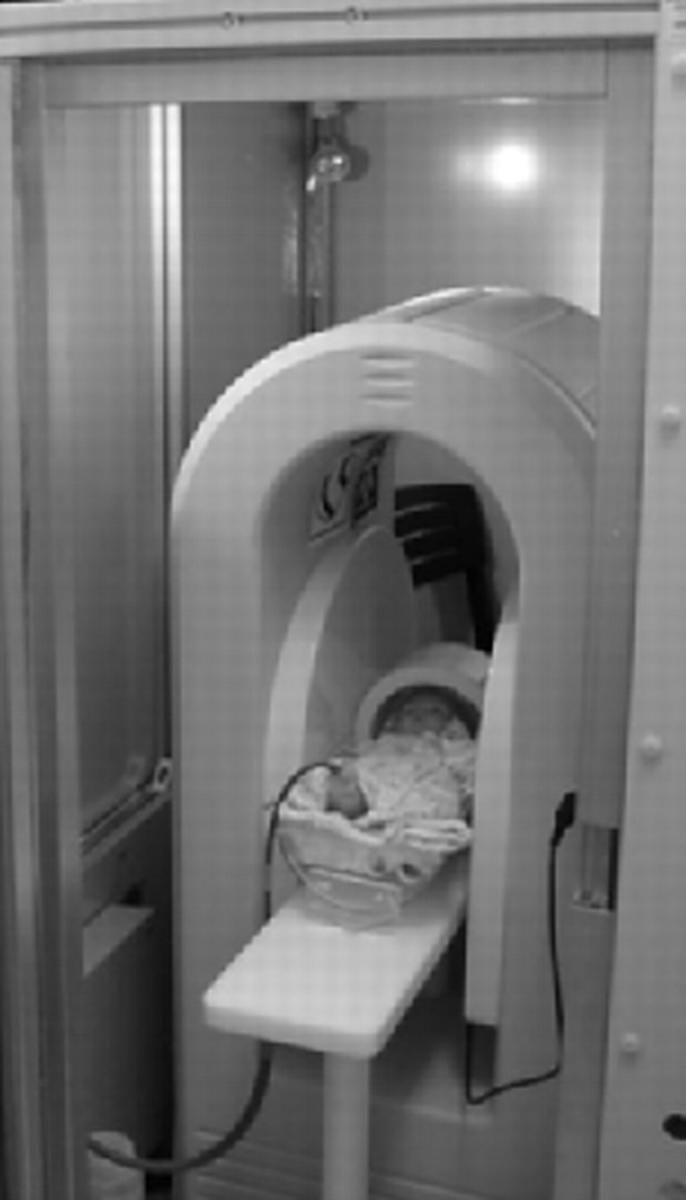
The magnetic resonance scanner.
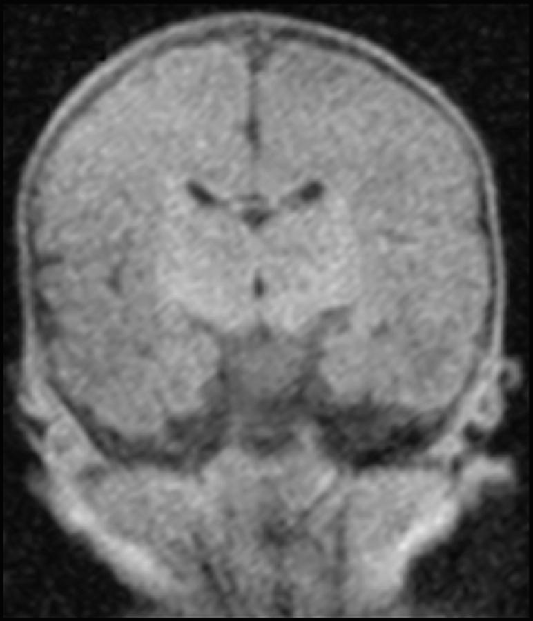
Figure 2 .
T1 weighted coronal scan of the brain of a normal term baby.
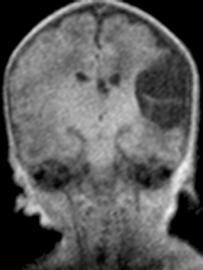
Figure 3 .
Coronal T1 weighted scan showing an established middle cerebral artery territory infarct.
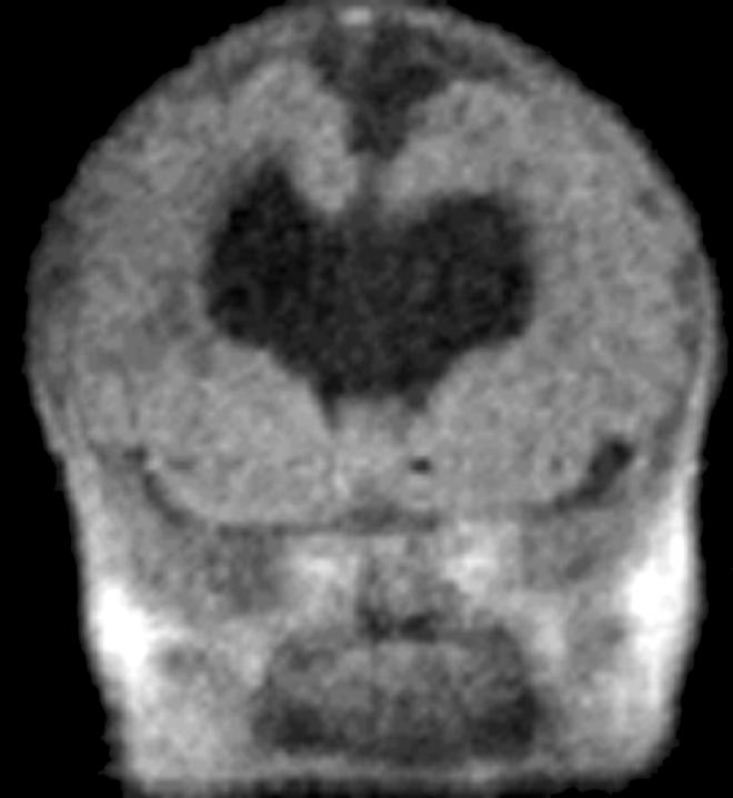
Figure 4 .
T1 weighted coronal image showing holoprosencephaly. Magnetic resonance showed the brain abnormality clearly over several image slices including other features of holoprosencephaly not seen on this slice.
Figure 5 .
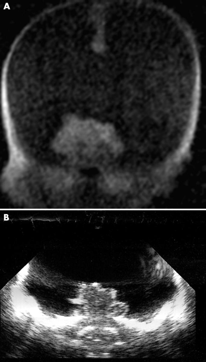
T1 weighted coronal image (A) and ultrasound image (B). Hydranencephaly is seen on both imaging modalities, but was easily detected by magnetic resonance. Massive hydrocephalus could not be completely excluded by ultrasound (although it was less likely).
Figure 6 .
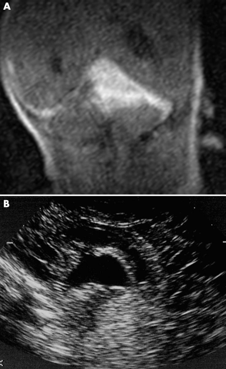
T1 weighted coronal image (A) and ultrasound sagittal image (B). A posterior fossa bleed is clearly seen on the magnetic resonance image but is poorly visualised using ultrasound.
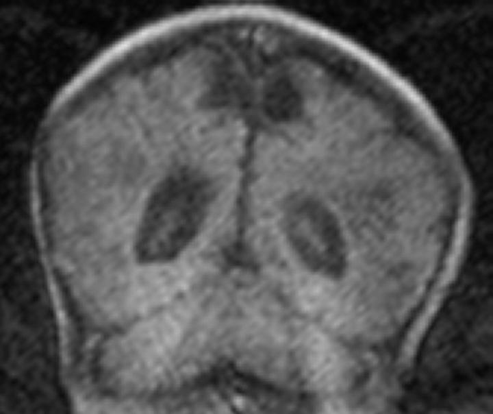
Figure 7 .
T1 weighted coronal image. Late hypoxic-ischaemic changes can be seen (on both ultrasound and magnetic resonance imaging).
Figure 8 .
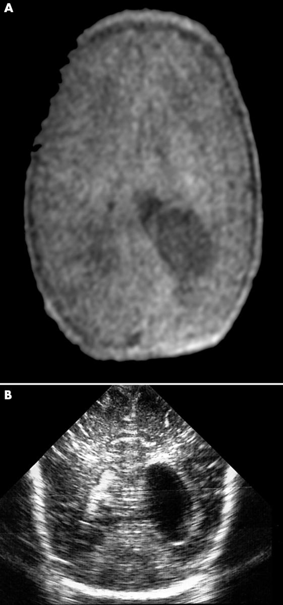
T1 weighted axial magnetic resonance image (A) and ultrasound image (B). An intraventricular cyst can be seen equally well on both images.
Selected References
These references are in PubMed. This may not be the complete list of references from this article.
- Barkovich A. J., Truwit C. L. Brain damage from perinatal asphyxia: correlation of MR findings with gestational age. AJNR Am J Neuroradiol. 1990 Nov-Dec;11(6):1087–1096. [PMC free article] [PubMed] [Google Scholar]
- Blankenberg F. G., Norbash A. M., Lane B., Stevenson D. K., Bracci P. M., Enzmann D. R. Neonatal intracranial ischemia and hemorrhage: diagnosis with US, CT, and MR imaging. Radiology. 1996 Apr;199(1):253–259. doi: 10.1148/radiology.199.1.8633155. [DOI] [PubMed] [Google Scholar]
- Blickman J. G., Jaramillo D., Cleveland R. H. Neonatal cranial ultrasonography. Curr Probl Diagn Radiol. 1991 May-Jun;20(3):91–119. doi: 10.1016/0363-0188(91)90023-u. [DOI] [PubMed] [Google Scholar]
- Boyer R. S. Neuroimaging of premature infants. Neuroimaging Clin N Am. 1994 May;4(2):241–261. [PubMed] [Google Scholar]
- Cohen H. L., Haller J. O. Advances in perinatal neurosonography. AJR Am J Roentgenol. 1994 Oct;163(4):801–810. doi: 10.2214/ajr.163.4.7916529. [DOI] [PubMed] [Google Scholar]
- Keeney S. E., Adcock E. W., McArdle C. B. Prospective observations of 100 high-risk neonates by high-field (1.5 Tesla) magnetic resonance imaging of the central nervous system. II. Lesions associated with hypoxic-ischemic encephalopathy. Pediatrics. 1991 Apr;87(4):431–438. [PubMed] [Google Scholar]
- Latchaw R. E., Truwit C. E. Imaging of perinatal hypoxic-ischemic brain injury. Semin Pediatr Neurol. 1995 Mar;2(1):72–89. doi: 10.1016/s1071-9091(05)80006-3. [DOI] [PubMed] [Google Scholar]
- McArdle C. B., Richardson C. J., Hayden C. K., Nicholas D. A., Amparo E. G. Abnormalities of the neonatal brain: MR imaging. Part II. Hypoxic-ischemic brain injury. Radiology. 1987 May;163(2):395–403. doi: 10.1148/radiology.163.2.3550882. [DOI] [PubMed] [Google Scholar]
- O'Shea T. M., Volberg F., Dillard R. G. Reliability of interpretation of cranial ultrasound examinations of very low-birthweight neonates. Dev Med Child Neurol. 1993 Feb;35(2):97–101. doi: 10.1111/j.1469-8749.1993.tb11611.x. [DOI] [PubMed] [Google Scholar]
- Taber K. H., Hayman L. A., Northrup S. R., Maturi L. Vital sign changes during infant magnetic resonance examinations. J Magn Reson Imaging. 1998 Nov-Dec;8(6):1252–1256. doi: 10.1002/jmri.1880080612. [DOI] [PubMed] [Google Scholar]


