

Abstract
Objectives: To compare the accuracy of ultrasonography (US) and magnetic resonance imaging (MRI) in diagnosing white matter abnormalities in preterm infants and to determine the specific indications for MRI.
Design: Prospective cohort study.
Setting: A neonatal intensive care unit in France.
Patients: All preterm infants (≤ 33 weeks gestation) without severe respiratory distress syndrome precluding MRI.
Main outcome measures: US and MRI performed contemporaneously during the third postnatal week were analysed by an independent observer. The findings were compared with those of a term MRI scan, the results of which were taken as the final diagnosis. Statistical analysis was performed to determine which early imaging study best predicted the term MRI findings.
Results: The early US and MRI findings (79 infants) correlated closely for severe lesions (cystic periventricular leucomalacia and parenchymal infarction; κ coefficient = 0.86) but not for moderate lesions (non-cystic leucomalacia and parenchymal punctate haemorrhages; κ = 0.62). Overall, early MRI findings predicted late MRI findings in 98% of patients (95% confidence interval (CI) 89.5 to 99.9) compared with only 68% for early US (95% CI 52.1 to 79.2).
Conclusions: US is highly effective in detecting severe lesions of the white matter in preterm infants, but MRI seems to be necessary for the diagnosis of less severe damage. MRI performed at about the third week of life is highly predictive of the final diagnosis at term.
Full Text
The Full Text of this article is available as a PDF (144.1 KB).
Figure 1 .

Comparison between the results of early ultrasonography (US) and early magnetic resonance imaging (MRI) performed on the same day in 79 preterm infants. PVL, Periventricular leucomalacia; HM, haemorrhage.
Figure 2 .
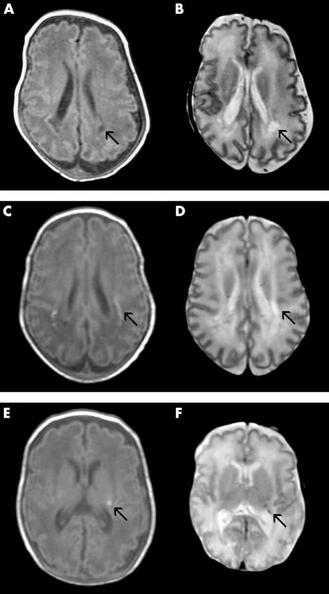
(A, B) Cystic periventricular leucomalacia. (A) T1 weighted image in the transverse plane low signal in the white matter (arrow) and (B) corresponding T2 weighted image in the same area showing high signal (arrow). Bilateral cysts in the white matter. (C, D) Non-cystic periventricular leucomalacia. (C) T1 weighted image in the transverse plane and (D) corresponding T2 weighted image in the same area showing high signal intensity in the white matter (arrows). (E, F) Parenchymal punctate haemorrhage. (E) T1 weighted image in the transverse plane showing circular high signal (arrow) and (F) corresponding T2 weighted image in the same area showing low signal (arrow).
Figure 3 .

Comparison of the cerebral lesion diagnoses from early and late imaging in 51 preterm infants. US, Ultrasonography; MRI, magnetic resonance imaging; PVL, periventricular leucomalacia; HM, haemorrhage.
Selected References
These references are in PubMed. This may not be the complete list of references from this article.
- Bass W. T., Jones M. A., White L. E., Montgomery T. R., Aiello F., 3rd, Karlowicz M. G. Ultrasonographic differential diagnosis and neurodevelopmental outcome of cerebral white matter lesions in premature infants. J Perinatol. 1999 Jul-Aug;19(5):330–336. doi: 10.1038/sj.jp.7200190. [DOI] [PubMed] [Google Scholar]
- Carson S. C., Hertzberg B. S., Bowie J. D., Burger P. C. Value of sonography in the diagnosis of intracranial hemorrhage and periventricular leukomalacia: a postmortem study of 35 cases. AJNR Am J Neuroradiol. 1990 Jul-Aug;11(4):677–683. [PMC free article] [PubMed] [Google Scholar]
- Childs A. M., Cornette L., Ramenghi L. A., Tanner S. F., Arthur R. J., Martinez D., Levene M. I. Magnetic resonance and cranial ultrasound characteristics of periventricular white matter abnormalities in newborn infants. Clin Radiol. 2001 Aug;56(8):647–655. doi: 10.1053/crad.2001.0754. [DOI] [PubMed] [Google Scholar]
- Cornette L. G., Tanner S. F., Ramenghi L. A., Miall L. S., Childs A. M., Arthur R. J., Martinez D., Levene M. I. Magnetic resonance imaging of the infant brain: anatomical characteristics and clinical significance of punctate lesions. Arch Dis Child Fetal Neonatal Ed. 2002 May;86(3):F171–F177. doi: 10.1136/fn.86.3.F171. [DOI] [PMC free article] [PubMed] [Google Scholar]
- Gibson J. Y., Massingale T. W., Graves G. R., LeBlanc M. H., Meydrech E. F. Relationship of cranial midline shift to outcome of very-low-birth-weight infants with periventricular hemorrhagic infarction. J Neuroimaging. 1994 Oct;4(4):212–217. doi: 10.1111/jon199444212. [DOI] [PubMed] [Google Scholar]
- Hope P. L., Gould S. J., Howard S., Hamilton P. A., Costello A. M., Reynolds E. O. Precision of ultrasound diagnosis of pathologically verified lesions in the brains of very preterm infants. Dev Med Child Neurol. 1988 Aug;30(4):457–471. doi: 10.1111/j.1469-8749.1988.tb04773.x. [DOI] [PubMed] [Google Scholar]
- Landis J. R., Koch G. G. The measurement of observer agreement for categorical data. Biometrics. 1977 Mar;33(1):159–174. [PubMed] [Google Scholar]
- Maalouf E. F., Duggan P. J., Counsell S. J., Rutherford M. A., Cowan F., Azzopardi D., Edwards A. D. Comparison of findings on cranial ultrasound and magnetic resonance imaging in preterm infants. Pediatrics. 2001 Apr;107(4):719–727. doi: 10.1542/peds.107.4.719. [DOI] [PubMed] [Google Scholar]
- Maalouf E. F., Duggan P. J., Rutherford M. A., Counsell S. J., Fletcher A. M., Battin M., Cowan F., Edwards A. D. Magnetic resonance imaging of the brain in a cohort of extremely preterm infants. J Pediatr. 1999 Sep;135(3):351–357. doi: 10.1016/s0022-3476(99)70133-2. [DOI] [PubMed] [Google Scholar]
- Paneth N. Classifying brain damage in preterm infants. J Pediatr. 1999 May;134(5):527–529. doi: 10.1016/s0022-3476(99)70231-3. [DOI] [PubMed] [Google Scholar]
- Pierrat V., Duquennoy C., van Haastert I. C., Ernst M., Guilley N., de Vries L. S. Ultrasound diagnosis and neurodevelopmental outcome of localised and extensive cystic periventricular leucomalacia. Arch Dis Child Fetal Neonatal Ed. 2001 May;84(3):F151–F156. doi: 10.1136/fn.84.3.F151. [DOI] [PMC free article] [PubMed] [Google Scholar]
- Rademaker K. J., Groenendaal F., Jansen G. H., Eken P., de Vries L. S. Unilateral haemorrhagic parenchymal lesions in the preterm infant: shape, site and prognosis. Acta Paediatr. 1994 Jun;83(6):602–608. doi: 10.1111/j.1651-2227.1994.tb13089.x. [DOI] [PubMed] [Google Scholar]
- Roelants-van Rijn A. M., Groenendaal F., Beek F. J., Eken P., van Haastert I. C., de Vries L. S. Parenchymal brain injury in the preterm infant: comparison of cranial ultrasound, MRI and neurodevelopmental outcome. Neuropediatrics. 2001 Apr;32(2):80–89. doi: 10.1055/s-2001-13875. [DOI] [PubMed] [Google Scholar]
- Rogers B., Msall M., Owens T., Guernsey K., Brody A., Buck G., Hudak M. Cystic periventricular leukomalacia and type of cerebral palsy in preterm infants. J Pediatr. 1994 Jul;125(1):S1–S8. doi: 10.1016/s0022-3476(94)70169-5. [DOI] [PubMed] [Google Scholar]
- Sie L. T., van der Knaap M. S., van Wezel-Meijler G., Taets van Amerongen A. H., Lafeber H. N., Valk J. Early MR features of hypoxic-ischemic brain injury in neonates with periventricular densities on sonograms. AJNR Am J Neuroradiol. 2000 May;21(5):852–861. [PMC free article] [PubMed] [Google Scholar]
- Zupan V., Gonzalez P., Lacaze-Masmonteil T., Boithias C., d'Allest A. M., Dehan M., Gabilan J. C. Periventricular leukomalacia: risk factors revisited. Dev Med Child Neurol. 1996 Dec;38(12):1061–1067. doi: 10.1111/j.1469-8749.1996.tb15068.x. [DOI] [PubMed] [Google Scholar]
- de Vries L. S., Eken P., Dubowitz L. M. The spectrum of leukomalacia using cranial ultrasound. Behav Brain Res. 1992 Jul 31;49(1):1–6. doi: 10.1016/s0166-4328(05)80189-5. [DOI] [PubMed] [Google Scholar]
- de Vries L. S., Eken P., Groenendaal F., van Haastert I. C., Meiners L. C. Correlation between the degree of periventricular leukomalacia diagnosed using cranial ultrasound and MRI later in infancy in children with cerebral palsy. Neuropediatrics. 1993 Oct;24(5):263–268. doi: 10.1055/s-2008-1071554. [DOI] [PubMed] [Google Scholar]
- de Vries L. S., Roelants-van Rijn A. M., Rademaker K. J., Van Haastert I. C., Beek F. J., Groenendaal F. Unilateral parenchymal haemorrhagic infarction in the preterm infant. Eur J Paediatr Neurol. 2001;5(4):139–149. doi: 10.1053/ejpn.2001.0494. [DOI] [PubMed] [Google Scholar]