Abstract
BACKGROUND—This study evaluated the ability of laser scanning tomography to distinguish between normal and glaucomatous optic nerve heads, and between glaucomatous subjects with and without field loss. METHODS—57 subjects were classified into three diagnostic groups: subjects with elevated intraocular pressure, normal optic nerve heads, and normal visual fields (n=10); subjects with glaucomatous optic neuropathy and normal visual fields (n=30); and subjects with glaucomatous optic neuropathy and repeatable visual field abnormality (n=17). Three 10 degree image series were acquired on each subject using the Heidelberg retina tomograph (HRT). From the 14 HRT stereometric variables, three were selected a priori for evaluation: (1) volume above reference (neuroretinal rim volume), (2) third moment in contour (cup shape), and (3) height variation contour (variation in relative nerve fibre layer height at the disc margin). Data were analysed using analysis of covariance, with age as the covariate. RESULTS—Volume above reference, third moment in contour, and mean height contour were significantly different between each of the three diagnostic groups (p<0.001). Height variation contour showed no significant difference among the three diagnostic groups (p=0.906). CONCLUSIONS—The HRT variables measuring rim volume, cup shape, and mean nerve fibre layer height distinguished between (1) subjects with elevated intraocular pressures and normal nerve heads, and glaucomatous optic nerve heads, and (2) glaucomatous optic nerve heads with and without repeatable visual field abnormality. This study did not directly assess the ability of the HRT to identify patients at risk of developing glaucoma. It is hypothesised that the greatest potential benefit of laser scanning tomography will be in the documentation of change within an individual over time.
Full Text
The Full Text of this article is available as a PDF (131.4 KB).
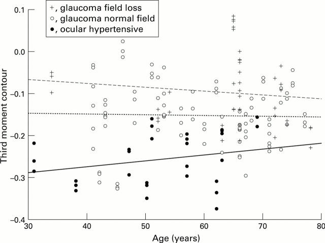
Figure 1 .
Scatter plot of third moment contour versus age (showing no significant interaction).
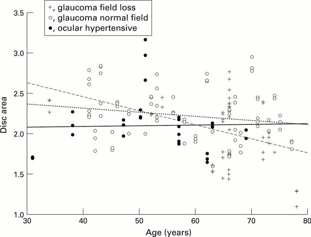
Figure 2 .
Scatter plot of disc area versus age (showing significant interaction).
Selected References
These references are in PubMed. This may not be the complete list of references from this article.
- Airaksinen P. J., Alanko H. I. Effect of retinal nerve fibre loss on the optic nerve head configuration in early glaucoma. Graefes Arch Clin Exp Ophthalmol. 1983;220(4):193–196. doi: 10.1007/BF02186668. [DOI] [PubMed] [Google Scholar]
- Airaksinen P. J., Drance S. M., Schulzer M. Neuroretinal rim area in early glaucoma. Am J Ophthalmol. 1985 Jan 15;99(1):1–4. doi: 10.1016/s0002-9394(14)75856-8. [DOI] [PubMed] [Google Scholar]
- Balazsi A. G., Drance S. M., Schulzer M., Douglas G. R. Neuroretinal rim area in suspected glaucoma and early chronic open-angle glaucoma. Correlation with parameters of visual function. Arch Ophthalmol. 1984 Jul;102(7):1011–1014. doi: 10.1001/archopht.1984.01040030813022. [DOI] [PubMed] [Google Scholar]
- Bickler-Bluth M., Trick G. L., Kolker A. E., Cooper D. G. Assessing the utility of reliability indices for automated visual fields. Testing ocular hypertensives. Ophthalmology. 1989 May;96(5):616–619. doi: 10.1016/s0161-6420(89)32840-5. [DOI] [PubMed] [Google Scholar]
- Brigatti L., Caprioli J. Correlation of visual field with scanning confocal laser optic disc measurements in glaucoma. Arch Ophthalmol. 1995 Sep;113(9):1191–1194. doi: 10.1001/archopht.1995.01100090117032. [DOI] [PubMed] [Google Scholar]
- Burk R. O., Rohrschneider K., Noack H., Völcker H. E. Are large optic nerve heads susceptible to glaucomatous damage at normal intraocular pressure? A three-dimensional study by laser scanning tomography. Graefes Arch Clin Exp Ophthalmol. 1992;230(6):552–560. doi: 10.1007/BF00181778. [DOI] [PubMed] [Google Scholar]
- Caprioli J., Miller J. M. Measurement of relative nerve fiber layer surface height in glaucoma. Ophthalmology. 1989 May;96(5):633–641. doi: 10.1016/s0161-6420(89)32837-5. [DOI] [PubMed] [Google Scholar]
- Caprioli J., Miller J. M. Videographic measurements of optic nerve topography in glaucoma. Invest Ophthalmol Vis Sci. 1988 Aug;29(8):1294–1298. [PubMed] [Google Scholar]
- Caprioli J. The contour of the juxtapapillary nerve fiber layer in glaucoma. Ophthalmology. 1990 Mar;97(3):358–366. doi: 10.1016/s0161-6420(90)32581-2. [DOI] [PubMed] [Google Scholar]
- Caprioli J. The contour of the juxtapapillary nerve fiber layer in glaucoma. Ophthalmology. 1990 Mar;97(3):358–366. doi: 10.1016/s0161-6420(90)32581-2. [DOI] [PubMed] [Google Scholar]
- Chauhan B. C., LeBlanc R. P., McCormick T. A., Rogers J. B. Test-retest variability of topographic measurements with confocal scanning laser tomography in patients with glaucoma and control subjects. Am J Ophthalmol. 1994 Jul 15;118(1):9–15. doi: 10.1016/s0002-9394(14)72836-3. [DOI] [PubMed] [Google Scholar]
- Concato J., Feinstein A. R., Holford T. R. The risk of determining risk with multivariable models. Ann Intern Med. 1993 Feb 1;118(3):201–210. doi: 10.7326/0003-4819-118-3-199302010-00009. [DOI] [PubMed] [Google Scholar]
- Damms T., Dannheim F. Sensitivity and specificity of optic disc parameters in chronic glaucoma. Invest Ophthalmol Vis Sci. 1993 Jun;34(7):2246–2250. [PubMed] [Google Scholar]
- Dreher A. W., Tso P. C., Weinreb R. N. Reproducibility of topographic measurements of the normal and glaucomatous optic nerve head with the laser tomographic scanner. Am J Ophthalmol. 1991 Feb 15;111(2):221–229. doi: 10.1016/s0002-9394(14)72263-9. [DOI] [PubMed] [Google Scholar]
- Gao H., Hollyfield J. G. Aging of the human retina. Differential loss of neurons and retinal pigment epithelial cells. Invest Ophthalmol Vis Sci. 1992 Jan;33(1):1–17. [PubMed] [Google Scholar]
- Jaffe G. J., Alvarado J. A., Juster R. P. Age-related changes of the normal visual field. Arch Ophthalmol. 1986 Jul;104(7):1021–1025. doi: 10.1001/archopht.1986.01050190079043. [DOI] [PubMed] [Google Scholar]
- Jonas J. B., Gusek G. C., Naumann G. O. Optic disc morphometry in chronic primary open-angle glaucoma. II. Correlation of the intrapapillary morphometric data to visual field indices. Graefes Arch Clin Exp Ophthalmol. 1988;226(6):531–538. doi: 10.1007/BF02169200. [DOI] [PubMed] [Google Scholar]
- Kruse F. E., Burk R. O., Völcker H. E., Zinser G., Harbarth U. Reproducibility of topographic measurements of the optic nerve head with laser tomographic scanning. Ophthalmology. 1989 Sep;96(9):1320–1324. doi: 10.1016/s0161-6420(89)32719-9. [DOI] [PubMed] [Google Scholar]
- Lichter P. R. Variability of expert observers in evaluating the optic disc. Trans Am Ophthalmol Soc. 1976;74:532–572. [PMC free article] [PubMed] [Google Scholar]
- Miller J. M., Caprioli J. An optimal reference plane to detect glaucomatous nerve fiber layer abnormalities with computerized image analysis. Graefes Arch Clin Exp Ophthalmol. 1992;230(2):124–128. doi: 10.1007/BF00164649. [DOI] [PubMed] [Google Scholar]
- Quigley H. A., Enger C., Katz J., Sommer A., Scott R., Gilbert D. Risk factors for the development of glaucomatous visual field loss in ocular hypertension. Arch Ophthalmol. 1994 May;112(5):644–649. doi: 10.1001/archopht.1994.01090170088028. [DOI] [PubMed] [Google Scholar]
- Quigley H. A., Katz J., Derick R. J., Gilbert D., Sommer A. An evaluation of optic disc and nerve fiber layer examinations in monitoring progression of early glaucoma damage. Ophthalmology. 1992 Jan;99(1):19–28. doi: 10.1016/s0161-6420(92)32018-4. [DOI] [PubMed] [Google Scholar]
- Ransohoff D. F., Feinstein A. R. Problems of spectrum and bias in evaluating the efficacy of diagnostic tests. N Engl J Med. 1978 Oct 26;299(17):926–930. doi: 10.1056/NEJM197810262991705. [DOI] [PubMed] [Google Scholar]
- Rohrschneider K., Burk R. O., Kruse F. E., Völcker H. E. Reproducibility of the optic nerve head topography with a new laser tomographic scanning device. Ophthalmology. 1994 Jun;101(6):1044–1049. doi: 10.1016/s0161-6420(94)31220-6. [DOI] [PubMed] [Google Scholar]
- Rohrschneider K., Burk R. O., Völcker H. E. Reproducibility of topometric data acquisition in normal and glaucomatous optic nerve heads with the laser tomographic scanner. Graefes Arch Clin Exp Ophthalmol. 1993 Aug;231(8):457–464. doi: 10.1007/BF02044232. [DOI] [PubMed] [Google Scholar]
- Sommer A., Katz J., Quigley H. A., Miller N. R., Robin A. L., Richter R. C., Witt K. A. Clinically detectable nerve fiber atrophy precedes the onset of glaucomatous field loss. Arch Ophthalmol. 1991 Jan;109(1):77–83. doi: 10.1001/archopht.1991.01080010079037. [DOI] [PubMed] [Google Scholar]
- Tielsch J. M., Katz J., Quigley H. A., Miller N. R., Sommer A. Intraobserver and interobserver agreement in measurement of optic disc characteristics. Ophthalmology. 1988 Mar;95(3):350–356. doi: 10.1016/s0161-6420(88)33177-5. [DOI] [PubMed] [Google Scholar]
- Tuulonen A., Airaksinen P. J. Initial glaucomatous optic disk and retinal nerve fiber layer abnormalities and their progression. Am J Ophthalmol. 1991 Apr 15;111(4):485–490. doi: 10.1016/s0002-9394(14)72385-2. [DOI] [PubMed] [Google Scholar]
- Varma R., Steinmann W. C., Scott I. U. Expert agreement in evaluating the optic disc for glaucoma. Ophthalmology. 1992 Feb;99(2):215–221. doi: 10.1016/s0161-6420(92)31990-6. [DOI] [PubMed] [Google Scholar]
- Weinreb R. N., Shakiba S., Sample P. A., Shahrokni S., van Horn S., Garden V. S., Asawaphureekorn S., Zangwill L. Association between quantitative nerve fiber layer measurement and visual field loss in glaucoma. Am J Ophthalmol. 1995 Dec;120(6):732–738. doi: 10.1016/s0002-9394(14)72726-6. [DOI] [PubMed] [Google Scholar]


