Abstract
AIMS/BACKGROUND—To examine the relative contributions of non-specific (for example, spectacle correction) and specific (that is, occlusion therapy) treatment effects on children with ametropic amblyopia. To assess the importance and practicality of objectively confirming the prescribed occlusion dose. METHODS—Subjects were entered into a two phase trial. In the first (`pretreatment') subjects were provided with spectacle correction and underwent repeat visual acuity (VA) and contrast sensitivity (CS) testing until acuity in their amblyopic eye had stabilised. Subjects then progressed to the second phase (`treatment') in which they underwent direct, unilateral occlusion for 1 hour per day for 4 weeks. Patching was objectively monitored using an occlusion dose monitor. RESULTS—Eight subjects completed the trial, all but one of whom achieved >80% concordance with the occlusion regimen. Within the pretreatment phase, mean amblyopic eye VA improved by 0.19 log units (p=0.008) while mean CS gained 0.09 log units (p=0.01). An identical improvement in mean VA was recorded in the fellow eyes (p=0.03) while mean CS gained 0.11 log units (p=0.02). Within the treatment phase, mean VA further improved (0.12 log units, p=0.009) although this gain had halved by the end of treatment and was no longer statistically significant (p=0.09). CONCLUSIONS—Visual performance improved significantly during pretreatment whereas further gains seen during occlusion were not sustained. Evaluation of occlusion regimens must take into consideration the potentially confounding influence of `pretreatment effects' and the necessity to confirm objectively the occlusion dose a child receives.
Full Text
The Full Text of this article is available as a PDF (125.6 KB).
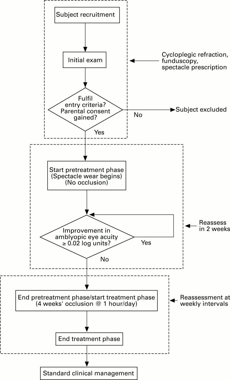
Figure 1 .

Flow diagram indicating subjects' progression through trial. Boxes (in broken lines) (top to bottom): enrolment, pretreatment phase, treatment phase.
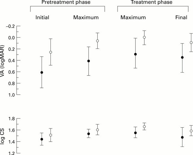
Figure 2 .
Mean (SD) visual acuity and letter contrast sensitivity of subjects' amblyopic eye (filled circles) and fellow eye (open circles). NB Error bars show variation in scores at the specified points within the trial but do not indicate changes attributable to interventions. The acuity scale has been inverted in order that improvements in visual acuity (VA) and contrast sensitivity (CS) each correspond to vertical increments on the ordinate.
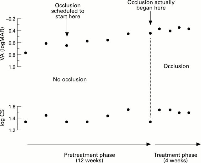
Figure 3 .
Consecutive performance measures obtained from subject RK during trial. Occlusion was scheduled to begin on the third pretreatment visit (left arrow) but did not in fact commence until 8 weeks later (right arrow). The acuity scale has been inverted in order that improvements in visual acuity (VA) and contrast sensitivity (CS) each correspond to vertical increments on the ordinate.
Selected References
These references are in PubMed. This may not be the complete list of references from this article.
- Fielder A. R., Irwin M., Auld R., Cocker K. D., Jones H. S., Moseley M. J. Compliance in amblyopia therapy: objective monitoring of occlusion. Br J Ophthalmol. 1995 Jun;79(6):585–589. doi: 10.1136/bjo.79.6.585. [DOI] [PMC free article] [PubMed] [Google Scholar]
- Grounds A. R., Holliday I. E., Ruddock K. H. Two spatio-temporal filters in human vision. 2. Selective modification in amblyopia, albinism, and hemianopia. Biol Cybern. 1983;47(3):191–201. doi: 10.1007/BF00337008. [DOI] [PubMed] [Google Scholar]
- Hiscox F., Strong N., Thompson J. R., Minshull C., Woodruff G. Occlusion for amblyopia: a comprehensive survey of outcome. Eye (Lond) 1992;6(Pt 3):300–304. doi: 10.1038/eye.1992.59. [DOI] [PubMed] [Google Scholar]
- Kivlin J. D., Flynn J. T. Therapy of anisometropic amblyopia. J Pediatr Ophthalmol Strabismus. 1981 Sep-Oct;18(5):47–56. doi: 10.3928/0191-3913-19810901-12. [DOI] [PubMed] [Google Scholar]
- Lithander J., Sjöstrand J. Anisometropic and strabismic amblyopia in the age group 2 years and above: a prospective study of the results of treatment. Br J Ophthalmol. 1991 Feb;75(2):111–116. doi: 10.1136/bjo.75.2.111. [DOI] [PMC free article] [PubMed] [Google Scholar]
- Mullen P. D. Compliance becomes concordance. BMJ. 1997 Mar 8;314(7082):691–692. doi: 10.1136/bmj.314.7082.691. [DOI] [PMC free article] [PubMed] [Google Scholar]
- Nucci P., Alfarano R., Piantanida A., Brancato R. Compliance in antiamblyopia occlusion therapy. Acta Ophthalmol (Copenh) 1992 Feb;70(1):128–131. doi: 10.1111/j.1755-3768.1992.tb02104.x. [DOI] [PubMed] [Google Scholar]
- Oliver M., Neumann R., Chaimovitch Y., Gotesman N., Shimshoni M. Compliance and results of treatment for amblyopia in children more than 8 years old. Am J Ophthalmol. 1986 Sep 15;102(3):340–345. doi: 10.1016/0002-9394(86)90008-5. [DOI] [PubMed] [Google Scholar]
- Sagi D., Tanne D. Perceptual learning: learning to see. Curr Opin Neurobiol. 1994 Apr;4(2):195–199. doi: 10.1016/0959-4388(94)90072-8. [DOI] [PubMed] [Google Scholar]
- Simons K. Preschool vision screening: rationale, methodology and outcome. Surv Ophthalmol. 1996 Jul-Aug;41(1):3–30. doi: 10.1016/s0039-6257(97)81990-x. [DOI] [PubMed] [Google Scholar]
- Wali N., Leguire L. E., Rogers G. L., Bremer D. L. CSF interocular interactions in childhood ambylopia. Optom Vis Sci. 1991 Feb;68(2):81–87. doi: 10.1097/00006324-199102000-00001. [DOI] [PubMed] [Google Scholar]
- Watson P. G., Sanac A. S., Pickering M. S. A comparison of various methods of treatment of amblyopia. A block study. Trans Ophthalmol Soc U K. 1985;104(Pt 3):319–328. [PubMed] [Google Scholar]
- Woodruff G., Hiscox F., Thompson J. R., Smith L. K. Factors affecting the outcome of children treated for amblyopia. Eye (Lond) 1994;8(Pt 6):627–631. doi: 10.1038/eye.1994.157. [DOI] [PubMed] [Google Scholar]


