Full Text
The Full Text of this article is available as a PDF (208.8 KB).
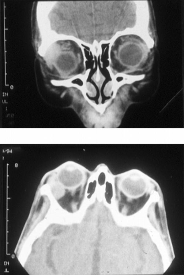
Figure 1 .
(Top) Sagittal computerised tomography scan showing diffuse right lacrimal gland enlargement and globe depression without bone or globe involvement. (Bottom) Axial computerised tomography scan showing diffuse right lacrimal gland enlargement and proptosis without bone or globe abnormality.
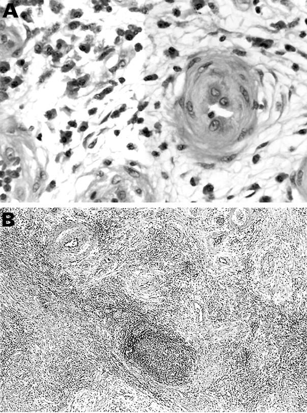
Figure 2 .

(A) Thick walled blood vessels are seen against a myxoid stroma which contains abundant plasma cells. Haematoxylin and eosin; × 40. (B) Plump endothelial cells are evident in the blood vessels; plasma cells and a few eosinophils surround the vessels. Haematoxylin and eosin; × 165.


