Abstract
AIM—To compare the performance of PROGRESSOR (pointwise linear regression) and STATPAC 2 (comparison with baseline values) in detecting early deterioration in the visual fields of glaucoma patients. METHODS—Visual field series from 19 untreated normal tension glaucoma eyes which were deteriorating on clinical grounds were analysed by PROGRESSOR and STATPAC 2. Progression criteria for PROGRESSOR were (1) inner points: slope < −1 dB/year, p < 0.05 and (2) edge points: slope < −2 dB/year, p < 0.05. Criteria for STATPAC 2 were p < 0.05 change probability for any point on three consecutive fields. Detection time was defined as the time interval between the initial field and the first field in which at least one progressing point was identified. Detection times produced by the two techniques were compared. RESULTS—PROGRESSOR and STATPAC 2 agreed on progression in all 19 eyes. Mean detection time for PROGRESSOR was 1.077 (SD 0.985) years and for STATPAC 2 was 2.161 (1.357) years. PROGRESSOR detected progression sooner than STATPAC 2 in 18 eyes (p < 0.01, Wilcoxon matched pairs signed rank test). PROGRESSOR detected progression earlier by a mean of 1.085 (0.936) years. CONCLUSIONS—PROGRESSOR consistently detected progression earlier than STATPAC 2. The PROGRESSOR software is a useful tool for the early detection of visual field deterioration in glaucoma.
Full Text
The Full Text of this article is available as a PDF (630.1 KB).
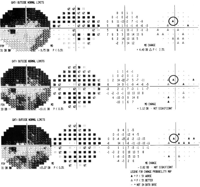
Figure 1 .
Example of a section of the STATPAC 2 glaucoma change probability analysis. The test location highlighted is labelled as showing significant deterioration in each of the three consecutive fields.
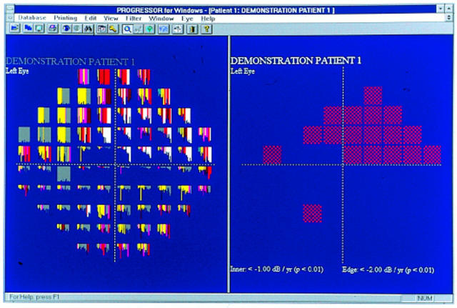
Figure 2A .
Example of the cumulative graphical output of PROGRESSOR for Windows for the left eye of an untreated normal tension glaucoma patient with visual field progression. The left pane shows the bar graph output. The right pane shows the locations which satisfy the progression criteria.
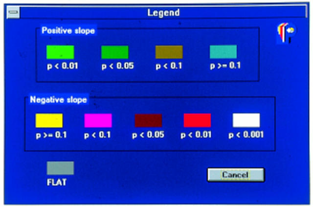
Figure 2B .
Legend for PROGRESSOR for Windows.
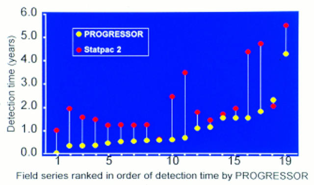
Figure 3 .
Drop line graph of detection times for each field series for PROGRESSOR and STATPAC 2. The field series are ranked in order of detection time by PROGRESSOR.
Figure 4 .
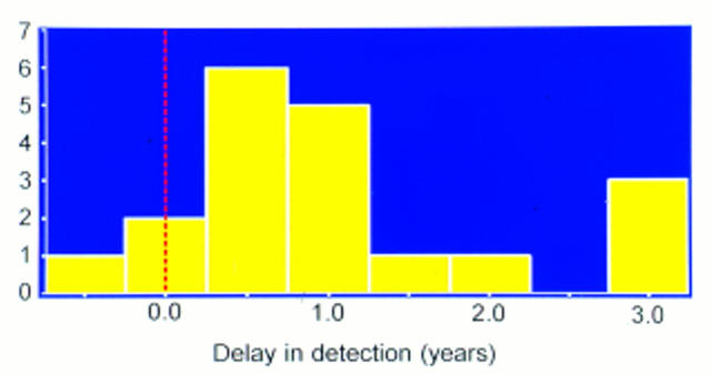
Histogram of delay in detection associated with STATPAC 2. The mean delay is 1.085 (SD 0.936) years.
Selected References
These references are in PubMed. This may not be the complete list of references from this article.
- Boeglin R. J., Caprioli J., Zulauf M. Long-term fluctuation of the visual field in glaucoma. Am J Ophthalmol. 1992 Apr 15;113(4):396–400. doi: 10.1016/s0002-9394(14)76161-6. [DOI] [PubMed] [Google Scholar]
- Chauhan B. C., Drance S. M., Douglas G. R. The use of visual field indices in detecting changes in the visual field in glaucoma. Invest Ophthalmol Vis Sci. 1990 Mar 1;31(3):512–520. [PubMed] [Google Scholar]
- Fitzke F. W., Hitchings R. A., Poinoosawmy D., McNaught A. I., Crabb D. P. Analysis of visual field progression in glaucoma. Br J Ophthalmol. 1996 Jan;80(1):40–48. doi: 10.1136/bjo.80.1.40. [DOI] [PMC free article] [PubMed] [Google Scholar]
- Fitzke F. W., McNaught A. I. The diagnosis of visual field progression in glaucoma. Curr Opin Ophthalmol. 1994 Apr;5(2):110–115. doi: 10.1097/00055735-199404000-00016. [DOI] [PubMed] [Google Scholar]
- Flammer J., Drance S. M., Zulauf M. Differential light threshold. Short- and long-term fluctuation in patients with glaucoma, normal controls, and patients with suspected glaucoma. Arch Ophthalmol. 1984 May;102(5):704–706. doi: 10.1001/archopht.1984.01040030560017. [DOI] [PubMed] [Google Scholar]
- Heijl A., Lindgren G., Olsson J. Normal variability of static perimetric threshold values across the central visual field. Arch Ophthalmol. 1987 Nov;105(11):1544–1549. doi: 10.1001/archopht.1987.01060110090039. [DOI] [PubMed] [Google Scholar]
- Hitchings K. Psychophysical testing in glaucoma. Br J Ophthalmol. 1993 Aug;77(8):471–472. doi: 10.1136/bjo.77.8.471. [DOI] [PMC free article] [PubMed] [Google Scholar]
- Holmin C., Krakau C. E. Regression analysis of the central visual field in chronic glaucoma cases. A follow-up study using automatic perimetry. Acta Ophthalmol (Copenh) 1982 Apr;60(2):267–274. doi: 10.1111/j.1755-3768.1982.tb08381.x. [DOI] [PubMed] [Google Scholar]
- Johnson C. A. Modern developments in clinical perimetry. Curr Opin Ophthalmol. 1993 Apr;4(2):7–13. [PubMed] [Google Scholar]
- Landis J. R., Koch G. G. The measurement of observer agreement for categorical data. Biometrics. 1977 Mar;33(1):159–174. [PubMed] [Google Scholar]
- McNaught A. I., Crabb D. P., Fitzke F. W., Hitchings R. A. Modelling series of visual fields to detect progression in normal-tension glaucoma. Graefes Arch Clin Exp Ophthalmol. 1995 Dec;233(12):750–755. doi: 10.1007/BF00184085. [DOI] [PubMed] [Google Scholar]
- McNaught A. I., Crabb D. P., Fitzke F. W., Hitchings R. A. Visual field progression: comparison of Humphrey Statpac2 and pointwise linear regression analysis. Graefes Arch Clin Exp Ophthalmol. 1996 Jul;234(7):411–418. doi: 10.1007/BF02539406. [DOI] [PubMed] [Google Scholar]
- Mikelberg F. S. Do computerised visual fields and automated optic disc analysis assist in the choice of therapy in glaucoma? Eye (Lond) 1992;6(Pt 1):47–49. doi: 10.1038/eye.1992.8. [DOI] [PubMed] [Google Scholar]
- Mikelberg F. S., Drance S. M. The mode of progression of visual field defects in glaucoma. Am J Ophthalmol. 1984 Oct 15;98(4):443–445. doi: 10.1016/0002-9394(84)90128-4. [DOI] [PubMed] [Google Scholar]
- Noureddin B. N., Poinoosawmy D., Fietzke F. W., Hitchings R. A. Regression analysis of visual field progression in low tension glaucoma. Br J Ophthalmol. 1991 Aug;75(8):493–495. doi: 10.1136/bjo.75.8.493. [DOI] [PMC free article] [PubMed] [Google Scholar]
- O'Brien C., Schwartz B., Takamoto T., Wu D. C. Intraocular pressure and the rate of visual field loss in chronic open-angle glaucoma. Am J Ophthalmol. 1991 Apr 15;111(4):491–500. doi: 10.1016/s0002-9394(14)72386-4. [DOI] [PubMed] [Google Scholar]
- O'Brien C., Schwartz B. The visual field in chronic open angle glaucoma: the rate of change in different regions of the field. Eye (Lond) 1990;4(Pt 4):557–562. doi: 10.1038/eye.1990.77. [DOI] [PubMed] [Google Scholar]
- Schulzer M. Errors in the diagnosis of visual field progression in normal-tension glaucoma. Ophthalmology. 1994 Sep;101(9):1589–1595. doi: 10.1016/s0161-6420(94)31133-x. [DOI] [PubMed] [Google Scholar]
- Smith S. D., Katz J., Quigley H. A. Analysis of progressive change in automated visual fields in glaucoma. Invest Ophthalmol Vis Sci. 1996 Jun;37(7):1419–1428. [PubMed] [Google Scholar]
- Weber J., Koll W., Krieglstein G. K. Intraocular pressure and visual field decay in chronic glaucoma. Ger J Ophthalmol. 1993 May;2(3):165–169. [PubMed] [Google Scholar]
- Werner E. B., Adelson A., Krupin T. Effect of patient experience on the results of automated perimetry in clinically stable glaucoma patients. Ophthalmology. 1988 Jun;95(6):764–767. doi: 10.1016/s0161-6420(88)33111-8. [DOI] [PubMed] [Google Scholar]
- Werner E. B., Bishop K. I., Koelle J., Douglas G. R., LeBlanc R. P., Mills R. P., Schwartz B., Whalen W. R., Wilensky J. T. A comparison of experienced clinical observers and statistical tests in detection of progressive visual field loss in glaucoma using automated perimetry. Arch Ophthalmol. 1988 May;106(5):619–623. doi: 10.1001/archopht.1988.01060130673024. [DOI] [PubMed] [Google Scholar]
- Werner E. B., Krupin T., Adelson A., Feitl M. E. Effect of patient experience on the results of automated perimetry in glaucoma suspect patients. Ophthalmology. 1990 Jan;97(1):44–48. doi: 10.1016/s0161-6420(90)32628-3. [DOI] [PubMed] [Google Scholar]
- Wild J. M., Hussey M. K., Flanagan J. G., Trope G. E. Pointwise topographical and longitudinal modeling of the visual field in glaucoma. Invest Ophthalmol Vis Sci. 1993 May;34(6):1907–1916. [PubMed] [Google Scholar]


