Abstract
AIMS—Scanning laser polarimetry is a new technique allowing quantitative analysis of the retinal nerve fibre layer in vivo. This technique was employed to investigate the variation of the retinal nerve fibre layer thickness in a group of normal subjects of different ages and ethnic groups. METHODS—150 normal volunteers of different ages and ethnic groups were recruited for this study. Three consecutive 15 degree polarimetric maps were acquired for each subjects. Nerve fibre layer thickness measurements were obtained at 1.5 disc diameters from the optic nerve. Four 90 degree quadrants were identified. RESULTS—The mean nerve fibre layer thickness varied from a minimum of 55.4 µm to a maximum of 105.3 µm, with a mean thickness value of 78.2 (SD 10.6) µm. Superior and inferior quadrants showed a comparatively thicker nerve fibre layer than nasal and temporal quadrants. Retinal nerve fibre layer thickness is inversely correlated with age (p < 0.001). White people showed thicker nerve fibre layers than Afro-Caribbeans (p = 0.002). CONCLUSION—The results indicate a progressive reduction of the nerve fibre layer thickness with increasing age. This may be due to a progressive loss of ganglion axons with age as suggested in postmortem studies. A racial difference in nerve fibre layer thickness is present between whites and Afro-Caribbeans.
Full Text
The Full Text of this article is available as a PDF (142.1 KB).
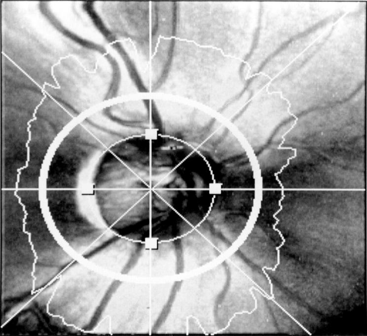
Figure 1 .

Fifteen degree retardation map of the retinal nerve fibre layer thickness of the right eye of a 35 year old subject. In the grey scale image thicker parts of retinal nerve fibre layer are indicated with lighter shades and thinner parts with darker shades. A circle band of 10 pixels width is centred around the optic disc at 1.5 disc diameter distance. The topographic map is divided in four 90 degree quadrants. Retinal nerve fibre layer thickness is measured within the circle band.
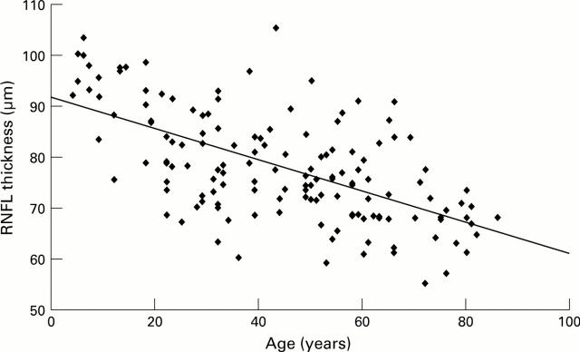
Figure 2 .
Linear regression analysis of the retinal nerve fibre layer thickness and age. R = 0.5938, R2 = 0.3526, SE 8.59324, p = <0.001, n = 150. The analysis indicates a retinal nerve fibre layer thickness decay of 0.38 µm/year.
Figure 3 .
Mean retinal nerve fibre layer thickness by quadrants for the different age groups.
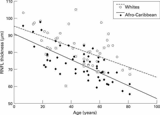
Figure 4 .
Linear regression analysis of the retinal nerve fibre layer thickness and age for white and Afro-Caribbean groups. R = 0.6499, R2 = 0.4224, SE 8.20026, p < 0.001, n = 59 for whites and R = 0.7588, R2 = 0.5758, SE 6.37953, p < 0.001, n = 51 for Afro-Caribbeans.
Selected References
These references are in PubMed. This may not be the complete list of references from this article.
- Airaksinen P. J., Drance S. M., Douglas G. R., Mawson D. K., Nieminen H. Diffuse and localized nerve fiber loss in glaucoma. Am J Ophthalmol. 1984 Nov;98(5):566–571. doi: 10.1016/0002-9394(84)90242-3. [DOI] [PubMed] [Google Scholar]
- Airaksinen P. J., Nieminen H. Retinal nerve fiber layer photography in glaucoma. Ophthalmology. 1985 Jul;92(7):877–879. doi: 10.1016/s0161-6420(85)33941-6. [DOI] [PubMed] [Google Scholar]
- Balazsi A. G., Rootman J., Drance S. M., Schulzer M., Douglas G. R. The effect of age on the nerve fiber population of the human optic nerve. Am J Ophthalmol. 1984 Jun;97(6):760–766. doi: 10.1016/0002-9394(84)90509-9. [DOI] [PubMed] [Google Scholar]
- Dolman C. L., McCormick A. Q., Drance S. M. Aging of the optic nerve. Arch Ophthalmol. 1980 Nov;98(11):2053–2058. doi: 10.1001/archopht.1980.01020040905024. [DOI] [PubMed] [Google Scholar]
- Heijl A., Lindgren G., Olsson J. Normal variability of static perimetric threshold values across the central visual field. Arch Ophthalmol. 1987 Nov;105(11):1544–1549. doi: 10.1001/archopht.1987.01060110090039. [DOI] [PubMed] [Google Scholar]
- Jonas J. B., Müller-Bergh J. A., Schlötzer-Schrehardt U. M., Naumann G. O. Histomorphometry of the human optic nerve. Invest Ophthalmol Vis Sci. 1990 Apr;31(4):736–744. [PubMed] [Google Scholar]
- Jonas J. B., Nguyen N. X., Naumann G. O. The retinal nerve fiber layer in normal eyes. Ophthalmology. 1989 May;96(5):627–632. doi: 10.1016/s0161-6420(89)32838-7. [DOI] [PubMed] [Google Scholar]
- Jonas J. B., Schmidt A. M., Müller-Bergh J. A., Schlötzer-Schrehardt U. M., Naumann G. O. Human optic nerve fiber count and optic disc size. Invest Ophthalmol Vis Sci. 1992 May;33(6):2012–2018. [PubMed] [Google Scholar]
- Katz J., Sommer A. Asymmetry and variation in the normal hill of vision. Arch Ophthalmol. 1986 Jan;104(1):65–68. doi: 10.1001/archopht.1986.01050130075023. [DOI] [PubMed] [Google Scholar]
- Miglior S., Rossetti L., Brigatti L., Bujtar E., Orzalesi N. Reproducibility of retinal nerve fiber layer evaluation by dynamics scanning laser ophthalmoscopy. Am J Ophthalmol. 1994 Jul 15;118(1):16–23. doi: 10.1016/s0002-9394(14)72837-5. [DOI] [PubMed] [Google Scholar]
- Mikelberg F. S., Drance S. M., Schulzer M., Yidegiligne H. M., Weis M. M. The normal human optic nerve. Axon count and axon diameter distribution. Ophthalmology. 1989 Sep;96(9):1325–1328. doi: 10.1016/s0161-6420(89)32718-7. [DOI] [PubMed] [Google Scholar]
- Mikelberg F. S., Yidegiligne H. M., White V. A., Schulzer M. Relation between optic nerve axon number and axon diameter to scleral canal area. Ophthalmology. 1991 Jan;98(1):60–63. doi: 10.1016/s0161-6420(91)32341-8. [DOI] [PubMed] [Google Scholar]
- Miller N. R., George T. W. Monochromatic (red-free) photography and ophthalmoscopy of the peripapillary retinal nerve fiber layer. Invest Ophthalmol Vis Sci. 1978 Nov;17(11):1121–1124. [PubMed] [Google Scholar]
- Quigley H. A., Addicks E. M. Quantitative studies of retinal nerve fiber layer defects. Arch Ophthalmol. 1982 May;100(5):807–814. doi: 10.1001/archopht.1982.01030030811018. [DOI] [PubMed] [Google Scholar]
- Quigley H. A., Green W. R. The histology of human glaucoma cupping and optic nerve damage: clinicopathologic correlation in 21 eyes. Ophthalmology. 1979 Oct;86(10):1803–1830. doi: 10.1016/s0161-6420(79)35338-6. [DOI] [PubMed] [Google Scholar]
- Quigley H. A., Reacher M., Katz J., Strahlman E., Gilbert D., Scott R. Quantitative grading of nerve fiber layer photographs. Ophthalmology. 1993 Dec;100(12):1800–1807. doi: 10.1016/s0161-6420(93)31395-3. [DOI] [PubMed] [Google Scholar]
- Radius R. L. Thickness of the retinal nerve fiber layer in primate eyes. Arch Ophthalmol. 1980 Sep;98(9):1625–1629. doi: 10.1001/archopht.1980.01020040477018. [DOI] [PubMed] [Google Scholar]
- Repka M. X., Quigley H. A. The effect of age on normal human optic nerve fiber number and diameter. Ophthalmology. 1989 Jan;96(1):26–32. doi: 10.1016/s0161-6420(89)32928-9. [DOI] [PubMed] [Google Scholar]
- Sommer A., D'Anna S. A., Kues H. A., George T. High-resolution photography of the retinal nerve fiber layer. Am J Ophthalmol. 1983 Oct;96(4):535–539. doi: 10.1016/s0002-9394(14)77918-8. [DOI] [PubMed] [Google Scholar]
- Sommer A., Katz J., Quigley H. A., Miller N. R., Robin A. L., Richter R. C., Witt K. A. Clinically detectable nerve fiber atrophy precedes the onset of glaucomatous field loss. Arch Ophthalmol. 1991 Jan;109(1):77–83. doi: 10.1001/archopht.1991.01080010079037. [DOI] [PubMed] [Google Scholar]
- Varma R., Tielsch J. M., Quigley H. A., Hilton S. C., Katz J., Spaeth G. L., Sommer A. Race-, age-, gender-, and refractive error-related differences in the normal optic disc. Arch Ophthalmol. 1994 Aug;112(8):1068–1076. doi: 10.1001/archopht.1994.01090200074026. [DOI] [PubMed] [Google Scholar]
- Weinreb R. N., Dreher A. W., Coleman A., Quigley H., Shaw B., Reiter K. Histopathologic validation of Fourier-ellipsometry measurements of retinal nerve fiber layer thickness. Arch Ophthalmol. 1990 Apr;108(4):557–560. doi: 10.1001/archopht.1990.01070060105058. [DOI] [PubMed] [Google Scholar]
- Weinreb R. N., Shakiba S., Zangwill L. Scanning laser polarimetry to measure the nerve fiber layer of normal and glaucomatous eyes. Am J Ophthalmol. 1995 May;119(5):627–636. doi: 10.1016/s0002-9394(14)70221-1. [DOI] [PubMed] [Google Scholar]


