Abstract
AIM—To determine the role of Langerhans cells (LCs) found to bear IgE in patients with atopic dermatitis (AD) by evaluating the surface distribution of these cells in the conjunctival epithelium and epidermis of skin lesions in patients with AD. METHODS—The double labelling method was used to evaluate IgE positive cells that were positive for anti-CD1a or anti-CD23 antibody in an epithelial sheet of the conjunctival limbus. Specimens of conjunctiva were obtained from 12 men, six of whom had AD and ocular complications. Five patients without atopic disease served as controls, plus one additional patient with asthma but no AD. A similar study was conducted using epidermal sheets obtained from two patients with AD and from one without AD. RESULTS—The number of CD1a+ cells present in the conjunctival epithelium of the patients with AD significantly exceeded that of the patients without AD. Most CD1a+ cells in the conjunctival epithelium and epidermis from the patients with AD bore IgE on their surfaces. Few such cells from patients without AD bore IgE. No CD23+ cells were found in the patients with or without AD. CONCLUSIONS—The presence of an increased number of LCs bearing IgE on their surfaces in the conjunctival epithelium of patients with AD suggests that these cells may be involved in eliciting the hypersensitivity reaction and participate in ocular inflammation.
Full Text
The Full Text of this article is available as a PDF (140.0 KB).
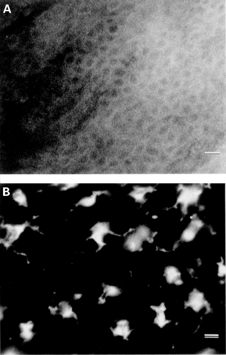
Figure 1 .
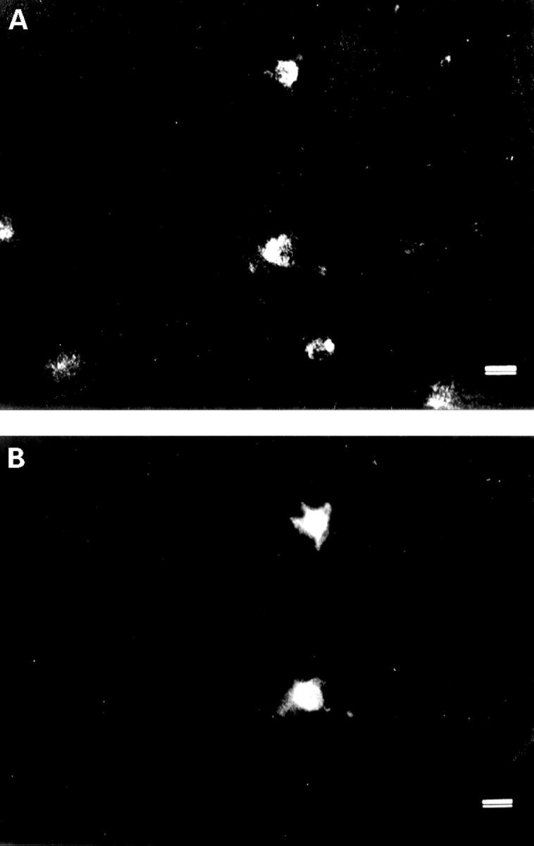
Epithelial sheet of conjunctival limbus from patient no 4 with AD and ocular complications. Cells were double stained for IgE (A) and CD1a (B). Numerous IgE positive cells (396 cells/mm2) (A) were found in the same area as the CD1a positive cells (B). Some IgE positive cells were CD1a negative. Bars = 10 µm.
Figure 2 .
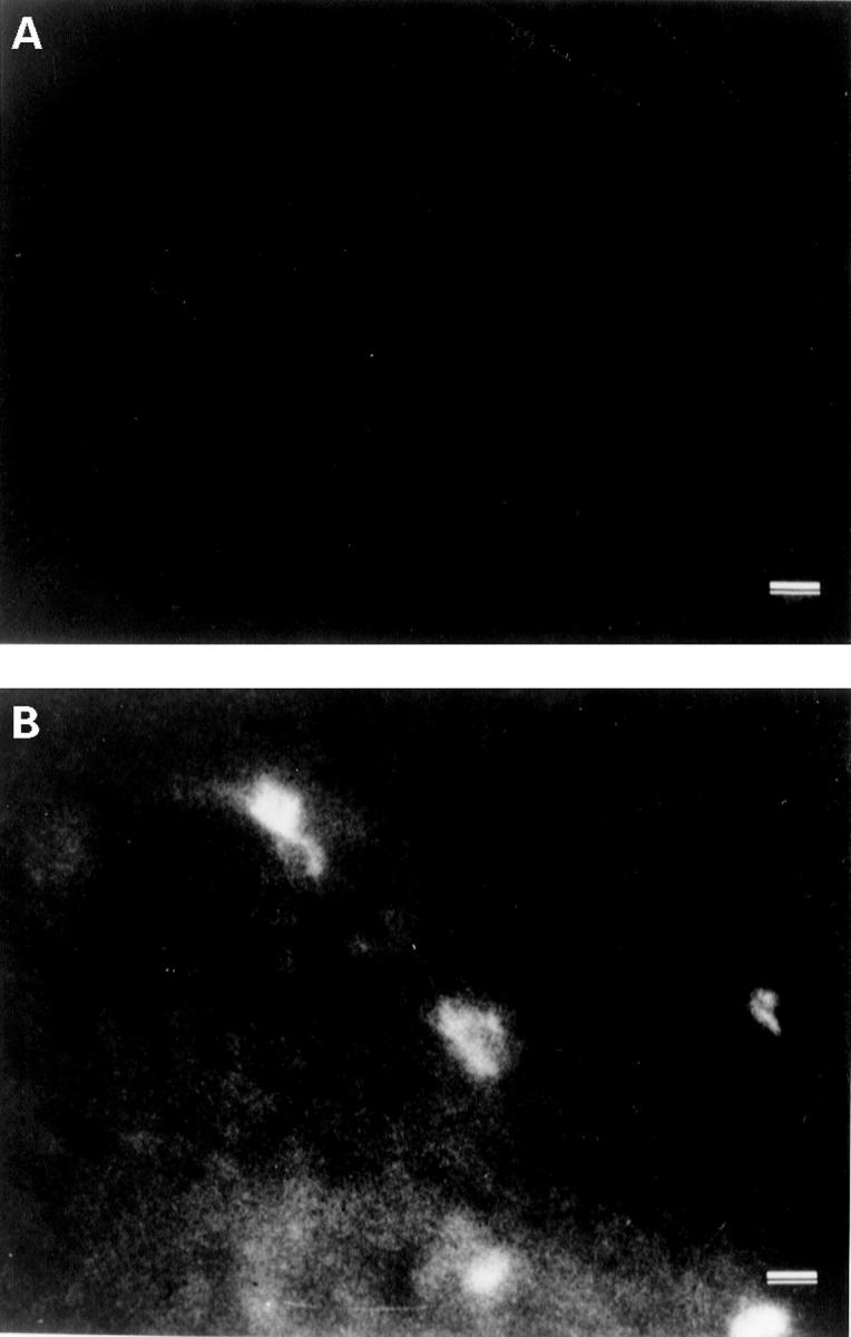
Epithelial sheet of conjunctival limbus from patient no 11 without AD. Cells were stained for IgE (A) and CD1a (B). Although no IgE positive cells (A) were observed, CD1a positive cells (B) were present (294 cells/mm2). Bars = 10 µm.
Figure 3 .
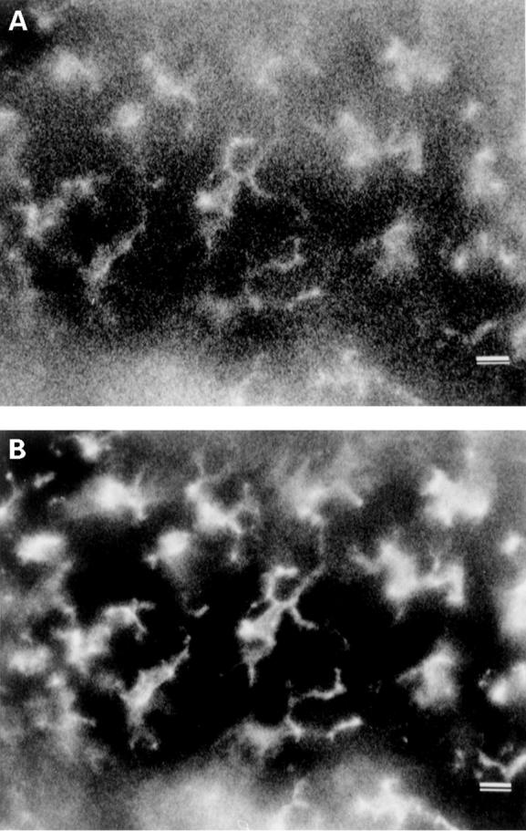
Epidermal sheet of abdominal skin from patient no 5 with AD and ocular complications. Cells were stained for IgE (A) and CD1a (B). Numerous IgE and CD1a positive cells (628 cells/mm2)were present. Bars = 10 µm.
Figure 4 .
Epidermal sheet of normal abdominal skin from a patient without AD stained for IgE (A) and CD1a (B). CD1a positive, IgE negative cells (913 cells/mm2) were present. Bars = 10 µm.
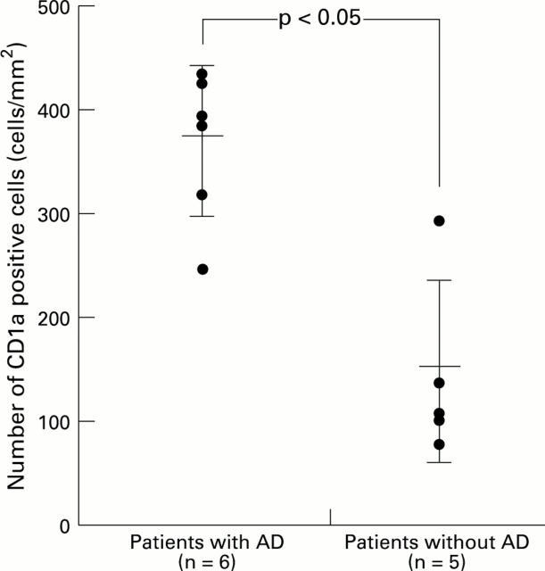
Figure 5 .
Number of CD1a positive cells in the conjunctival epithelium of patients with and without AD. The number of such cells in the patients with AD and ocular complications significantly exceeded that in the patients without AD (p<0.05).
Selected References
These references are in PubMed. This may not be the complete list of references from this article.
- Barker J. N., Alegre V. A., MacDonald D. M. Surface-bound immunoglobulin E on antigen-presenting cells in cutaneous tissue of atopic dermatitis. J Invest Dermatol. 1988 Feb;90(2):117–121. doi: 10.1111/1523-1747.ep12462074. [DOI] [PubMed] [Google Scholar]
- Bieber T., Braun-Falco O. IgE-bearing Langerhans cells are not specific to atopic eczema but are found in inflammatory skin diseases. J Am Acad Dermatol. 1991 Apr;24(4):658–659. doi: 10.1016/s0190-9622(08)80168-5. [DOI] [PubMed] [Google Scholar]
- Bieber T., Dannenberg B., Prinz J. C., Rieber E. P., Stolz W., Braun-Falco O., Ring J. Occurrence of IgE-bearing epidermal Langerhans cells in atopic eczema: a study of the time course of the lesions and with regard to the IgE serum level. J Invest Dermatol. 1989 Aug;93(2):215–219. doi: 10.1111/1523-1747.ep12277574. [DOI] [PubMed] [Google Scholar]
- Bieber T., Rieger A., Neuchrist C., Prinz J. C., Rieber E. P., Boltz-Nitulescu G., Scheiner O., Kraft D., Ring J., Stingl G. Induction of Fc epsilon R2/CD23 on human epidermal Langerhans cells by human recombinant interleukin 4 and gamma interferon. J Exp Med. 1989 Jul 1;170(1):309–314. doi: 10.1084/jem.170.1.309. [DOI] [PMC free article] [PubMed] [Google Scholar]
- Bieber T., de la Salle H., Wollenberg A., Hakimi J., Chizzonite R., Ring J., Hanau D., de la Salle C. Human epidermal Langerhans cells express the high affinity receptor for immunoglobulin E (Fc epsilon RI). J Exp Med. 1992 May 1;175(5):1285–1290. doi: 10.1084/jem.175.5.1285. [DOI] [PMC free article] [PubMed] [Google Scholar]
- Bruynzeel-Koomen C., van Wichen D. F., Toonstra J., Berrens L., Bruynzeel P. L. The presence of IgE molecules on epidermal Langerhans cells in patients with atopic dermatitis. Arch Dermatol Res. 1986;278(3):199–205. doi: 10.1007/BF00412924. [DOI] [PubMed] [Google Scholar]
- Buckley C. C., Ivison C., Poulter L. W., Rustin M. H. Fc epsilon R11/CD23 receptor distribution in patch test reactions to aeroallergens in atopic dermatitis. J Invest Dermatol. 1992 Aug;99(2):184–188. doi: 10.1111/1523-1747.ep12616813. [DOI] [PubMed] [Google Scholar]
- Buckley C., Ivison C., Poulter L. W., Rustin M. H. CD23/Fc epsilon R11 expression in contact sensitivity reactions: a comparison between aeroallergen patch test reactions in atopic dermatitis and the nickel patch test reaction in non-atopic individuals. Clin Exp Immunol. 1993 Mar;91(3):357–361. doi: 10.1111/j.1365-2249.1993.tb05909.x. [DOI] [PMC free article] [PubMed] [Google Scholar]
- Fithian E., Kung P., Goldstein G., Rubenfeld M., Fenoglio C., Edelson R. Reactivity of Langerhans cells with hybridoma antibody. Proc Natl Acad Sci U S A. 1981 Apr;78(4):2541–2544. doi: 10.1073/pnas.78.4.2541. [DOI] [PMC free article] [PubMed] [Google Scholar]
- Fokkens W. J., Vroom T. M., Rijntjes E., Mulder P. G. CD-1 (T6), HLA-DR-expressing cells, presumably Langerhans cells, in nasal mucosa. Allergy. 1989 Apr;44(3):167–172. doi: 10.1111/j.1398-9995.1989.tb02257.x. [DOI] [PubMed] [Google Scholar]
- Imayama S., Hashizume T., Miyahara H., Tanahashi T., Takeishi M., Kubota Y., Koga T., Hori Y., Fukuda H. Combination of patch test and IgE for dust mite antigens differentiates 130 patients with atopic dermatitis into four groups. J Am Acad Dermatol. 1992 Oct;27(4):531–538. doi: 10.1016/0190-9622(92)70218-5. [DOI] [PubMed] [Google Scholar]
- Imayama S., Shimozono Y., Hoashi M., Yasumoto S., Ohta S., Yoneyama K., Hori Y. Reduced secretion of IgA to skin surface of patients with atopic dermatitis. J Allergy Clin Immunol. 1994 Aug;94(2 Pt 1):195–200. doi: 10.1016/0091-6749(94)90040-x. [DOI] [PubMed] [Google Scholar]
- Imayama S., Yashima Y., Hori Y. Differing cell surface distribution of human leukocyte antigen-DR molecules on epidermal Langerhans cells and eccrine duct cells. J Histochem Cytochem. 1992 Aug;40(8):1191–1196. doi: 10.1177/40.8.1619281. [DOI] [PubMed] [Google Scholar]
- Katsushima H., Miyazaki I., Sekine N., Nishio C., Matsuda M. [Incidence of cataract and retinal detachment associated with atopic dermatitis]. Nippon Ganka Gakkai Zasshi. 1994 May;98(5):495–500. [PubMed] [Google Scholar]
- Matsuo T., Shiraga F., Matsuo N. Intraoperative observation of the vitreous base in patients with atopic dermatitis and retinal detachment. Retina. 1995;15(4):286–290. doi: 10.1097/00006982-199515040-00003. [DOI] [PubMed] [Google Scholar]
- Miyazaki I., Katsushima H., Suzuki J., Nakagawa T. [Pigmentation on the anterior chamber angle in retinal detachment associated with atopic dermatitis]. Nippon Ganka Gakkai Zasshi. 1994 Oct;98(10):998–1004. [PubMed] [Google Scholar]
- Mudde G. C., Van Reijsen F. C., Boland G. J., de Gast G. C., Bruijnzeel P. L., Bruijnzeel-Koomen C. A. Allergen presentation by epidermal Langerhans' cells from patients with atopic dermatitis is mediated by IgE. Immunology. 1990 Mar;69(3):335–341. [PMC free article] [PubMed] [Google Scholar]
- Mudde G. C., van Reijsen F. C., Bruijnzeel-Koomen C. A. IgE-positive Langerhans cells and Th2 allergen-specific T cells in atopic dermatitis. J Invest Dermatol. 1992 Nov;99(5):103S–103S. doi: 10.1111/1523-1747.ep12669981. [DOI] [PubMed] [Google Scholar]
- Ring J., Bieber T., Vieluf D., Kunz B., Przybilla B. Atopic eczema, Langerhans cells and allergy. Int Arch Allergy Appl Immunol. 1991;94(1-4):194–201. doi: 10.1159/000235361. [DOI] [PubMed] [Google Scholar]
- Schmitt D. A., Bieber T., Cazenave J. P., Hanau D. Fc receptors of human Langerhans cells. J Invest Dermatol. 1990 Jun;94(6 Suppl):15S–21S. doi: 10.1111/1523-1747.ep12874984. [DOI] [PubMed] [Google Scholar]
- Sugiura H., Uehara M., Maeda T. IgE-positive epidermal Langerhans cells in allergic contact dermatitis lesions provoked in patients with atopic dermatitis. Arch Dermatol Res. 1990;282(5):295–299. doi: 10.1007/BF00375722. [DOI] [PubMed] [Google Scholar]
- Tagawa Y., Takeuchi T., Matsuda H. [The distribution of Langerhans cells in the ocular surface epithelium and its role in the corneal immunopathology (author's transl)]. Nippon Ganka Gakkai Zasshi. 1981 Aug 10;85(8):1053–1059. [PubMed] [Google Scholar]
- Takeuchi T., Tagawa Y., Higuchi M., Matsuda H. [Langerhans cells in allergic diseases of the conjunctiva]. Nippon Ganka Gakkai Zasshi. 1983 Mar 10;87(3):209–218. [PubMed] [Google Scholar]
- Tigalonowa M., Braathen L. R., Lea T. IgE on Langerhans cells in the skin of patients with atopic dermatitis and birch allergy. Allergy. 1988 Aug;43(6):464–468. doi: 10.1111/j.1398-9995.1988.tb00920.x. [DOI] [PubMed] [Google Scholar]
- Wang B., Rieger A., Kilgus O., Ochiai K., Maurer D., Födinger D., Kinet J. P., Stingl G. Epidermal Langerhans cells from normal human skin bind monomeric IgE via Fc epsilon RI. J Exp Med. 1992 May 1;175(5):1353–1365. doi: 10.1084/jem.175.5.1353. [DOI] [PMC free article] [PubMed] [Google Scholar]
- Wollenberg A., de la Salle H., Hanau D., Liu F. T., Bieber T. Human keratinocytes release the endogenous beta-galactoside-binding soluble lectin immunoglobulin E (IgE-binding protein) which binds to Langerhans cells where it modulates their binding capacity for IgE glycoforms. J Exp Med. 1993 Sep 1;178(3):777–785. doi: 10.1084/jem.178.3.777. [DOI] [PMC free article] [PubMed] [Google Scholar]


