Full Text
The Full Text of this article is available as a PDF (234.2 KB).
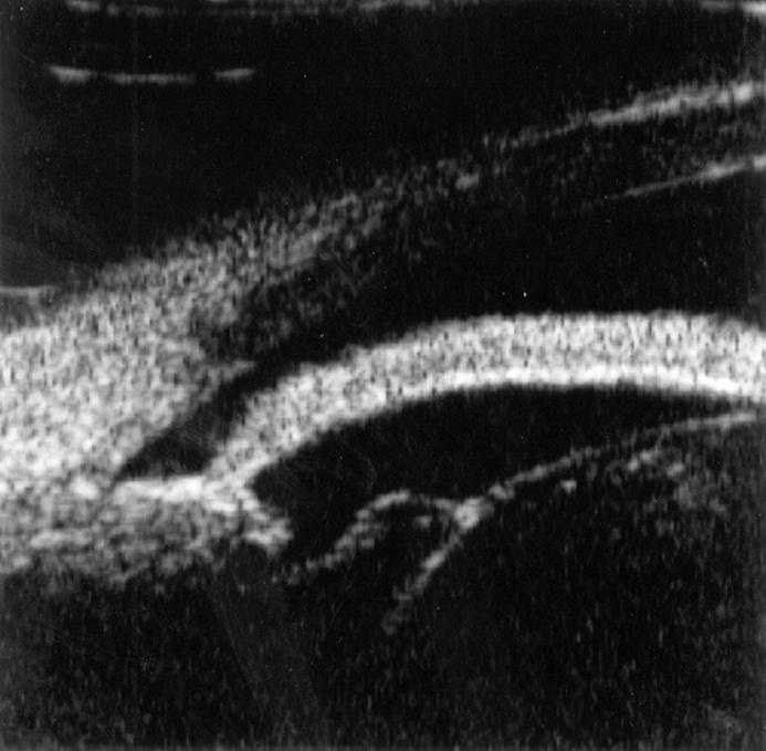
Figure 1 .
Ultrasound biomicroscopic image of the anterior segment of the inferior section in the right eye. Zonule is loose and the lens has a spherical shape. The iris shows a marked anterior bowing with the presence of pupillary block. The angle is narrow at the mid peripheral anterior chamber.
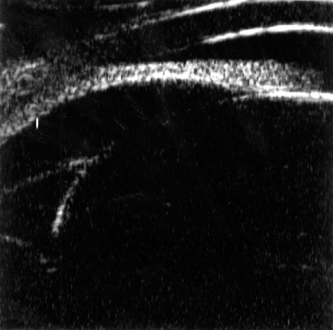
Figure 2 .
Ultrasound biomicroscopic image of the anterior segment of the inferior section in the left eye. Zonules are thick and well defined, and the lens has a spherical shape. The angle is closed with pupillary block.


