Abstract
BACKGROUND—Optic nerve head drusen may present diagnostic difficulties in cases of disc swelling. Imaging of the nerve in a search for drusen is often inconclusive, especially in children, where drusen may be buried below the surface of the nerve head. METHODS—A small study was carried out using a scanning laser ophthalmoscope (SLO) with an infrared confocal facility to scan deep within optic discs in an attempt to image drusen. RESULTS—The SLO was able to demonstrate superficial and buried drusen (using the infrared confocal facility). The superiority of the SLO over ultrasound in the presence of lens opacity was revealed, as the SLO simultaneously demonstrated both drusen and the associated anomalous disc features which are not detected by ultrasound. CONCLUSION—The SLO can help in the diagnosis of optic disc drusen especially in difficult cases where lens opacity or buried drusen hinders their definitive diagnosis.
Full Text
The Full Text of this article is available as a PDF (104.3 KB).
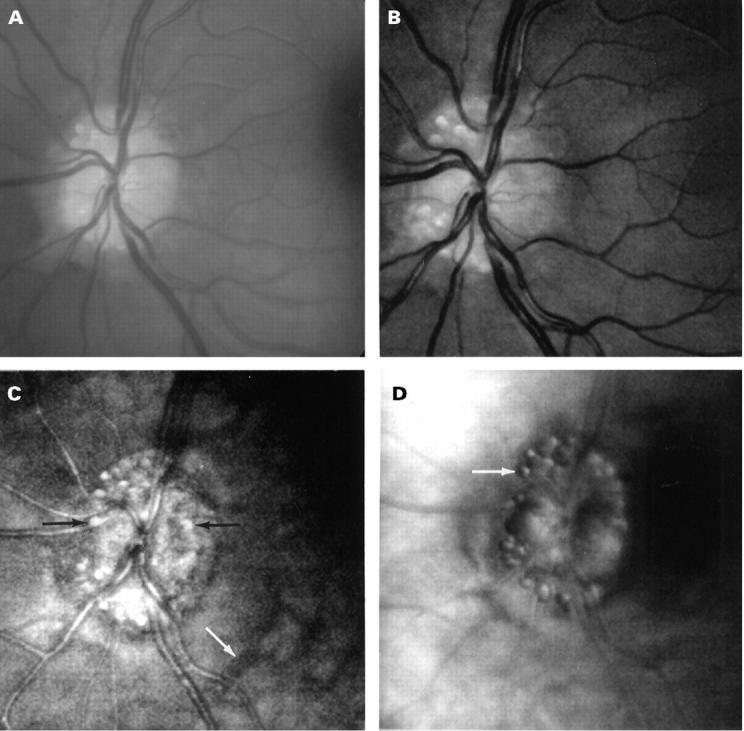
Figure 1 .

Fundus images of a patient with optic disc drusen. (A) Fundus photograph showing superficial disc drusen. (B) Short wavelength (540 nm, green) SLO images showing superficial structures including disc drusen, retinal vessels, and the nerve fibre layer. (C) Long wavelength (780 nm, infrared) confocal mode SLO images showing more drusen including buried ones (black arrows) which are not visible in (A) or (B). Note the retinal vessels appear out of focus as they lie superficial to the plane of focus, whereas deep choroidal vessels are now visible (white arrow). (D) Indirect mode (780 nm, infrared) SLO images of disc drusen showing the distinctive `bubble' appearance.
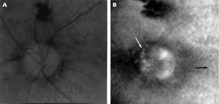
Figure 2 .
(A) Fundus photograph showing the poor view of disc drusen in a patient with significant lens opacity. (B) Indirect mode SLO image of the same patient demonstrating disc drusen (white arrow) and retinal drusen (black arrow) in the presence of lens opacity.
Selected References
These references are in PubMed. This may not be the complete list of references from this article.
- Boyce S. W., Platia E. V., Green W. R. Drusen of the optic nerve head. Ann Ophthalmol. 1978 Jun;10(6):695–704. [PubMed] [Google Scholar]
- Cohen D. N. Drusen of the optic disc and the development of field defects. Arch Ophthalmol. 1971 Feb;85(2):224–226. doi: 10.1001/archopht.1971.00990050226018. [DOI] [PubMed] [Google Scholar]
- Kheterpal S., Good P. A., Beale D. J., Kritzinger E. E. Imaging of optic disc drusen: a comparative study. Eye (Lond) 1995;9(Pt 1):67–69. doi: 10.1038/eye.1995.10. [DOI] [PubMed] [Google Scholar]
- Kirkpatrick J. N., Spencer T., Manivannan A., Sharp P. F., Forrester J. V. Quantitative image analysis of macular drusen from fundus photographs and scanning laser ophthalmoscope images. Eye (Lond) 1995;9(Pt 1):48–55. doi: 10.1038/eye.1995.7. [DOI] [PubMed] [Google Scholar]
- Manivannan A., Kirkpatrick J. N., Sharp P. F., Forrester J. V. Clinical investigation of an infrared digital scanning laser ophthalmoscope. Br J Ophthalmol. 1994 Feb;78(2):84–90. doi: 10.1136/bjo.78.2.84. [DOI] [PMC free article] [PubMed] [Google Scholar]
- Manivannan A., Sharp P. F., Forrester J. V. Performance measurements of an infrared digital scanning laser ophthalmoscope. Physiol Meas. 1994 Aug;15(3):317–324. doi: 10.1088/0967-3334/15/3/010. [DOI] [PubMed] [Google Scholar]
- Manivannan A., Sharp P. F., Phillips R. P., Forrester J. V. Digital fundus imaging using a scanning laser ophthalmoscope. Physiol Meas. 1993 Feb;14(1):43–56. doi: 10.1088/0967-3334/14/1/006. [DOI] [PubMed] [Google Scholar]
- Savino P. J., Glaser J. S., Rosenberg M. A. A clinical analysis of pseudopapilledema. II. Visual field defects. Arch Ophthalmol. 1979 Jan;97(1):71–75. doi: 10.1001/archopht.1979.01020010011002. [DOI] [PubMed] [Google Scholar]


