Abstract
AIM—A study was designed to investigate whether measurements of the optic nerve diameter (OND) and cross sectional area (ONCSA), as measured by B-scan ultrasonography, are altered in glaucoma. The reproducibility and test-retest variability of echographic estimates of retrobulbar optic nerve dimensions was also tested. METHODS—One eye of 49 glaucoma patients and 90 control subjects underwent five repeated echographic measurements of the maximal interpial diameter and cross sectional area of the orbital optic nerve on two separate occasions. All measurements were taken by one experienced ultrasonographer. RESULTS—Mean optic nerve diameter (SD) for the control group was 2.86 (0.46) mm, and was independent of height (multiple regression analysis: p = 0.21), axial length (p = 0.74), spherical equivalent (p = 0.97), sex (ANOVA: p=0.36), or race (p=0.14), but was inversely related to age (p = 0.01). Reproducibility of OND readings in control subjects was 0.149 mm (coefficient of repeatability). Test-retest variability of interpial diameter was −0.02 (0.29) mm. Mean interpial diameter of the optic nerve was significantly smaller among glaucomatous eyes (2.58 (0.501) mm) than controls (Mann-Whitney U test: p < 0.0001). Glaucomatous optic nerves also had a significantly smaller cross sectional area (6.68 (2.58) mm2) than those of healthy volunteers (8.25 (1.67) mm2) (p = 0.004). CONCLUSION—Echographic measurements of the orbital optic nerve are highly reproducible and not subject to clinically meaningful test-retest variability. Optic nerve interpial diameter and cross sectional area are reduced in glaucomatous eyes, reflecting nerve fibre loss. This technique may be useful in distinguishing between normal and glaucomatous eyes where optic disc morphometry is inconclusive or impossible as a result of opaque media. Keywords: glaucoma; optic nerve; ultrasound
Full Text
The Full Text of this article is available as a PDF (133.6 KB).
Figure 1 .
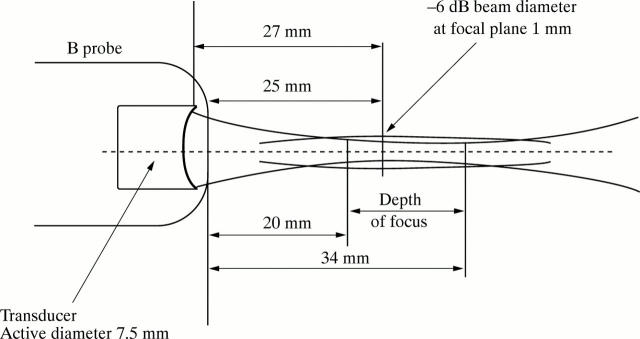
Diagrammatic representation of the B-scan probe. The radius of concavity of the transducer is approximately equal to its focal distance (27 mm). As the transducer is 2 mm inside the probe, this ensures an external focus of 25 mm.
Figure 2 .

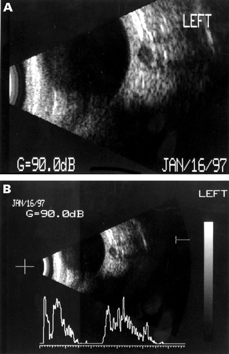
(A) Cross sectional B-scan echograph of the retrobulbar optic nerve. The interpial space appears dark and the dural sheath can be clearly seen. (B) Superimposed A-scan confirms the scan through the nerve is perpendicular, as the distal dural echo is within 20% of the height of the proximal echo.
Figure 3 .
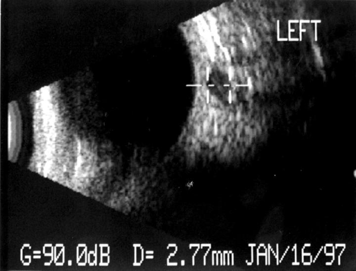
The interpial diameter is calculated by the instrument as the distance between two cursors placed on the dural sheath by the operator.
Figure 4 .
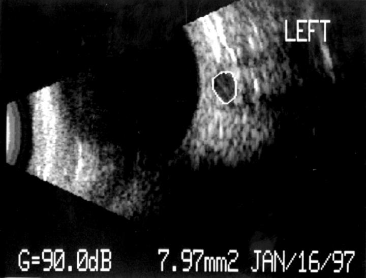
The optic nerve cross sectional area is calculated by the instrument as the area enclosed by the dural sheath, which is marked with a cursor by the operator.
Selected References
These references are in PubMed. This may not be the complete list of references from this article.
- Airaksinen P. J., Drance S. M., Douglas G. R., Schulzer M., Wijsman K. Visual field and retinal nerve fiber layer comparisons in glaucoma. Arch Ophthalmol. 1985 Feb;103(2):205–207. doi: 10.1001/archopht.1985.01050020057019. [DOI] [PubMed] [Google Scholar]
- Atta H. R. Imaging of the optic nerve with standardised echography. Eye (Lond) 1988;2(Pt 4):358–366. doi: 10.1038/eye.1988.66. [DOI] [PubMed] [Google Scholar]
- Autzen T., Pugesgaard T., Work K. Automated static perimetry and nerve fibre layer defects in glaucoma and controls. Acta Ophthalmol (Copenh) 1990 Dec;68(6):677–680. doi: 10.1111/j.1755-3768.1990.tb01693.x. [DOI] [PubMed] [Google Scholar]
- Balazsi A. G., Rootman J., Drance S. M., Schulzer M., Douglas G. R. The effect of age on the nerve fiber population of the human optic nerve. Am J Ophthalmol. 1984 Jun;97(6):760–766. doi: 10.1016/0002-9394(84)90509-9. [DOI] [PubMed] [Google Scholar]
- Dees C., Buimer R., Dick A. D., Atta H. R. Ultrasonographic investigation of optic neuritis. Eye (Lond) 1995;9(Pt 4):488–494. doi: 10.1038/eye.1995.113. [DOI] [PubMed] [Google Scholar]
- Dichtl A., Jonas J. B. Echographic measurement of optic nerve thickness correlated with neuroretinal rim area and visual field defect in glaucoma. Am J Ophthalmol. 1996 Oct;122(4):514–519. doi: 10.1016/s0002-9394(14)72111-7. [DOI] [PubMed] [Google Scholar]
- Gaasterland D., Tanishima T., Kuwabara T. Axoplasmic flow during chronic experimental glaucoma. 1. Light and electron microscopic studies of the monkey optic nervehead during development of glaucomatous cupping. Invest Ophthalmol Vis Sci. 1978 Sep;17(9):838–846. [PubMed] [Google Scholar]
- Jonas J. B., Fernandez M. C., Naumann G. O. Glaucomatous optic nerve atrophy in small discs with low cup-to-disc ratios. Ophthalmology. 1990 Sep;97(9):1211–1215. doi: 10.1016/s0161-6420(90)32434-x. [DOI] [PubMed] [Google Scholar]
- Jonas J. B., Gusek G. C., Naumann G. O. Optic disk morphometry in high myopia. Graefes Arch Clin Exp Ophthalmol. 1988;226(6):587–590. doi: 10.1007/BF02169209. [DOI] [PubMed] [Google Scholar]
- Jonas J. B., Königsreuther K. A. Optic disk appearance in ocular hypertensive eyes. Am J Ophthalmol. 1994 Jun 15;117(6):732–740. doi: 10.1016/s0002-9394(14)70316-2. [DOI] [PubMed] [Google Scholar]
- Jonas J. B., Schmidt A. M., Müller-Bergh J. A., Naumann G. O. Optic nerve fiber count and diameter of the retrobulbar optic nerve in normal and glaucomatous eyes. Graefes Arch Clin Exp Ophthalmol. 1995 Jul;233(7):421–424. doi: 10.1007/BF00180945. [DOI] [PubMed] [Google Scholar]
- Parravano J. G., Toledo A., Kucharczyk W. Dimensions of the optic nerves, chiasm, and tracts: MR quantitative comparison between patients with optic atrophy and normals. J Comput Assist Tomogr. 1993 Sep-Oct;17(5):688–690. doi: 10.1097/00004728-199309000-00003. [DOI] [PubMed] [Google Scholar]
- Schulzer M., Drance S. M., Carter C. J., Brooks D. E., Douglas G. R., Lau W. Biostatistical evidence for two distinct chronic open angle glaucoma populations. Br J Ophthalmol. 1990 Apr;74(4):196–200. doi: 10.1136/bjo.74.4.196. [DOI] [PMC free article] [PubMed] [Google Scholar]
- Scimeca H. A. Optic disc changes in glaucoma. Int Ophthalmol Clin. 1979 Spring;19(1):127–154. doi: 10.1097/00004397-197901910-00009. [DOI] [PubMed] [Google Scholar]
- Sommer A. Doyne Lecture. Glaucoma: facts and fancies. Eye (Lond) 1996;10(Pt 3):295–301. doi: 10.1038/eye.1996.63. [DOI] [PubMed] [Google Scholar]
- Zangwill L. M., van Horn S., de Souza Lima M., Sample P. A., Weinreb R. N. Optic nerve head topography in ocular hypertensive eyes using confocal scanning laser ophthalmoscopy. Am J Ophthalmol. 1996 Oct;122(4):520–525. doi: 10.1016/s0002-9394(14)72112-9. [DOI] [PubMed] [Google Scholar]


