Full Text
The Full Text of this article is available as a PDF (191.6 KB).
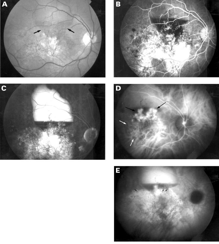
Figure 1 .
(A) Fundus photograph of the right eye showing marked atrophy of the retinal pigment epithelium and an exudative pigment epithelial detachment with a meniscus of subretinal blood (arrows). (B) and (C) Fluorescein angiogram reveals a window defect and filling of the pigment epithelial detachment with a blood-fluid level. (D) Early phase of indocyanine green angiogram reveals a branching vascular pattern (white arrows) with polypoidal elements (black arrows) at their border in the area of the subretinal blood. (E) There is late staining of the vascular network and filling of the pigment epithelium detachment. The core of the polypoidal elements became hypofluorescent (arrows).
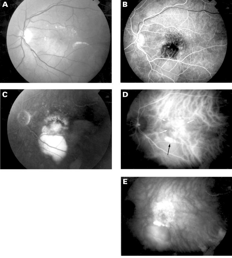
Figure 2 .
(A) Fundus photograph of the left eye showing a serous detachment of the neurosensory retina and subretinal lipid deposits. (B) Early phase fluorescein angiogram reveals mottled hyperfluorescence. (C) Late phase fluorescein angiogram demonstrating staining of the subfoveal lesion and filling of the two pigment epithelial detachments. (D) Early phase indocyanine green angiogram reveals a branching vascular pattern (white arrows) with polypoidal elements (black arrow) at the inferior border. (E) There is late staining of the vascular network.


