Abstract
AIM—To observe pathophysiological changes in patients with Vogt-Koyanagi-Harada (VKH) syndrome using the scanning laser ophthalmoscope (SLO) to perform videofunduscopy, and indocyanine green (ICG) and fluorescein videoangiography. METHODS—18 patients diagnosed with VKH syndrome were studied. 15 patients were examined in both acute and convalescent phases, and three patients were examined in the convalescent phase only. Retrospective review of charts was performed to obtain data on visual acuities, clinical findings, and results of cerebrospinal fluid cytology and histocompatibility leucocyte antigen D locus testing. RESULTS—Videofunduscopy revealed abnormalities in the central macula at the level of the retinal pigment epithelium or choroid in 40% of patients in the acute phase and 83% of patients in the convalescent phase. ICG videoangiography showed irregular hypofluorescence (80%), slow or patchy filling (67%), indistinct or fewer choroidal vessels (73%), filling defects (73%), and focal leakage (33%) in patients in the acute phase. These changes improved with systemic corticosteroid treatment, although at least one ICG videoangiographic abnormality persisted into the convalescent phase in 83% of patients in the absence of clinical inflammation. In contrast, fluorescein videoangiography revealed fewer and decreased severity of abnormalities, with greater resolution in the convalescent phase when compared with ICG videoangiography. CONCLUSIONS—SLO videofunduscopy and videoangiography revealed numerous abnormalities in patients with VKH syndrome, many of which persisted well after clinical recovery, suggesting the development of permanent morphological changes in the fundus. ICG videoangiography was more sensitive than fluorescein videoangiography in delineating abnormalities in layers deep to the sensory retina. Keywords: fluorescein; fundus angiography; indocyanine green; Vogt-Koyanagi-Harada syndrome
Full Text
The Full Text of this article is available as a PDF (168.6 KB).
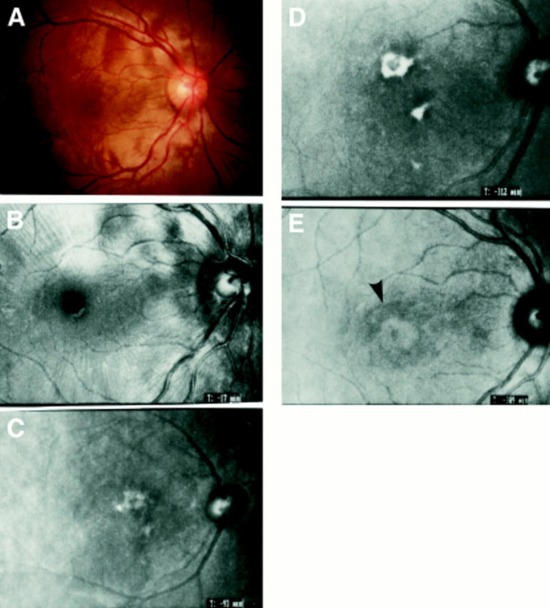
Figure 1 .

(A) Colour photograph of patient 6 at presentation showing marked serous retinal detachment. (B) Videofunduscopy (argon) performed at the same time showed radiating retinal folds about the macula. (C) Videofunduscopy (infrared) performed on patient 2 after 6 weeks of systemic steroid therapy revealed granular hard exudates at the level of the RPE and choroid that were not visible by clinical examination. (D) Videofunduscopy (infrared) performed on patient 2 after 9 months of treatment showed that the RPE/choroidal exudates had become plaque-like and increased in size. This was now faintly visible clinically, although much less prominent. (E) Videofunduscopy (infrared) of patient 6 after 6 months of systemic corticosteroid therapy showed a target-shaped abnormality in the central macula (arrow) that was not visible by clinical examination.
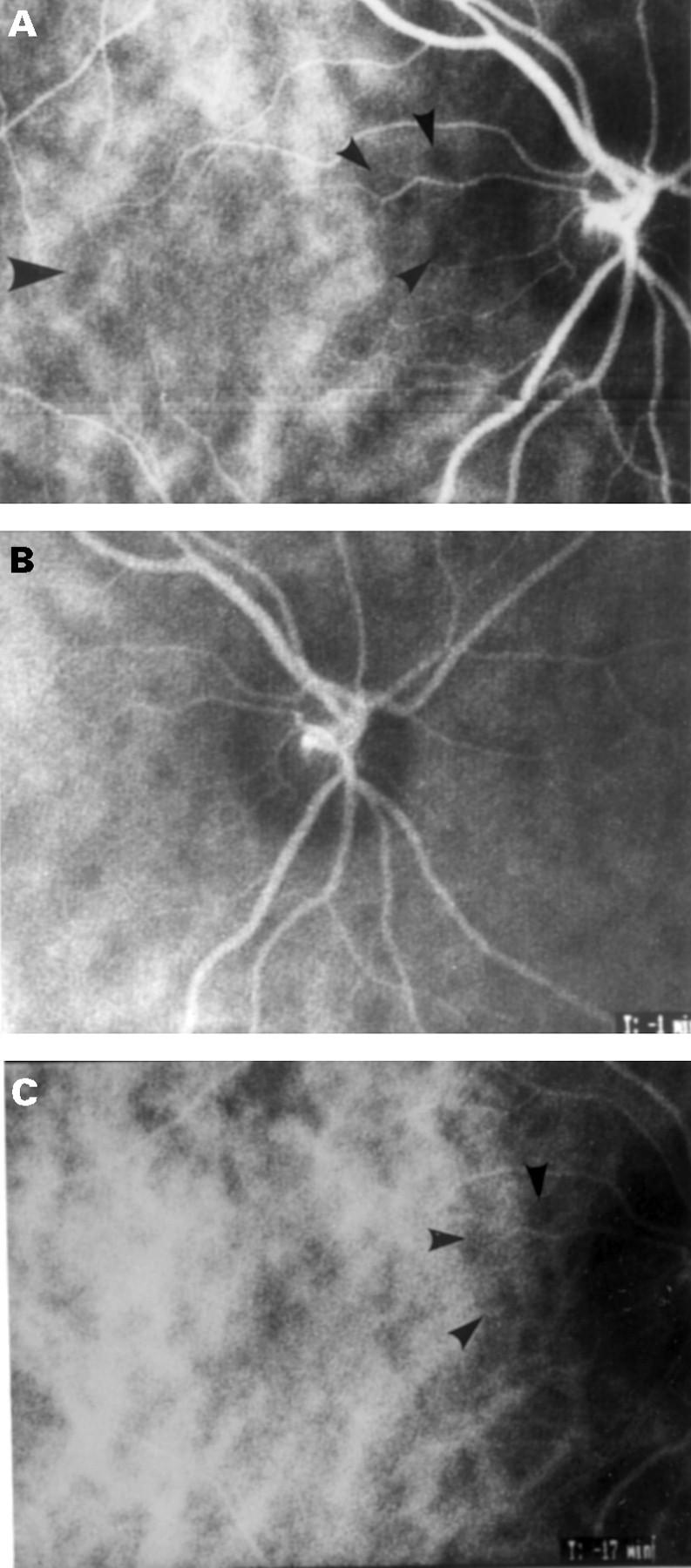
Figure 2 .

(A) ICG videoangiography performed on patient 6 at presentation revealed irregular hypofluorescence of the central macula (large arrow), corresponding to areas of chorioretinal thickening observed clinically. Numerous filling defects approximately 1/5 disc diameter in size (small arrows) were also present. (B) The filling defects were most prominent surrounding the optic disc. (C) After 6 months of treatment, ICG videoangiography showed marked improvement of the hypofluorescence of the macula but persistence of some filling defects (arrows).
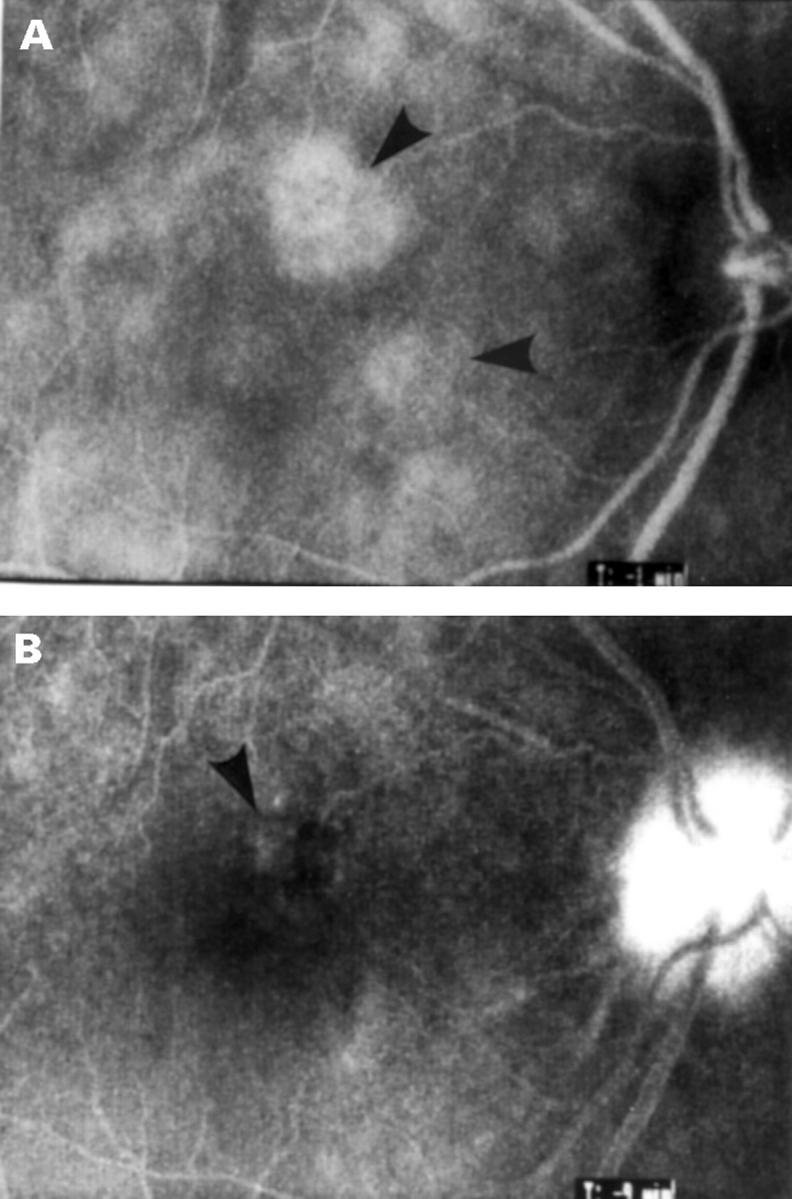
Figure 3 .

(A) ICG videoangiography of patient 2 in the acute phase showed focal hyperfluorescent areas (arrows). (B) Fluorescein videoangiography taken at the same time showed only faint RPE staining in one area (arrow).
Selected References
These references are in PubMed. This may not be the complete list of references from this article.
- Bartsch D. U., Weinreb R. N., Zinser G., Freeman W. R. Confocal scanning infrared laser ophthalmoscopy for indocyanine green angiography. Am J Ophthalmol. 1995 Nov;120(5):642–651. doi: 10.1016/s0002-9394(14)72211-1. [DOI] [PubMed] [Google Scholar]
- Chan C. C., Palestine A. G., Kuwabara T., Nussenblatt R. B. Immunopathologic study of Vogt-Koyanagi-Harada syndrome. Am J Ophthalmol. 1988 Jun 15;105(6):607–611. doi: 10.1016/0002-9394(88)90052-9. [DOI] [PubMed] [Google Scholar]
- Guyer D. R., Puliafito C. A., Monés J. M., Friedman E., Chang W., Verdooner S. R. Digital indocyanine-green angiography in chorioretinal disorders. Ophthalmology. 1992 Feb;99(2):287–291. doi: 10.1016/s0161-6420(92)31981-5. [DOI] [PubMed] [Google Scholar]
- Guyer D. R., Yannuzzi L. A., Slakter J. S., Sorenson J. A., Ho A., Orlock D. Digital indocyanine green videoangiography of central serous chorioretinopathy. Arch Ophthalmol. 1994 Aug;112(8):1057–1062. doi: 10.1001/archopht.1994.01090200063023. [DOI] [PubMed] [Google Scholar]
- Hayreh S. S. In vivo choroidal circulation and its watershed zones. Eye (Lond) 1990;4(Pt 2):273–289. doi: 10.1038/eye.1990.39. [DOI] [PubMed] [Google Scholar]
- Hayreh S. S. Physiological anatomy of the choroidal vascular bed. Int Ophthalmol. 1983 Feb;6(2):85–93. doi: 10.1007/BF00127636. [DOI] [PubMed] [Google Scholar]
- Howe L. J., Woon H., Graham E. M., Fitzke F., Bhandari A., Marshall J. Choroidal hypoperfusion in acute posterior multifocal placoid pigment epitheliopathy. An indocyanine green angiography study. Ophthalmology. 1995 May;102(5):790–798. doi: 10.1016/s0161-6420(95)30955-4. [DOI] [PubMed] [Google Scholar]
- Ie D., Glaser B. M., Murphy R. P., Gordon L. W., Sjaarda R. N., Thompson J. T. Indocyanine green angiography in multiple evanescent white-dot syndrome. Am J Ophthalmol. 1994 Jan 15;117(1):7–12. doi: 10.1016/s0002-9394(14)73008-9. [DOI] [PubMed] [Google Scholar]
- Iida T., Hagimura N., Otani T., Ikeda F., Muraoka K. [Choroidal vascular lesions in serous retinal detachment viewed with indocyanine green angiography]. Nippon Ganka Gakkai Zasshi. 1996 Oct;100(10):817–824. [PubMed] [Google Scholar]
- Joko S., Numaga J., Fujino Y., Masuda K., Hirata R., Maeda H. [HLA and uveitis in leprosy]. Nippon Ganka Gakkai Zasshi. 1995 Oct;99(10):1181–1185. [PubMed] [Google Scholar]
- Matsunaga H., Matsubara T., Fukushima I., Uyama M. [Indocyanine green fluorescence angiography in Harada disease]. Nippon Ganka Gakkai Zasshi. 1994 Sep;98(9):852–857. [PubMed] [Google Scholar]
- Oshima Y., Harino S., Hara Y., Tano Y. Indocyanine green angiographic findings in Vogt-Koyanagi-Harada disease. Am J Ophthalmol. 1996 Jul;122(1):58–66. doi: 10.1016/s0002-9394(14)71964-6. [DOI] [PubMed] [Google Scholar]
- Pece A., Bolognesi G., Introini U., Brancato R. Indocyanine green angiography in Vogt-Koyanagi-Harada-type disease. Arch Ophthalmol. 1997 Jun;115(6):804–806. doi: 10.1001/archopht.1997.01100150806023. [DOI] [PubMed] [Google Scholar]
- Perry H. D., Font R. L. Clinical and histopathologic observations in severe Vogt-Koyanagi-Harada syndrome. Am J Ophthalmol. 1977 Feb;83(2):242–254. doi: 10.1016/0002-9394(77)90623-7. [DOI] [PubMed] [Google Scholar]
- Scheider A., Kaboth A., Neuhauser L. Detection of subretinal neovascular membranes with indocyanine green and an infrared scanning laser ophthalmoscope. Am J Ophthalmol. 1992 Jan 15;113(1):45–51. doi: 10.1016/s0002-9394(14)75752-6. [DOI] [PubMed] [Google Scholar]
- Scheider A., Nasemann J. E., Lund O. E. Fluorescein and indocyanine green angiographies of central serous choroidopathy by scanning laser ophthalmoscopy. Am J Ophthalmol. 1993 Jan;115(1):50–56. doi: 10.1016/s0002-9394(14)73524-x. [DOI] [PubMed] [Google Scholar]
- Scheider A., Schroedel C. High resolution indocyanine green angiography with a scanning laser ophthalmoscope. Am J Ophthalmol. 1989 Oct 15;108(4):458–459. doi: 10.1016/s0002-9394(14)73325-2. [DOI] [PubMed] [Google Scholar]
- Sharp P. F., Manivannan A. The scanning laser ophthalmoscope. Phys Med Biol. 1997 May;42(5):951–966. doi: 10.1088/0031-9155/42/5/014. [DOI] [PubMed] [Google Scholar]
- Snyder D. A., Tessler H. H. Vogt-Koyanagi-Harada syndrome. Am J Ophthalmol. 1980 Jul;90(1):69–75. [PubMed] [Google Scholar]
- Wolf S., Wald K. J., Elsner A. E., Staurenghi G. Indocyanine green choroidal videoangiography: a comparison of imaging analysis with the scanning laser ophthalmoscope and the fundus camera. Retina. 1993;13(3):266–269. doi: 10.1097/00006982-199313030-00025. [DOI] [PubMed] [Google Scholar]
- Yannuzzi L. A., Slakter J. S., Sorenson J. A., Guyer D. R., Orlock D. A. Digital indocyanine green videoangiography and choroidal neovascularization. Retina. 1992;12(3):191–223. [PubMed] [Google Scholar]
- Yokoi H., Goto H., Sakai J., Takano S., Usui M. [Incidence of uveitis at Tokyo Medical College Hospital]. Nippon Ganka Gakkai Zasshi. 1995 Jun;99(6):710–714. [PubMed] [Google Scholar]
- Yuzawa M., Kawamura A., Matsui M. Indocyanine green video-angiographic findings in Harada's disease. Jpn J Ophthalmol. 1993;37(4):456–466. [PubMed] [Google Scholar]


