Abstract
AIMS—The vertical cup/disc ratio (CDR) has long been used in the assessment of the glaucoma suspect, though the wide range of CDR values in the normal population limits its use. Cup size is related physiologically to disc size and pathologically to glaucomatous damage. Disc size can be measured at the slit lamp as the vertical disc diameter (DD). The ability of the CDR, in relation to DD, to identify glaucomatous optic discs was investigated. METHODS—88 normal, 53 early glaucoma, and 59 ocular hypertensive subjects underwent stereoscopic optic disc photography and clinical biometry. Photographs were analysed in a masked fashion by computer assisted planimetry. The relation between vertical cup diameter and DD was explored by linear regression, and expressed in terms of CDR. The upper limit of normal was defined by the 95% prediction intervals of this regression (method 1) and by the upper 97.5 percentile for CDR (method 2). The sensitivity and specificity of CDR to identify an optic disc as glaucomatous was tested with these disc size dependent and disc size independent cut offs in small, medium, and large discs. RESULTS—The CDR was related to DD by the equation CDR = (−1.31 + (1.194 × DD))/DD. The sensitivity in small, medium, and large discs was 80%, 60%, and 38% respectively for method 1 and 33%, 67%, and 63% respectively for method 2. Specificity was 98.9% (method 1) and 97.7% (method 2). CONCLUSIONS—The CDR, relative to disc size, is useful clinically, especially to assist in identifying small glaucomatous discs. Keywords: cup/disc ratio; glaucoma; imaging
Full Text
The Full Text of this article is available as a PDF (141.1 KB).
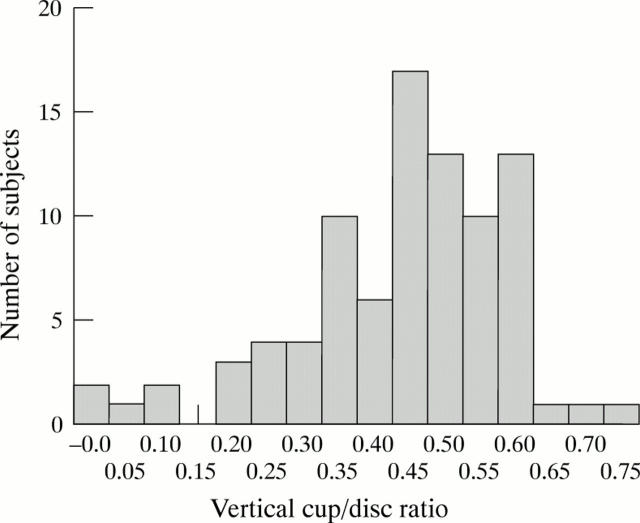
Figure 1 .
Distribution of cup/disc ratio values in the control group.
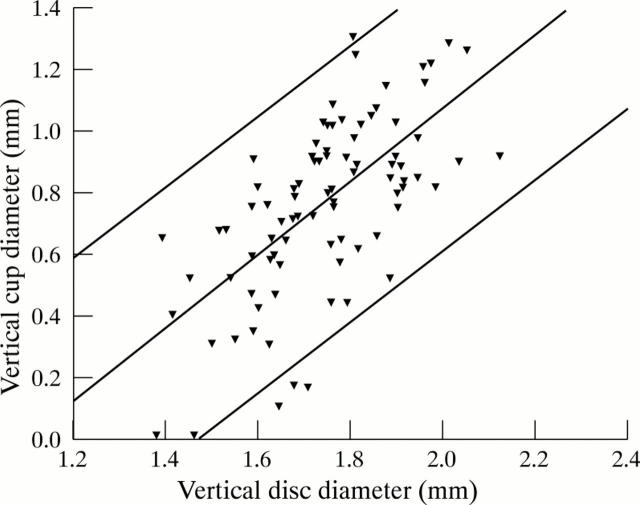
Figure 2 .
Plot of vertical cup diameter against vertical disc diameter. Mean (regression line) (R2 = 0.42, p <0.000) and 95% prediction intervals shown.
Figure 3 .
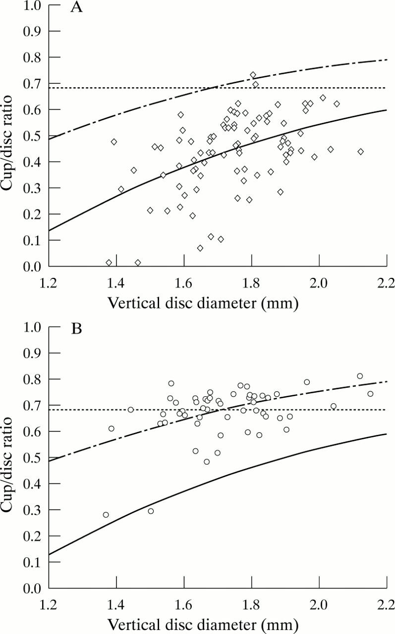
Plot of vertical cup/disc ratio against vertical disc diameter. (A) Control subjects. (B) Glaucoma patients. Solid line, mean (disc size dependent); broken line, upper limit of normal (disc size dependent); dotted line, upper limit of normal (disc size independent).(The vertical disc diameter is calculated from measurements of optic disc photographs using the Littmann correction for ocular magnification. When measuring the optic disc at the slit lamp, the correction factor for the 78 D lens is 1.11 and for the 90 D lens 1.39 (unpublished data presented at Glaucoma Society (UK & Eire) Annual Meeting, November 1996)).
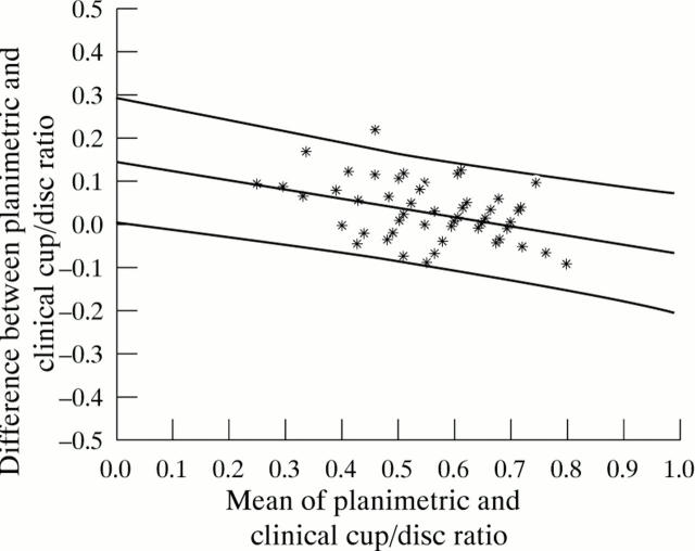
Figure 4 .
Agreement between clinical and planimetric estimation of cup/disc ratio (ocular hypertensive patients). Mean and 95% prediction intervals shown.
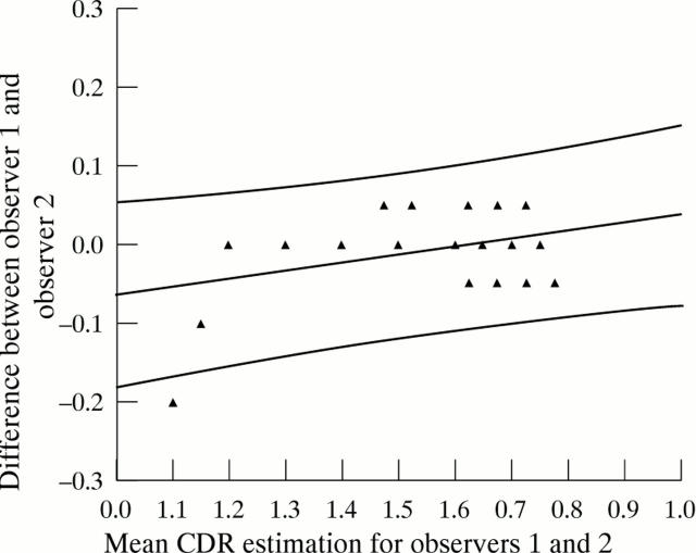
Figure 5 .
Agreement between two clinicians' estimation of CDR (ocular hypertensive patients). Mean and 95% prediction intervals shown.
Selected References
These references are in PubMed. This may not be the complete list of references from this article.
- Advanced Glaucoma Intervention Study. 2. Visual field test scoring and reliability. Ophthalmology. 1994 Aug;101(8):1445–1455. [PubMed] [Google Scholar]
- Airaksinen P. J., Drance S. M. Neuroretinal rim area and retinal nerve fiber layer in glaucoma. Arch Ophthalmol. 1985 Feb;103(2):203–204. doi: 10.1001/archopht.1985.01050020055018. [DOI] [PubMed] [Google Scholar]
- Airaksinen P. J., Tuulonen A., Alanko H. I. Rate and pattern of neuroretinal rim area decrease in ocular hypertension and glaucoma. Arch Ophthalmol. 1992 Feb;110(2):206–210. doi: 10.1001/archopht.1992.01080140062028. [DOI] [PubMed] [Google Scholar]
- Armaly M. F. Genetic determination of cup/disc ratio of the optic nerve. Arch Ophthalmol. 1967 Jul;78(1):35–43. doi: 10.1001/archopht.1967.00980030037007. [DOI] [PubMed] [Google Scholar]
- Armaly M. F., Krueger D. E., Maunder L., Becker B., Hetherington J., Jr, Kolker A. E., Levene R. Z., Maumenee A. E., Pollack I. P., Shaffer R. N. Biostatistical analysis of the collaborative glaucoma study. I. Summary report of the risk factors for glaucomatous visual-field defects. Arch Ophthalmol. 1980 Dec;98(12):2163–2171. doi: 10.1001/archopht.1980.01020041015002. [DOI] [PubMed] [Google Scholar]
- Beck R. W., Messner D. K., Musch D. C., Martonyi C. L., Lichter P. R. Is there a racial difference in physiologic cup size? Ophthalmology. 1985 Jul;92(7):873–876. doi: 10.1016/s0161-6420(85)33942-8. [DOI] [PubMed] [Google Scholar]
- Bengtsson B. The alteration and asymmetry of cup and disc diameters. Acta Ophthalmol (Copenh) 1980 Oct;58(5):726–732. doi: 10.1111/j.1755-3768.1980.tb06685.x. [DOI] [PubMed] [Google Scholar]
- Bengtsson B. The variation and covariation of cup and disc diameters. Acta Ophthalmol (Copenh) 1976 Dec;54(6):804–818. doi: 10.1111/j.1755-3768.1976.tb01801.x. [DOI] [PubMed] [Google Scholar]
- Betz P., Camps F., Collignon-Brach J., Lavergne G., Weekers R. Biometric study of the disc cup in open-angle glaucoma. Graefes Arch Clin Exp Ophthalmol. 1982;218(2):70–74. doi: 10.1007/BF02153714. [DOI] [PubMed] [Google Scholar]
- Britton R. J., Drance S. M., Schulzer M., Douglas G. R., Mawson D. K. The area of the neuroretinal rim of the optic nerve in normal eyes. Am J Ophthalmol. 1987 Apr 15;103(4):497–504. doi: 10.1016/s0002-9394(14)74271-0. [DOI] [PubMed] [Google Scholar]
- COLENBRANDER M. C. Measurement of the excavation. Ophthalmologica. 1960 Jun;139:491–493. doi: 10.1159/000303746. [DOI] [PubMed] [Google Scholar]
- Carpel E. F., Engstrom P. F. The normal cup-disk ratio. Am J Ophthalmol. 1981 May;91(5):588–597. doi: 10.1016/0002-9394(81)90056-8. [DOI] [PubMed] [Google Scholar]
- Derrick Tilton Vail, 1898-1973, President of the Academy, 1951. Trans Am Acad Ophthalmol Otolaryngol. 1973 May-Jun;77(3):143–146. [PubMed] [Google Scholar]
- Fishman R. S. Optic disc asymmetry. A sign of ocular hypertension. Arch Ophthalmol. 1970 Nov;84(5):590–594. doi: 10.1001/archopht.1970.00990040592006. [DOI] [PubMed] [Google Scholar]
- Garway-Heath D. F., Hitchings R. A. Quantitative evaluation of the optic nerve head in early glaucoma. Br J Ophthalmol. 1998 Apr;82(4):352–361. doi: 10.1136/bjo.82.4.352. [DOI] [PMC free article] [PubMed] [Google Scholar]
- Garway-Heath D. F., Rudnicka A. R., Lowe T., Foster P. J., Fitzke F. W., Hitchings R. A. Measurement of optic disc size: equivalence of methods to correct for ocular magnification. Br J Ophthalmol. 1998 Jun;82(6):643–649. doi: 10.1136/bjo.82.6.643. [DOI] [PMC free article] [PubMed] [Google Scholar]
- Garway-Heath D. F., Wollstein G., Hitchings R. A. Aging changes of the optic nerve head in relation to open angle glaucoma. Br J Ophthalmol. 1997 Oct;81(10):840–845. doi: 10.1136/bjo.81.10.840. [DOI] [PMC free article] [PubMed] [Google Scholar]
- Gloster J. Quantitative relationship between cupping of the optic disc and visual field loss in chronic simple glaucoma. Br J Ophthalmol. 1978 Oct;62(10):665–669. doi: 10.1136/bjo.62.10.665. [DOI] [PMC free article] [PubMed] [Google Scholar]
- Healey P. R., Mitchell P., Smith W., Wang J. J. Relationship between cup-disc ratio and optic disc diameter: the Blue Mountains Eye Study. Aust N Z J Ophthalmol. 1997 May;25 (Suppl 1):S99–101. doi: 10.1111/j.1442-9071.1997.tb01771.x. [DOI] [PubMed] [Google Scholar]
- Heijl A., Mölder H. Optic disc diameter influences the ability to detect glaucomatous disc damage. Acta Ophthalmol (Copenh) 1993 Feb;71(1):122–129. doi: 10.1111/j.1755-3768.1993.tb04974.x. [DOI] [PubMed] [Google Scholar]
- Hitchings R. A., Genio C., Anderton S., Clark P. An optic disc grid: its evaluation in reproducibility studies on the cup/disc ratio. Br J Ophthalmol. 1983 Jun;67(6):356–361. doi: 10.1136/bjo.67.6.356. [DOI] [PMC free article] [PubMed] [Google Scholar]
- Hitchings R. A., Spaeth G. L. The optic disc in glaucoma. I: Classification. Br J Ophthalmol. 1976 Nov;60(11):778–785. doi: 10.1136/bjo.60.11.778. [DOI] [PMC free article] [PubMed] [Google Scholar]
- Hollows F. C., McGuiness R. The size of the optic cup. Trans Ophthalmol Soc Aust. 1966;25:33–38. [PubMed] [Google Scholar]
- Jonas J. B., Fernández M. C., Naumann G. O. Glaucomatous parapapillary atrophy. Occurrence and correlations. Arch Ophthalmol. 1992 Feb;110(2):214–222. doi: 10.1001/archopht.1992.01080140070030. [DOI] [PubMed] [Google Scholar]
- Jonas J. B., Gusek G. C., Naumann G. O. Optic disc, cup and neuroretinal rim size, configuration and correlations in normal eyes. Invest Ophthalmol Vis Sci. 1988 Jul;29(7):1151–1158. [PubMed] [Google Scholar]
- Jonas J. B., Königsreuther K. A. Optic disk appearance in ocular hypertensive eyes. Am J Ophthalmol. 1994 Jun 15;117(6):732–740. doi: 10.1016/s0002-9394(14)70316-2. [DOI] [PubMed] [Google Scholar]
- Jonas J. B., Nguyen N. X., Naumann G. O. Non-quantitative morphologic features in normal and glaucomatous optic discs. Acta Ophthalmol (Copenh) 1989 Aug;67(4):361–366. doi: 10.1111/j.1755-3768.1989.tb01615.x. [DOI] [PubMed] [Google Scholar]
- Jonas J. B., Papastathopoulos K. Ophthalmoscopic measurement of the optic disc. Ophthalmology. 1995 Jul;102(7):1102–1106. doi: 10.1016/s0161-6420(95)30905-0. [DOI] [PubMed] [Google Scholar]
- Kahn H. A., Leibowitz H., Ganley J. P., Kini M., Colton T., Nickerson R., Dawber T. R. Randomized controlled clinical trial. National Eye Institute workshop for ophthalmologists. Standardizing diagnostic procedures. Am J Ophthalmol. 1975 May;79(5):768–775. [PubMed] [Google Scholar]
- Kronfeld P. C. Normal variations of the optic disc as observed by conventional ophthalmoscopy and their anatomic correlations. Trans Sect Ophthalmol Am Acad Ophthalmol Otolaryngol. 1976 Mar-Apr;81(2):214–216. [PubMed] [Google Scholar]
- Lichter P. R. Variability of expert observers in evaluating the optic disc. Trans Am Ophthalmol Soc. 1976;74:532–572. [PMC free article] [PubMed] [Google Scholar]
- Lim C. S., O'Brien C., Bolton N. M. A simple clinical method to measure the optic disc size in glaucoma. J Glaucoma. 1996 Aug;5(4):241–245. [PubMed] [Google Scholar]
- Littmann H. Zur Bestimmung der wahren Grösse eines Objektes auf dem Hintergrund des lebenden Auges. Klin Monbl Augenheilkd. 1982 Apr;180(4):286–289. doi: 10.1055/s-2008-1055068. [DOI] [PubMed] [Google Scholar]
- Mikelberg F. S., Douglas G. R., Schulzer M., Cornsweet T. N., Wijsman K. Reliability of optic disk topographic measurements recorded with a video-ophthalmograph. Am J Ophthalmol. 1984 Jul 15;98(1):98–102. doi: 10.1016/0002-9394(84)90194-6. [DOI] [PubMed] [Google Scholar]
- Montgomery D. M. Clinical disc biometry in early glaucoma. Ophthalmology. 1993 Jan;100(1):52–56. doi: 10.1016/s0161-6420(13)31713-8. [DOI] [PubMed] [Google Scholar]
- Montgomery D. M. Measurement of optic disc and neuroretinal rim areas in normal and glaucomatous eyes. A new clinical method. Ophthalmology. 1991 Jan;98(1):50–59. doi: 10.1016/s0161-6420(91)32342-x. [DOI] [PubMed] [Google Scholar]
- Motolko M., Drance S. M. Features of the optic disc in preglaucomatous eyes. Arch Ophthalmol. 1981 Nov;99(11):1992–1994. doi: 10.1001/archopht.1981.03930020868010. [DOI] [PubMed] [Google Scholar]
- O'Connor D. J., Zeyen T., Caprioli J. Comparisons of methods to detect glaucomatous optic nerve damage. Ophthalmology. 1993 Oct;100(10):1498–1503. doi: 10.1016/s0161-6420(93)31450-8. [DOI] [PubMed] [Google Scholar]
- Odberg T., Riise D. Early diagnosis of glaucoma. The value of successive stereophotography of the optic disc. Acta Ophthalmol (Copenh) 1985 Jun;63(3):257–263. doi: 10.1111/j.1755-3768.1985.tb06801.x. [DOI] [PubMed] [Google Scholar]
- Pederson J. E., Anderson D. R. The mode of progressive disc cupping in ocular hypertension and glaucoma. Arch Ophthalmol. 1980 Mar;98(3):490–495. doi: 10.1001/archopht.1980.01020030486010. [DOI] [PubMed] [Google Scholar]
- Pickard R. THE ALTERATION IN SIZE OF THE NORMAL OPTIC DISC CUP. Br J Ophthalmol. 1948 Jun;32(6):355–361. doi: 10.1136/bjo.32.6.355. [DOI] [PMC free article] [PubMed] [Google Scholar]
- Quigley H. A., Brown A. E., Morrison J. D., Drance S. M. The size and shape of the optic disc in normal human eyes. Arch Ophthalmol. 1990 Jan;108(1):51–57. doi: 10.1001/archopht.1990.01070030057028. [DOI] [PubMed] [Google Scholar]
- Quigley H. A., Katz J., Derick R. J., Gilbert D., Sommer A. An evaluation of optic disc and nerve fiber layer examinations in monitoring progression of early glaucoma damage. Ophthalmology. 1992 Jan;99(1):19–28. doi: 10.1016/s0161-6420(92)32018-4. [DOI] [PubMed] [Google Scholar]
- Ruben S. Estimation of optic disc size using indirect biomicroscopy. Br J Ophthalmol. 1994 May;78(5):363–364. doi: 10.1136/bjo.78.5.363. [DOI] [PMC free article] [PubMed] [Google Scholar]
- Rudnicka A. R., Edgar D. F., Bennett A. G. Construction of a model eye and its applications. Ophthalmic Physiol Opt. 1992 Oct;12(4):485–490. [PubMed] [Google Scholar]
- SNYDACKER D. THE NORMAL OPTIC DISC. OPHTHALMOSCOPIC AND PHOTOGRAPHIC STUDIES. Am J Ophthalmol. 1964 Dec;58:958–964. [PubMed] [Google Scholar]
- Schwartz B. Optic disc changes in ocular hypertension. Surv Ophthalmol. 1980 Nov-Dec;25(3):148–154. doi: 10.1016/0039-6257(80)90091-0. [DOI] [PubMed] [Google Scholar]
- Sommer A., Pollack I., Maumenee A. E. Optic disc parameters and onset of glaucomatous field loss. I. Methods and progressive changes in disc morphology. Arch Ophthalmol. 1979 Aug;97(8):1444–1448. doi: 10.1001/archopht.1979.01020020106002. [DOI] [PubMed] [Google Scholar]
- Sommer A., Pollack I., Maumenee A. E. Optic disc parameters and onset of glaucomatous field loss. II. Static screening criteria. Arch Ophthalmol. 1979 Aug;97(8):1449–1454. doi: 10.1001/archopht.1979.01020020111003. [DOI] [PubMed] [Google Scholar]
- Spencer A. F., Vernon S. A. Optic disc measurement with the Zeiss four mirror contact lens. Br J Ophthalmol. 1994 Oct;78(10):775–780. doi: 10.1136/bjo.78.10.775. [DOI] [PMC free article] [PubMed] [Google Scholar]
- Stürmer J., Poinoosawmy D., Broadway D. C., Hitchings R. A. Intra- and inter-observer variation of optic nerve head measurements in glaucoma suspects using disc-data. Int Ophthalmol. 1992 Sep;16(4-5):227–233. doi: 10.1007/BF00917966. [DOI] [PubMed] [Google Scholar]
- Teal P. K., Morin J. D., McCulloch C. Assessment of the normal disc. Trans Am Ophthalmol Soc. 1972;70:164–177. [PMC free article] [PubMed] [Google Scholar]
- Tielsch J. M., Katz J., Quigley H. A., Miller N. R., Sommer A. Intraobserver and interobserver agreement in measurement of optic disc characteristics. Ophthalmology. 1988 Mar;95(3):350–356. doi: 10.1016/s0161-6420(88)33177-5. [DOI] [PubMed] [Google Scholar]
- Tuulonen A., Airaksinen P. J. Initial glaucomatous optic disk and retinal nerve fiber layer abnormalities and their progression. Am J Ophthalmol. 1991 Apr 15;111(4):485–490. doi: 10.1016/s0002-9394(14)72385-2. [DOI] [PubMed] [Google Scholar]
- Varma R., Spaeth G. L., Steinmann W. C., Katz L. J. Agreement between clinicians and an image analyzer in estimating cup-to-disc ratios. Arch Ophthalmol. 1989 Apr;107(4):526–529. doi: 10.1001/archopht.1989.01070010540027. [DOI] [PubMed] [Google Scholar]
- Varma R., Steinmann W. C., Scott I. U. Expert agreement in evaluating the optic disc for glaucoma. Ophthalmology. 1992 Feb;99(2):215–221. doi: 10.1016/s0161-6420(92)31990-6. [DOI] [PubMed] [Google Scholar]
- Varma R., Tielsch J. M., Quigley H. A., Hilton S. C., Katz J., Spaeth G. L., Sommer A. Race-, age-, gender-, and refractive error-related differences in the normal optic disc. Arch Ophthalmol. 1994 Aug;112(8):1068–1076. doi: 10.1001/archopht.1994.01090200074026. [DOI] [PubMed] [Google Scholar]
- Zangwill L., Shakiba S., Caprioli J., Weinreb R. N. Agreement between clinicians and a confocal scanning laser ophthalmoscope in estimating cup/disk ratios. Am J Ophthalmol. 1995 Apr;119(4):415–421. doi: 10.1016/s0002-9394(14)71226-7. [DOI] [PubMed] [Google Scholar]


