Abstract
AIM—To confirm the mutation of the keratoepithelin gene in patients with a severe form of superficial juvenile granular corneal dystrophy (GCD). METHODS—Five Japanese probands in whom GCD was diagnosed after histopathological examination and who developed severe manifestations of GCD in their first decade of life were investigated. Other affected family members of two probands were also examined. All probands were the offspring of consanguineous parents. DNA was extracted from their peripheral blood leucocytes and mutational analysis of the gene was performed by the polymerase chain reaction and direct sequencing. RESULTS—Four of the five probands underwent their first keratectomy or keratoplasty in their teens and subsequently underwent a second or third keratoplasty. Each proband had a homozygous G → A transition at codon 124, replacing Arg → His, of the keratoepithelin gene. Their moderately affected family members were heterozygous for the mutation. CONCLUSIONS—This finding suggests that the severity of the corneal phenotype depends on the dose effect of the mutant gene. Keywords: Avellino corneal dystrophy; keratoepithelin; R124H mutation; homozygote
Full Text
The Full Text of this article is available as a PDF (198.1 KB).
Figure 1 .
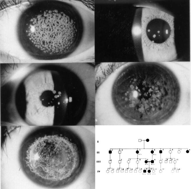
Slit lamp photographs of the corneas of affected family members in case 1. Top left, the proband at age 10 years before surgery (IV-10). Top right, the proband's brother at 21 years of age (IV-9). Second row left, the proband's father at 46 years (III-6). Second row right, the proband's mother at 48 years (III-5). Bottom left, the proband 9 year after the first surgery. Bottom right, Pedigree of the family of case 1. The genotype with regard to the R124H mutation in the BIGH3 gene is indicated: +/+, homozygous mutation; +/−, heterozygous mutation; −/−, wild type.
Figure 2 .
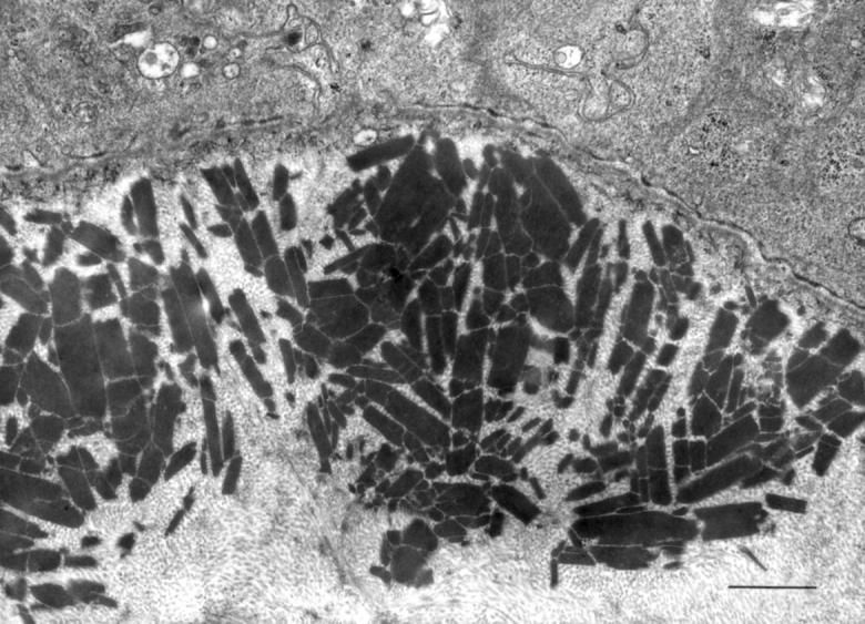
Electron microscopy of the cornea of case 1. Subepithelial and superficial stromal granules. Scale bar, 1 µm.
Figure 3 .
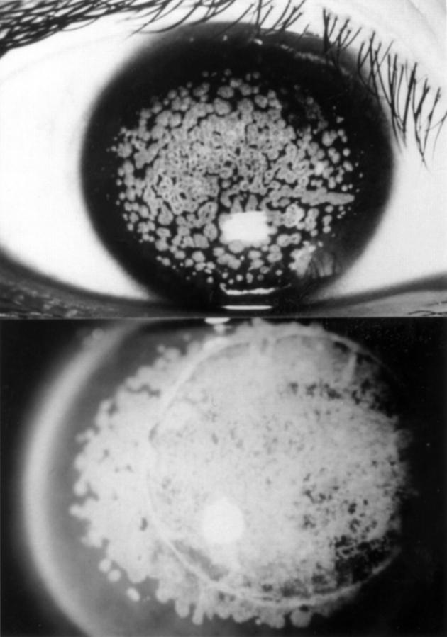
Slit lamp photographs of the cornea of case 2. Top, obtained preoperatively at the age of 13 years. Bottom, 14 years after the first surgery.
Figure 4 .
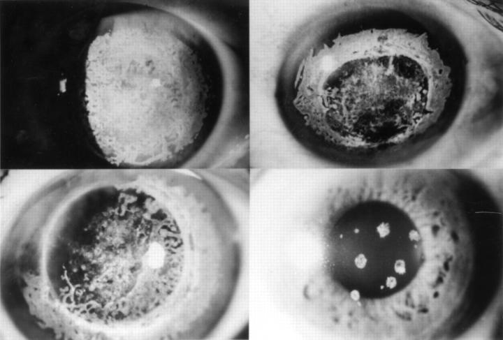
Slit lamp photograph of the corneas of recurrence. Untreated recipient corneas show dense subepithelial opacities in each case. Top left, case 3, 29 years after the second operation performed at the age of 21. Top right, case 4, 20 years after the first operation at the age of 25. Bottom left, case 5, 21 years after the second operation at the age of 53. Bottom right, daughter of case 5 at 48 years of age. Cornea shows only several granular opacities.
Figure 5 .
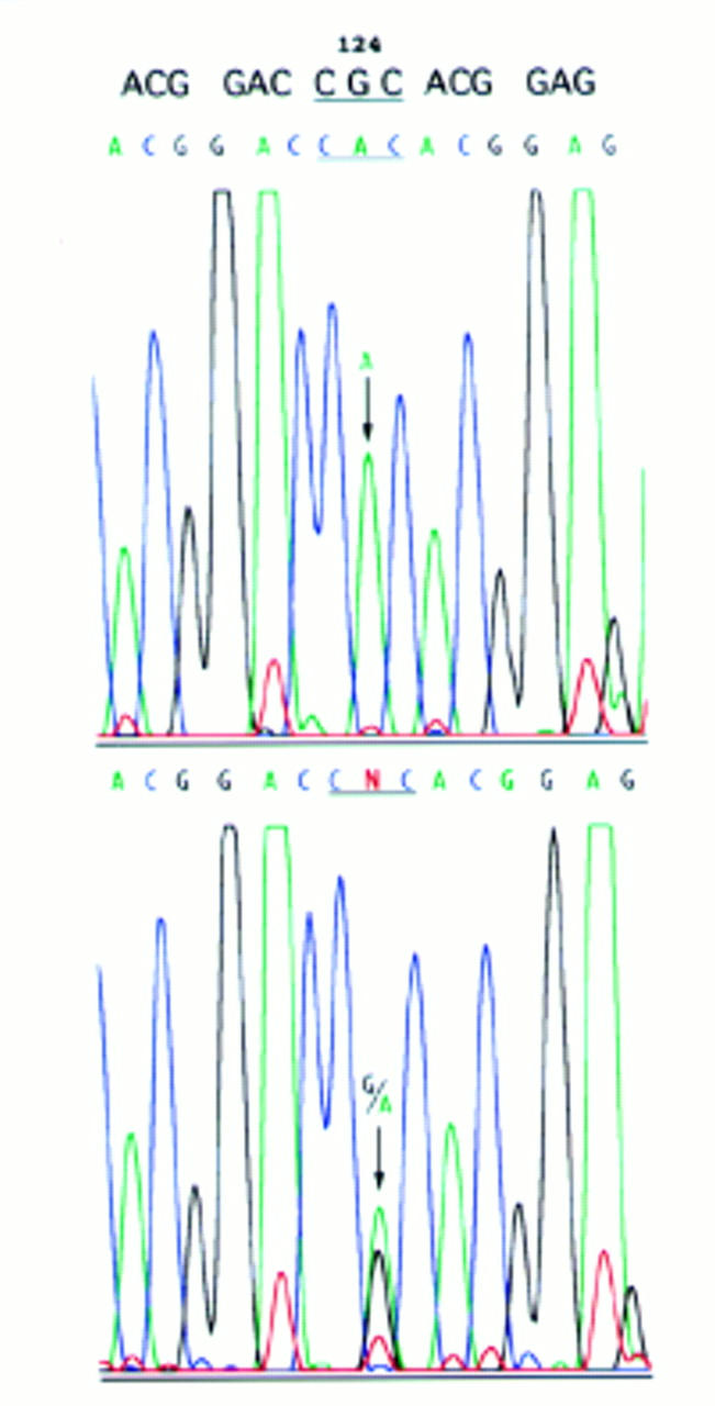
Direct sequencing analysis of the BIGH3 gene (exon 4) with an automated fluorimetric sequencer. Top, case 1. A homozygous G → A nucleotide substitution was detected at the second position of codon 124 (CGC) (arrow). Bottom, her mother. A heterozygous G → A nucleotide substitution was apparent at the second position of codon 124 (CGC). Two waves of G (black) and A (green) coexist within one peak (arrow). Top line shows normal DNA sequence.
Selected References
These references are in PubMed. This may not be the complete list of references from this article.
- Akiya S., Brown S. I. Granular dystrophy of the cornea. Characteristic electron microscopic lesion. Arch Ophthalmol. 1970 Aug;84(2):179–192. doi: 10.1001/archopht.1970.00990040181011. [DOI] [PubMed] [Google Scholar]
- Escribano J., Hernando N., Ghosh S., Crabb J., Coca-Prados M. cDNA from human ocular ciliary epithelium homologous to beta ig-h3 is preferentially expressed as an extracellular protein in the corneal epithelium. J Cell Physiol. 1994 Sep;160(3):511–521. doi: 10.1002/jcp.1041600314. [DOI] [PubMed] [Google Scholar]
- Folberg R., Alfonso E., Croxatto J. O., Driezen N. G., Panjwani N., Laibson P. R., Boruchoff S. A., Baum J., Malbran E. S., Fernandez-Meijide R. Clinically atypical granular corneal dystrophy with pathologic features of lattice-like amyloid deposits. A study of these families. Ophthalmology. 1988 Jan;95(1):46–51. doi: 10.1016/s0161-6420(88)33226-4. [DOI] [PubMed] [Google Scholar]
- Folberg R., Stone E. M., Sheffield V. C., Mathers W. D. The relationship between granular, lattice type 1, and Avellino corneal dystrophies. A histopathologic study. Arch Ophthalmol. 1994 Aug;112(8):1080–1085. doi: 10.1001/archopht.1994.01090200086027. [DOI] [PubMed] [Google Scholar]
- Garner A. Histochemistry of corneal granular dystrophy. Br J Ophthalmol. 1969 Dec;53(12):799–807. doi: 10.1136/bjo.53.12.799. [DOI] [PMC free article] [PubMed] [Google Scholar]
- Haddad R., Font R. L., Fine B. S. Unusual superficial variant of granular dystrophy of the cornea. Am J Ophthalmol. 1977 Feb;83(2):213–218. doi: 10.1016/0002-9394(77)90619-5. [DOI] [PubMed] [Google Scholar]
- Harada T., Kojima K., Hoshino M., Murakami M. [A light and electromicroscopic study of heredo-familial corneal dystrophy (Groenouw I) (author's transl)]. Nippon Ganka Gakkai Zasshi. 1977 Jan 10;81(1):48–61. [PubMed] [Google Scholar]
- Holland E. J., Daya S. M., Stone E. M., Folberg R., Dobler A. A., Cameron J. D., Doughman D. J. Avellino corneal dystrophy. Clinical manifestations and natural history. Ophthalmology. 1992 Oct;99(10):1564–1568. doi: 10.1016/s0161-6420(92)31766-x. [DOI] [PubMed] [Google Scholar]
- Kanai A., Yamaguchi T., Nakajima A. [The histochemical and analytical electron microscopy studies of the corneal granular dystrophy (author's transl)]. Nippon Ganka Gakkai Zasshi. 1977 Feb 10;81(2):145–154. [PubMed] [Google Scholar]
- Konishi M., Mashima Y., Nakamura Y., Yamada M., Sugiura H. Granular-lattice (Avellino) corneal dystrophy in Japanese patients. Cornea. 1997 Nov;16(6):635–638. [PubMed] [Google Scholar]
- Mashima Y., Imamura Y., Konishi M., Nagasawa A., Yamada M., Oguchi Y., Kudoh J., Shimizu N. Homogeneity of kerato-epithelin codon 124 mutations in Japanese patients with either of two types of corneal stromal dystrophy. Am J Hum Genet. 1997 Dec;61(6):1448–1450. doi: 10.1086/301649. [DOI] [PMC free article] [PubMed] [Google Scholar]
- Munier F. L., Korvatska E., Djemaï A., Le Paslier D., Zografos L., Pescia G., Schorderet D. F. Kerato-epithelin mutations in four 5q31-linked corneal dystrophies. Nat Genet. 1997 Mar;15(3):247–251. doi: 10.1038/ng0397-247. [DOI] [PubMed] [Google Scholar]
- Møller H. U., Ridgway A. E. Granular corneal dystrophy Groenouw type I. A report of a probable homozygous patient. Acta Ophthalmol (Copenh) 1990 Feb;68(1):97–101. doi: 10.1111/j.1755-3768.1990.tb01658.x. [DOI] [PubMed] [Google Scholar]
- Owens S. L., Sugar J., Edward D. P. Superficial granular corneal dystrophy with amyloid deposits. Arch Ophthalmol. 1992 Feb;110(2):175–176. doi: 10.1001/archopht.1992.01080140025015. [DOI] [PubMed] [Google Scholar]
- Rodrigues M. M., Gaster R. N., Pratt M. V. Unusual superficial confluent form of granular corneal dystrophy. Ophthalmology. 1983 Dec;90(12):1507–1511. [PubMed] [Google Scholar]
- Rosenwasser G. O., Sucheski B. M., Rosa N., Pastena B., Sebastiani A., Sassani J. W., Perry H. D. Phenotypic variation in combined granular-lattice (Avellino) corneal dystrophy. Arch Ophthalmol. 1993 Nov;111(11):1546–1552. doi: 10.1001/archopht.1993.01090110112035. [DOI] [PubMed] [Google Scholar]
- Sajjadi S. H., Javadi M. A. Superficial juvenile granular dystrophy. Ophthalmology. 1992 Jan;99(1):95–102. doi: 10.1016/s0161-6420(92)32004-4. [DOI] [PubMed] [Google Scholar]
- Takagi M., Ishizu M., Suzuki H. [An electron microscopic and histochemical study on a corneal granular dystrophy (Groenouw type I)]. Nihon Ganka Kiyo. 1971 Jul;22(7):479–484. [PubMed] [Google Scholar]


