Abstract
AIMS—To evaluate the relation between the optic disc size and the thickness of the peripapillary retinal nerve fibre layer (RNFL) in normal Japanese subjects by means of scanning laser polarimetry. METHODS—Scanning laser polarimetry was performed in 60 normal subjects. One eye of each subject was randomly selected for study. Using a scanning laser polarimeter, the integral of RNFL thickness was measured totally and regionally within a circular band located 1.75 disc diameters from the centre of the optic disc. The correlation between the optic disc size and the integral of RNFL thickness was examined. RESULTS—The optic disc size showed a significant correlation with the integral of RNFL thickness (R = 0.497, p <0.001). A significant negative correlation was observed between the optic disc size and the ratio of inferior integral to total integral of RNFL thickness (R = −0.274, p = 0.034). CONCLUSIONS—The cross sectional area occupied by the RNF, measured by scanning laser polarimetry increased significantly with an increase in optic disc size while the ratio of inferior to total cross sectional area decreased significantly. These facts should be considered when one evaluates the RNFL thickness in patients with progressive optic neuropathies such as glaucoma. Keywords: scanning laser polarimeter; normal subjects; optic disc; retinal nerve fibre layer
Full Text
The Full Text of this article is available as a PDF (109.7 KB).
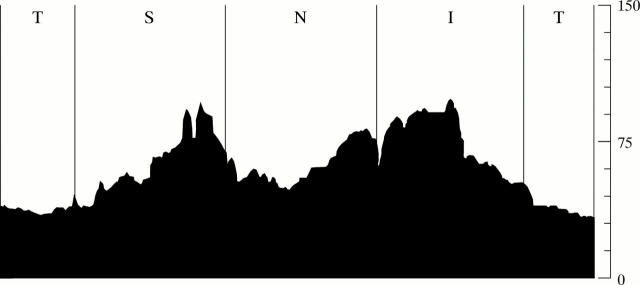
Figure 1 .
Actual image of the integral of retinal nerve fibre layer RNFL thickness in the nerve fibre analyser. The longitudinal axis indicates the thickness of the RNFL (µm). The integral of RNFL thickness indicates the sectional area which is the integration of the thickness of the RNFL calculated along the transverse axis. S=superior quadrant; T=temporal quadrant; I=inferior quadrant; N=nasal quadrant.
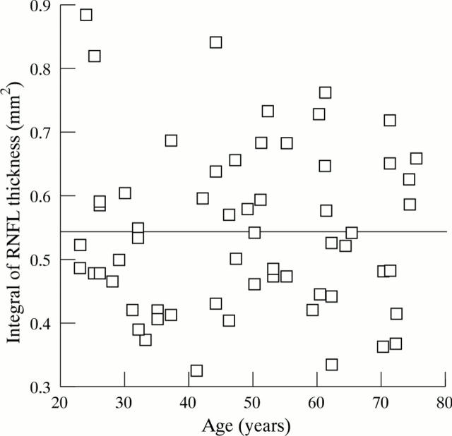
Figure 2 .
Correlation between age and the integral of thickness of the RNFL in 60 normal eyes (Y = 0.000x + 0.543, R = 0.002, R2 = 0.000, p = 0.988).
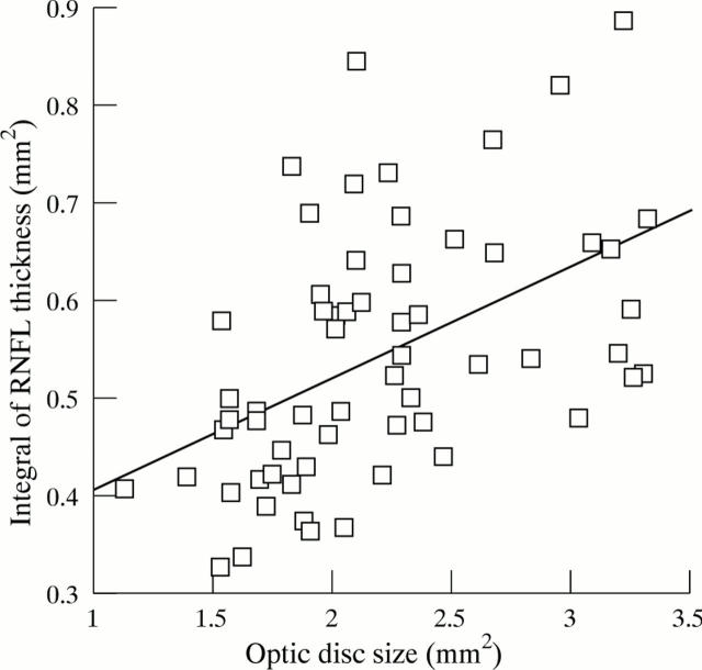
Figure 3 .
Correlation between size of the optic disc and the integral of thickness of the RNFL in 60 normal eyes (Y = 0.116x + 0.288, R = 0.497, R2 = 0.247, p <0.001).
Selected References
These references are in PubMed. This may not be the complete list of references from this article.
- Anton A., Zangwill L., Emdadi A., Weinreb R. N. Nerve fiber layer measurements with scanning laser polarimetry in ocular hypertension. Arch Ophthalmol. 1997 Mar;115(3):331–334. doi: 10.1001/archopht.1997.01100150333004. [DOI] [PubMed] [Google Scholar]
- Bengtsson B., Krakau C. E. Correction of optic disc measurements on fundus photographs. Graefes Arch Clin Exp Ophthalmol. 1992;230(1):24–28. doi: 10.1007/BF00166758. [DOI] [PubMed] [Google Scholar]
- Chi T., Ritch R., Stickler D., Pitman B., Tsai C., Hsieh F. Y. Racial differences in optic nerve head parameters. Arch Ophthalmol. 1989 Jun;107(6):836–839. doi: 10.1001/archopht.1989.01070010858029. [DOI] [PubMed] [Google Scholar]
- Jonas J. B., Fernández M. C., Naumann G. O. Correlation of the optic disc size to glaucoma susceptibility. Ophthalmology. 1991 May;98(5):675–680. doi: 10.1016/s0161-6420(91)32234-6. [DOI] [PubMed] [Google Scholar]
- Jonas J. B., Schmidt A. M., Müller-Bergh J. A., Schlötzer-Schrehardt U. M., Naumann G. O. Human optic nerve fiber count and optic disc size. Invest Ophthalmol Vis Sci. 1992 May;33(6):2012–2018. [PubMed] [Google Scholar]
- Junghardt A., Schmid M. K., Schipper I., Wildberger H., Seifert B. Reproducibility of the data determined by scanning laser polarimetry. Graefes Arch Clin Exp Ophthalmol. 1996 Oct;234(10):628–632. doi: 10.1007/BF00185296. [DOI] [PubMed] [Google Scholar]
- Mikelberg F. S., Yidegiligne H. M., White V. A., Schulzer M. Relation between optic nerve axon number and axon diameter to scleral canal area. Ophthalmology. 1991 Jan;98(1):60–63. doi: 10.1016/s0161-6420(91)32341-8. [DOI] [PubMed] [Google Scholar]
- Niessen A. G., Van Den Berg T. J., Langerhorst C. T., Greve E. L. Retinal nerve fiber layer assessment by scanning laser polarimetry and standardized photography. Am J Ophthalmol. 1996 May;121(5):484–493. doi: 10.1016/s0002-9394(14)75422-4. [DOI] [PubMed] [Google Scholar]
- Poinoosawmy D., Fontana L., Wu J. X., Fitzke F. W., Hitchings R. A. Variation of nerve fibre layer thickness measurements with age and ethnicity by scanning laser polarimetry. Br J Ophthalmol. 1997 May;81(5):350–354. doi: 10.1136/bjo.81.5.350. [DOI] [PMC free article] [PubMed] [Google Scholar]
- Provis J. M., van Driel D., Billson F. A., Russell P. Human fetal optic nerve: overproduction and elimination of retinal axons during development. J Comp Neurol. 1985 Aug 1;238(1):92–100. doi: 10.1002/cne.902380108. [DOI] [PubMed] [Google Scholar]
- Quigley H. A., Addicks E. M. Quantitative studies of retinal nerve fiber layer defects. Arch Ophthalmol. 1982 May;100(5):807–814. doi: 10.1001/archopht.1982.01030030811018. [DOI] [PubMed] [Google Scholar]
- Quigley H. A., Coleman A. L., Dorman-Pease M. E. Larger optic nerve heads have more nerve fibers in normal monkey eyes. Arch Ophthalmol. 1991 Oct;109(10):1441–1443. doi: 10.1001/archopht.1991.01080100121056. [DOI] [PubMed] [Google Scholar]
- Radius R. L. Thickness of the retinal nerve fiber layer in primate eyes. Arch Ophthalmol. 1980 Sep;98(9):1625–1629. doi: 10.1001/archopht.1980.01020040477018. [DOI] [PubMed] [Google Scholar]
- Rakic P., Riley K. P. Overproduction and elimination of retinal axons in the fetal rhesus monkey. Science. 1983 Mar 25;219(4591):1441–1444. doi: 10.1126/science.6828871. [DOI] [PubMed] [Google Scholar]
- Tjon-Fo-Sang M. J., de Vries J., Lemij H. G. Measurement by nerve fiber analyzer of retinal nerve fiber layer thickness in normal subjects and patients with ocular hypertension. Am J Ophthalmol. 1996 Aug;122(2):220–227. doi: 10.1016/s0002-9394(14)72013-6. [DOI] [PubMed] [Google Scholar]
- Varma R., Skaf M., Barron E. Retinal nerve fiber layer thickness in normal human eyes. Ophthalmology. 1996 Dec;103(12):2114–2119. doi: 10.1016/s0161-6420(96)30381-3. [DOI] [PubMed] [Google Scholar]
- Weinreb R. N., Dreher A. W., Coleman A., Quigley H., Shaw B., Reiter K. Histopathologic validation of Fourier-ellipsometry measurements of retinal nerve fiber layer thickness. Arch Ophthalmol. 1990 Apr;108(4):557–560. doi: 10.1001/archopht.1990.01070060105058. [DOI] [PubMed] [Google Scholar]
- Weinreb R. N., Shakiba S., Sample P. A., Shahrokni S., van Horn S., Garden V. S., Asawaphureekorn S., Zangwill L. Association between quantitative nerve fiber layer measurement and visual field loss in glaucoma. Am J Ophthalmol. 1995 Dec;120(6):732–738. doi: 10.1016/s0002-9394(14)72726-6. [DOI] [PubMed] [Google Scholar]
- Weinreb R. N., Shakiba S., Zangwill L. Scanning laser polarimetry to measure the nerve fiber layer of normal and glaucomatous eyes. Am J Ophthalmol. 1995 May;119(5):627–636. doi: 10.1016/s0002-9394(14)70221-1. [DOI] [PubMed] [Google Scholar]


