Abstract
AIMS—High hyperopia constitutes the majority of refractive errors in large scale visual screening at preschool ages. The authors aimed to assess the validity of the Retinomax hand held refractor to detect high hyperopia in a refractive screening performed without cycloplegia and carried out on children aged 9-36 months. They considered +1.5 D of manifest hyperopia to be the threshold value and abnormal absolute hyperopia to be above +3.5 D. METHODS—Of the 897 children screened without cycloplegia, 220 were refracted with cycloplegia. The validity of several thresholds of manifest hyperopia was estimated by receiver operating characteristic (ROC) curves using cycloplegic measures as a reference. The reproducibility of Retinomax measurements was assessed. Normal and quick mode measurements were compared using the Wilcoxon test. RESULTS—The manifest threshold of +1.5 D offered the best combination of sensitivity (70.2%), specificity (94.6%), positive predictive value (78.6%), and negative predictive value (91.9%) to disclose abnormal absolute hyperopia. A good agreement was obtained between the various measurements using Retinomax on the same subject. In the results of this survey, there is no evidence that accommodation is minimised in the normal mode of measurement compared with the quick mode. CONCLUSION—The Retinomax hand held infrared autorefractor is a suitable instrument to diagnose abnormal hyperopia (manifest hyperopia >+1.5 D) in non-cycloplegic refractive screening at preschool ages. It is suggested as the quick mode of measurement as it is more feasible in children (success rate 98.5%). Keywords: non-cycloplegic refractive screening; hyperopia; hand held infrared automated refractor; Retinomax
Full Text
The Full Text of this article is available as a PDF (124.7 KB).
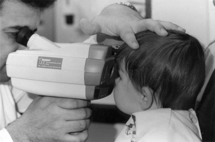
Figure 1 .
Measuring refraction with the Retinomax.
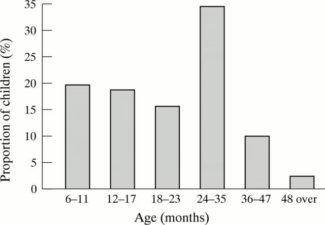
Figure 2 .
Age distribution of the 897 screened children.
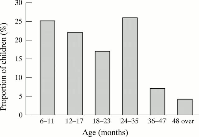
Figure 3 .
Age distribution of the 220 children with cycloplegia.
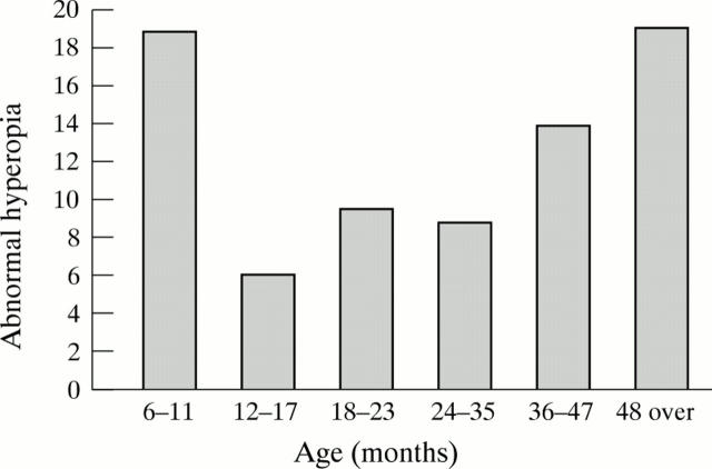
Figure 4 .
Age distribution of abnormal hyperopia.
Selected References
These references are in PubMed. This may not be the complete list of references from this article.
- Angi M. R., Pucci V., Forattini F., Formentin P. A. Results of photorefractometric screening for amblyogenic defects in children aged 20 months. Behav Brain Res. 1992 Jul 31;49(1):91–97. doi: 10.1016/s0166-4328(05)80198-6. [DOI] [PubMed] [Google Scholar]
- Atkinson J., Braddick O. J., Durden K., Watson P. G., Atkinson S. Screening for refractive errors in 6-9 month old infants by photorefraction. Br J Ophthalmol. 1984 Feb;68(2):105–112. doi: 10.1136/bjo.68.2.105. [DOI] [PMC free article] [PubMed] [Google Scholar]
- Atkinson J., Braddick O., Robier B., Anker S., Ehrlich D., King J., Watson P., Moore A. Two infant vision screening programmes: prediction and prevention of strabismus and amblyopia from photo- and videorefractive screening. Eye (Lond) 1996;10(Pt 2):189–198. doi: 10.1038/eye.1996.46. [DOI] [PubMed] [Google Scholar]
- Friedman Z., Neumann E., Hyams S. W., Peleg B. Ophthalmic screening of 38,000 children, age 1 to 2 1/2 years, in child welfare clinics. J Pediatr Ophthalmol Strabismus. 1980 Jul-Aug;17(4):261–267. doi: 10.3928/0191-3913-19800701-16. [DOI] [PubMed] [Google Scholar]
- Hanley J. A., McNeil B. J. The meaning and use of the area under a receiver operating characteristic (ROC) curve. Radiology. 1982 Apr;143(1):29–36. doi: 10.1148/radiology.143.1.7063747. [DOI] [PubMed] [Google Scholar]
- Ingram R. M. Refraction as a basis for screening children for squint and amblyopia. Br J Ophthalmol. 1977 Jan;61(1):8–15. doi: 10.1136/bjo.61.1.8. [DOI] [PMC free article] [PubMed] [Google Scholar]
- Kallay OP, Cordonnier MJ, Dramaix MM. Cycloplegic refractive errors in children: comparison of a standard and a hand-held refractor. Strabismus. 1998 Mar;6(1):3–7. doi: 10.1076/stra.6.1.3.668. [DOI] [PubMed] [Google Scholar]
- Metz C. E. Basic principles of ROC analysis. Semin Nucl Med. 1978 Oct;8(4):283–298. doi: 10.1016/s0001-2998(78)80014-2. [DOI] [PubMed] [Google Scholar]
- Olver J. M. Simple retinoscopic screening. Eye (Lond) 1988;2(Pt 3):309–313. doi: 10.1038/eye.1988.58. [DOI] [PubMed] [Google Scholar]
- Ottar W. L., Scott W. E., Holgado S. I. Photoscreening for amblyogenic factors. J Pediatr Ophthalmol Strabismus. 1995 Sep-Oct;32(5):289–295. doi: 10.3928/0191-3913-19950901-06. [DOI] [PubMed] [Google Scholar]
- Preslan M. W., Novak A. Baltimore Vision Screening Project. Ophthalmology. 1996 Jan;103(1):105–109. doi: 10.1016/s0161-6420(96)30753-7. [DOI] [PubMed] [Google Scholar]
- Saunders K. J. Early refractive development in humans. Surv Ophthalmol. 1995 Nov-Dec;40(3):207–216. doi: 10.1016/s0039-6257(95)80027-1. [DOI] [PubMed] [Google Scholar]
- Simons K. Preschool vision screening: rationale, methodology and outcome. Surv Ophthalmol. 1996 Jul-Aug;41(1):3–30. doi: 10.1016/s0039-6257(97)81990-x. [DOI] [PubMed] [Google Scholar]
- Williamson T. H., Andrews R., Dutton G. N., Murray G., Graham N. Assessment of an inner city visual screening programme for preschool children. Br J Ophthalmol. 1995 Dec;79(12):1068–1073. doi: 10.1136/bjo.79.12.1068. [DOI] [PMC free article] [PubMed] [Google Scholar]


