Full Text
The Full Text of this article is available as a PDF (153.6 KB).
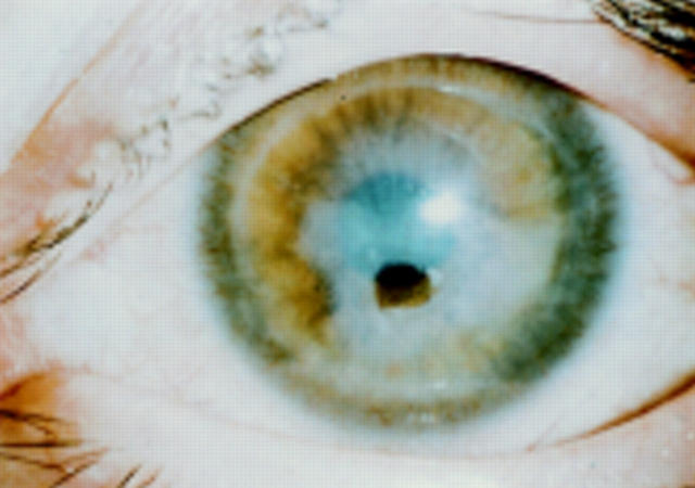
Figure 1 .
Paracentral corneal perforation (38 year old woman). There is little or no inflammation on the ocular surface. The anterior chamber is maintained by a therapeutic contact lens.
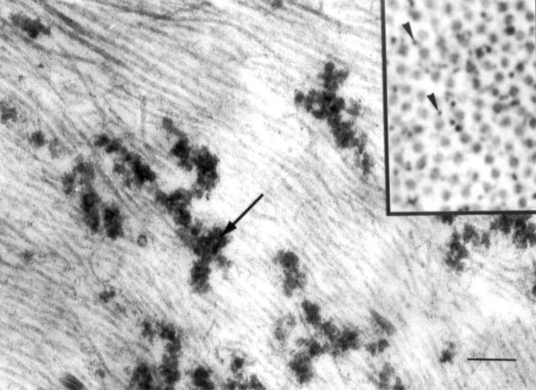
Figure 2 .
Corneal stroma surrounding the perforated area (76 year old woman). Electron dense material (arrow), possibly aggregated collagen, is located throughout the extracellular matrix. Also (inset), many atypical, thin fibres (arrowheads) are interspersed with collagen. Bar=200 nm (main figure) and 100 nm (inset).


