

Abstract
BACKGROUND/AIMS—Between June 1992 and July 1995, 29 uveal melanomas were treated radiosurgically with the Leksell gamma unit at the University of Graz. The aim of this retrospective study was to examine the pattern of regression and the extent and time period of the decrease in tumour size. METHODS—The Leksell gamma knife, model B, was used. Patients were divided into three groups according to marginal dose: group 1: eight patients with a marginal dose >50 Gy, group 2: 15 patients with a marginal dose = 50 Gy, and group 3: six patients with a marginal dose = 45 Gy. For the retrospective study two groups were examined: group A, tumours <5 mm and group B, tumours ⩾5 mm. RESULTS—No significant correlation was found between tumour regression and the marginal dose. Tumour shrinkage depends on the pretreatment height. In the group of eight patients with an initial tumour prominence of less than 5 mm, no prominence was found after therapy. In the group of patients with an original tumour prominence of 5 mm and more, only two tumours formed a flat scar while a residual prominence was found in 18 patients. Increase in reflectivity combined with a decrease in size appears to be a good criterion for the effectiveness of the treatment. In five patients with tumours showing low reflectivity, over a longer period of time metastases were found. An enucleation was performed in two patients because of uncertain tumour regression and in one patient as a result of an increase in tumour size. CONCLUSION—The pattern of echographic reflectivity and decrease in size is similar to brachytherapy and is one of the most important diagnostic variables for evaluation of tumour regression. An increase in reflectivity as well as a decrease in tumour size in the first 6-8 months can be considered a therapeutic success. Keywords: gamma knife; uveal melanoma; echographic examination; reflectivity
Full Text
The Full Text of this article is available as a PDF (143.5 KB).
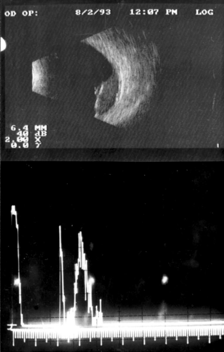
Figure 1 .
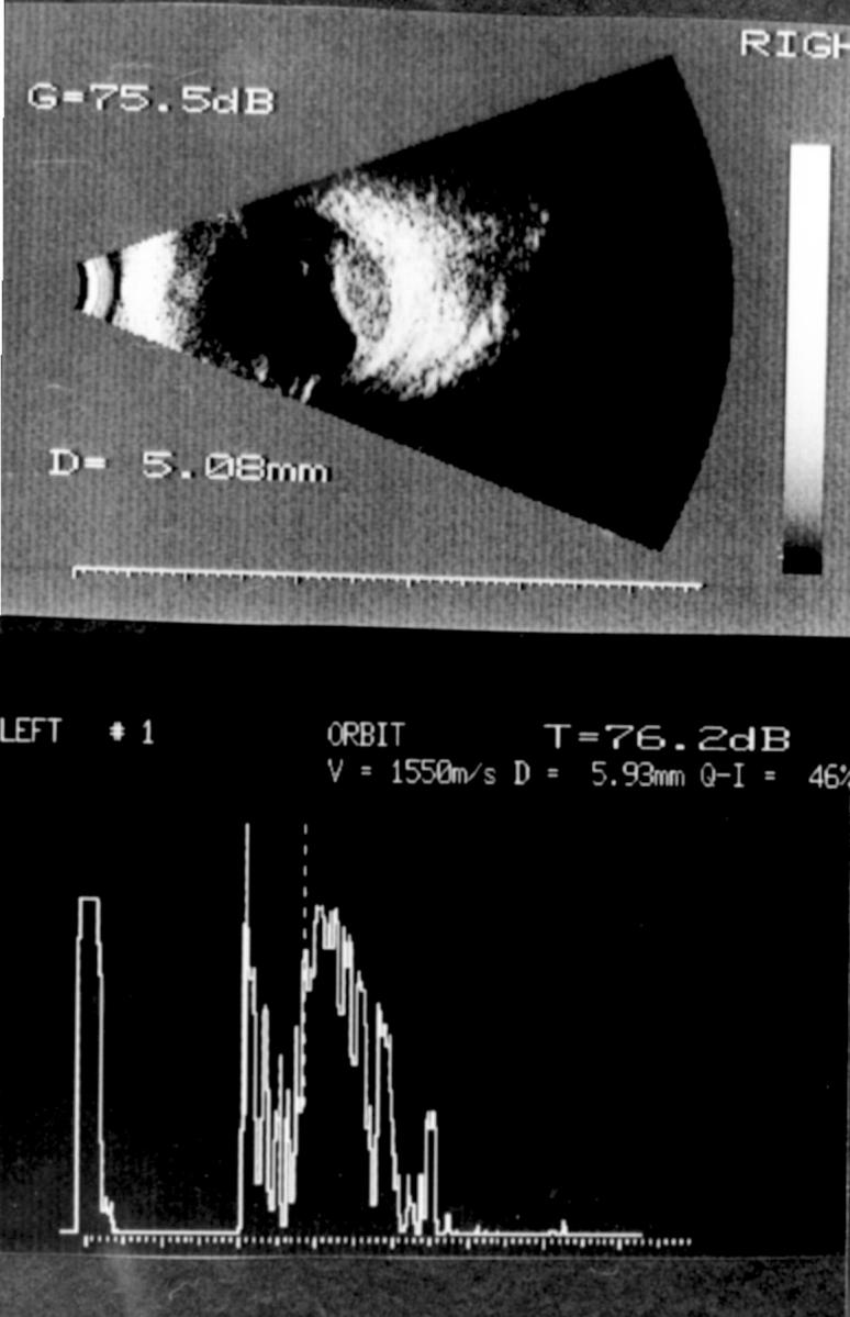
A 71 year old man. A-scan 5.9 mm solid tumour, low internal reflectivity, B-scan 5.1 × 12.2 mm.
Figure 2 .
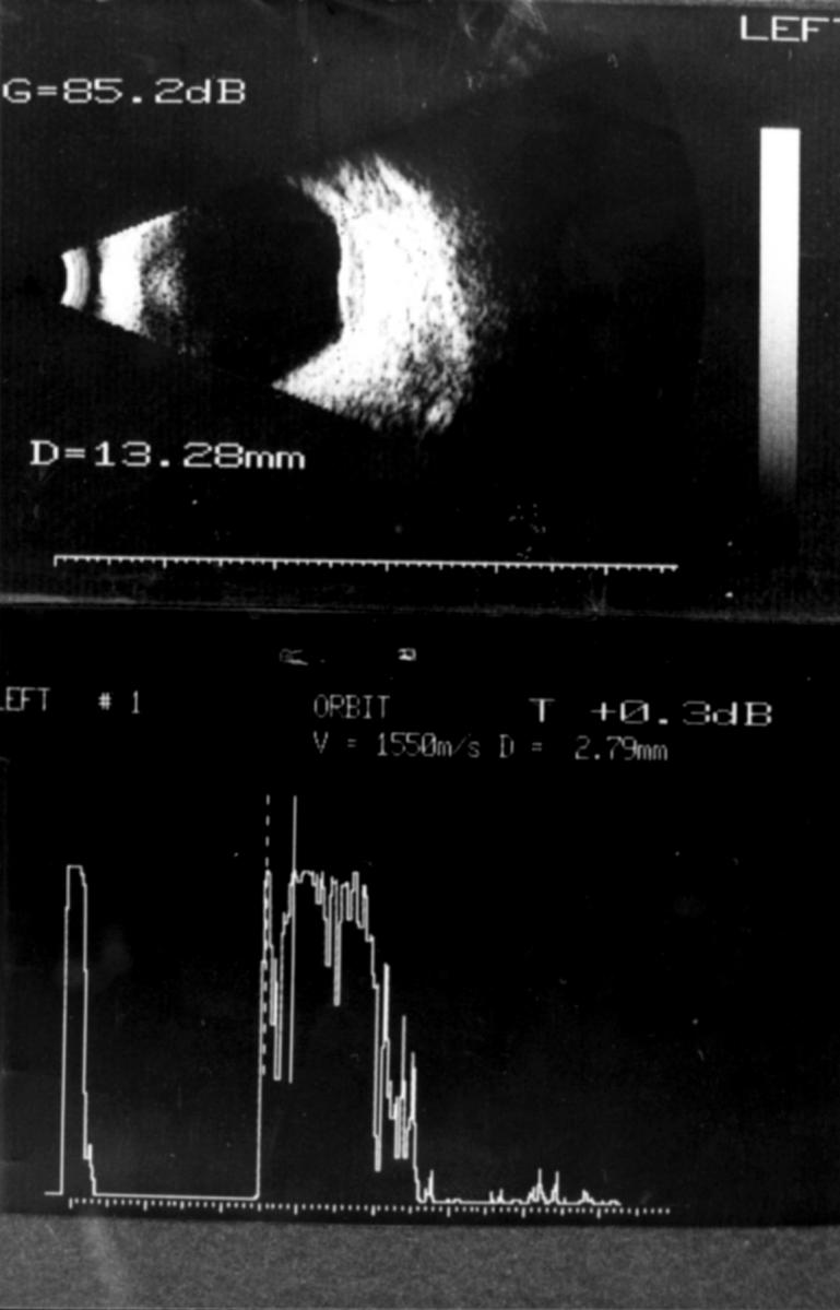
Same patient as in Figure 1, 12 months after stereotactic surgery. A scan 2.8 mm high prominence with high internal reflectivity.
Figure 3 .
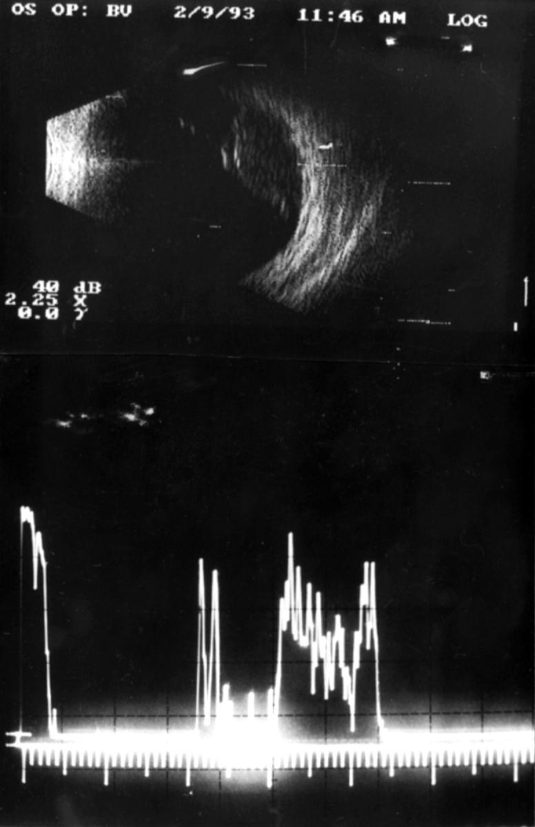
A 35 year old man with a solid tumour of 7.9 mm prominence with low internal reflectivity.
Figure 4 .
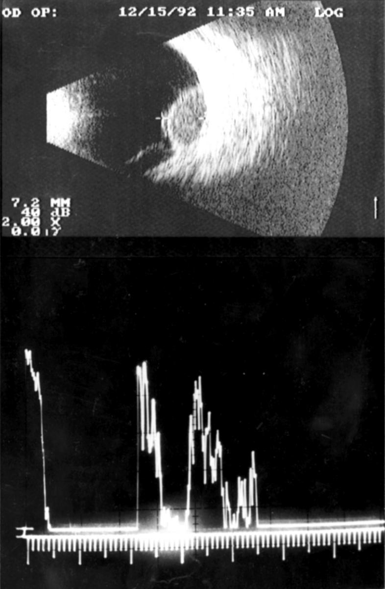
Same patient as in Figure 3, 4 months after stereotactic surgery. Tumour with low internal reflectivity and a height of 7.9 mm prominence.
Figure 5 .
Same patient as in Figure 3, 12 months after stereotactic surgery. Tumour with height of 5.9 mm and low internal reflectivity. Metastases were found 28 months after treatment.
Selected References
These references are in PubMed. This may not be the complete list of references from this article.
- Char D. H., Castro J. R. Helium ion therapy for choroidal melanoma. Arch Ophthalmol. 1982 Jun;100(6):935–938. doi: 10.1001/archopht.1982.01030030943008. [DOI] [PubMed] [Google Scholar]
- Coleman D. J., Silverman R. H., Rondeau M. J., Coleman J. A., Rosberger D., Ellsworth R. M., Lizzi F. L. Ultrasonic tissue characterization of uveal melanoma and prediction of patient survival after enucleation and brachytherapy. Am J Ophthalmol. 1991 Dec 15;112(6):682–688. doi: 10.1016/s0002-9394(14)77275-7. [DOI] [PubMed] [Google Scholar]
- Eichler C., Hertel A., Lommatzsch P. K., Fuhrmann P. Echographische Befunde vor und nach beta-Bestrahlung (106Ru/106Rh) von Aderhautmelanomen. Klin Monbl Augenheilkd. 1987 Jan;190(1):17–20. doi: 10.1055/s-2008-1050320. [DOI] [PubMed] [Google Scholar]
- Gragoudas E. S., Goitein M., Verhey L., Munzenreider J., Suit H. D., Koehler A. Proton beam irradiation. An alternative to enucleation for intraocular melanomas. Ophthalmology. 1980 Jun;87(6):571–581. doi: 10.1016/s0161-6420(80)35212-3. [DOI] [PubMed] [Google Scholar]
- Guthoff R., Haase J., von Domarus D., Draeger J., Lauritzen K. Das Regressionsverhalten des Aderhautmelanoms nach Strahlentherapie--ein neuer prognostischer Parameter? Klin Monbl Augenheilkd. 1990 Jan;196(1):6–10. doi: 10.1055/s-2008-1046119. [DOI] [PubMed] [Google Scholar]
- Guthoff R., von Domarus D., Schroeder W. Gegenüberstellung klinischer, echographischer und histologischer Befunde beim malignen Melanom der Aderhaut. Klin Monbl Augenheilkd. 1981 Nov;179(5):330–332. doi: 10.1055/s-2008-1057322. [DOI] [PubMed] [Google Scholar]
- Kindy-Degnan N. A., Char D. H., Castro J. R., Kroll S., Stone R. D., Quivey J. M., Phillips T. L., Irvine A. R. Effect of various doses of radiation for uveal melanoma on regression, visual acuity, complications, and survival. Am J Ophthalmol. 1989 Feb 15;107(2):114–120. doi: 10.1016/0002-9394(89)90208-0. [DOI] [PubMed] [Google Scholar]
- Kissinger A., Bornfeld N., Foerster M. H., Gerke E., Wessing A., Meyer-Schwickerath G. Echographische Beurteilung maligner Melanome der Uvea nach Ruthenium-Therapie. Fortschr Ophthalmol. 1986;83(6):721–725. [PubMed] [Google Scholar]
- Marchini G., Babighian S., Tomazzoli L., Gerosa M. A., Nicolato A., Bricolo A., Piovan E., Zampieri P. G., Alessandrini F., Benati A. Stereotactic radiosurgery of uveal melanomas: preliminary results with Gamma Knife treatment. Stereotact Funct Neurosurg. 1995;64 (Suppl 1):72–79. doi: 10.1159/000098766. [DOI] [PubMed] [Google Scholar]
- Menapace R., Binder W., Skorpik C., Gnad H. D. Assessment of echographic regression criteria in ruthenium-irradiated melanoma. Ophthalmologica. 1989;198(3):129–134. doi: 10.1159/000309975. [DOI] [PubMed] [Google Scholar]
- Moses K. C., LaPiana F. G. Controlled enucleation. Ophthalmic Surg. 1987 May;18(5):379–382. [PubMed] [Google Scholar]
- Pupyshev A. B., Korolenko T. A. Effektivnaia ékstraktsiia lisosomal'nykh fermentov s pomoshch'iu digitonina. Ukr Biokhim Zh (1978) 1989 Mar-Apr;61(2):49–54. [PubMed] [Google Scholar]
- Rennie I., Forster D., Kemeny A., Walton L., Kunkler I. The use of single fraction Leksell stereotactic radiosurgery in the treatment of uveal melanoma. Acta Ophthalmol Scand. 1996 Dec;74(6):558–562. doi: 10.1111/j.1600-0420.1996.tb00734.x. [DOI] [PubMed] [Google Scholar]
- Shields J. A., Augsburger J. J., Brady L. W., Day J. L. Cobalt plaque therapy of posterior uveal melanomas. Ophthalmology. 1982 Oct;89(10):1201–1207. doi: 10.1016/s0161-6420(82)34658-8. [DOI] [PubMed] [Google Scholar]