Abstract
AIMS—To evaluate the reproducibility of the retardation values (change in polarisation) obtained with the scanning laser polarimeter in a series of normal subjects and glaucoma patients. To improve the analysis of the raw data by devising and evaluating a blood vessel removal algorithm. METHODS—Scanning laser polarimetry was performed on 10 normal subjects and 10 glaucoma patients. A series of six images was obtained from each eye. The normal subjects were re-imaged 3 months after their initial assessment. The retardation values obtained from each eye were analysed using the authors' own methods, including the use of an algorithm to remove blood vessels from the polar profiles. The reproducibility of these measurements and the performance of the blood vessel removal algorithm were assessed. RESULTS—The "individual point" coefficient of variation was approximately 12.5% for normal subjects and 17.0% for glaucoma patients. The "integral" coefficient of variation for these groups was approximately 5.5% and 9.5% respectively. The reproducibility of the measurements did not improve with an increased number of measurements. There was no difference in the reproducibility of the measurements in normal subjects over time. The blood vessel removal algorithm improved the reproducibility of the measurements when the shape of the profile was assessed. CONCLUSION—The intraoperator reproducibility of retardation values obtained with the scanning laser polarimeter is satisfactory for its use as a clinical tool. The use of a blood vessel removal algorithm improves the reproducibility of the measurements and also assists the clinician in the interpretation of the polar profiles. Furthermore, it allows the construction of normal database polar profiles, thereby enabling the identification, location and quantification of retinal nerve fibre layer damage in an "at risk" individual's polar profile. Keywords: scanning laser polarimetry; glaucoma; reproducibility; algorithm
Full Text
The Full Text of this article is available as a PDF (138.4 KB).
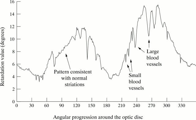
Figure 1 .
Example of a polar profile of a normal subject.
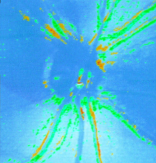
Figure 2 .
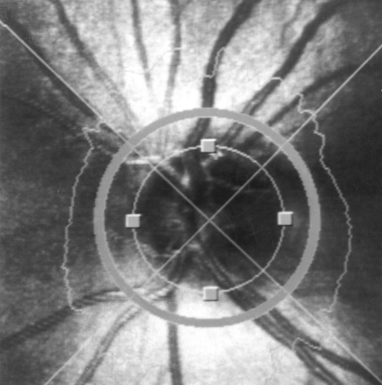
An illustration of a retardation image of a normal subject demonstrating the location and size of the concentric sampling zone used in this study. The circumferential polar profile is also demonstrated (see main text for details).
Figure 3 .
An illustration of a comparison map of compiled by the NFA I software of two retardation images of the same eye of a normal subject taken within 2 minutes of each other by the same operator (AW). The yellow and green regions reflect areas of statistically significant change in the retardation values obtained from these locations.
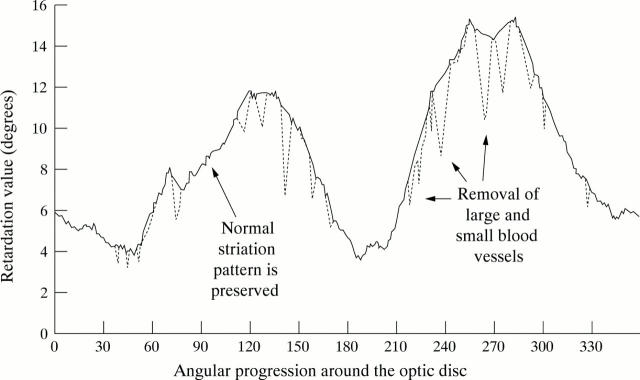
Figure 4 .
Example of a polar profile of a normal subject demonstrating the effect of the application of the blood vessel removal algorithm. The blood vessel removed polar profile is illustrated by the solid line, while the original polar profile containing the blood vessels is illustrated by the broken line.
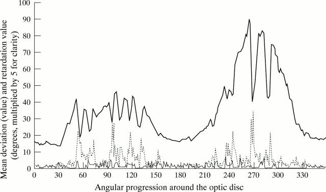
Figure 5 .
A polar profile of a normal subject with an illustration of the areas of greatest measurement variability in the shape of this profile. The polar profile (retardation values multiplied by five for clarity) is illustrated by the solid bold line. The variability of the profile shape at individual locations is illustrated by the broken line (before the application of the blood vessel removal algorithm) and by the fine line (after the application of the algorithm).
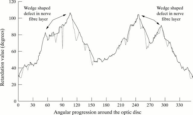
Figure 6 .
Example of a polar profile of a glaucoma patient with focal wedge-shaped nerve fibre layer defects in both hemiretinas. The blood vessel removed polar profile is illustrated by the solid line, while the original polar profile containing the blood vessels is illustrated by the broken line. The wedge-shaped defects have not been removed by the algorithm.
Selected References
These references are in PubMed. This may not be the complete list of references from this article.
- Airaksinen P. J., Nieminen H. Retinal nerve fiber layer photography in glaucoma. Ophthalmology. 1985 Jul;92(7):877–879. doi: 10.1016/s0161-6420(85)33941-6. [DOI] [PubMed] [Google Scholar]
- Niessen A. G., Van Den Berg T. J., Langerhorst C. T., Greve E. L. Retinal nerve fiber layer assessment by scanning laser polarimetry and standardized photography. Am J Ophthalmol. 1996 May;121(5):484–493. doi: 10.1016/s0002-9394(14)75422-4. [DOI] [PubMed] [Google Scholar]
- Quigley H. A., Addicks E. M., Green W. R. Optic nerve damage in human glaucoma. III. Quantitative correlation of nerve fiber loss and visual field defect in glaucoma, ischemic neuropathy, papilledema, and toxic neuropathy. Arch Ophthalmol. 1982 Jan;100(1):135–146. doi: 10.1001/archopht.1982.01030030137016. [DOI] [PubMed] [Google Scholar]
- Quigley H. A., Addicks E. M. Quantitative studies of retinal nerve fiber layer defects. Arch Ophthalmol. 1982 May;100(5):807–814. doi: 10.1001/archopht.1982.01030030811018. [DOI] [PubMed] [Google Scholar]
- Sommer A., Katz J., Quigley H. A., Miller N. R., Robin A. L., Richter R. C., Witt K. A. Clinically detectable nerve fiber atrophy precedes the onset of glaucomatous field loss. Arch Ophthalmol. 1991 Jan;109(1):77–83. doi: 10.1001/archopht.1991.01080010079037. [DOI] [PubMed] [Google Scholar]
- Sommer A., Miller N. R., Pollack I., Maumenee A. E., George T. The nerve fiber layer in the diagnosis of glaucoma. Arch Ophthalmol. 1977 Dec;95(12):2149–2156. doi: 10.1001/archopht.1977.04450120055003. [DOI] [PubMed] [Google Scholar]
- Weinreb R. N., Dreher A. W., Bille J. F. Quantitative assessment of the optic nerve head with the laser tomographic scanner. Int Ophthalmol. 1989 Jan;13(1-2):25–29. doi: 10.1007/BF02028633. [DOI] [PubMed] [Google Scholar]
- Weinreb R. N., Dreher A. W., Coleman A., Quigley H., Shaw B., Reiter K. Histopathologic validation of Fourier-ellipsometry measurements of retinal nerve fiber layer thickness. Arch Ophthalmol. 1990 Apr;108(4):557–560. doi: 10.1001/archopht.1990.01070060105058. [DOI] [PubMed] [Google Scholar]
- Weinreb R. N., Lusky M., Bartsch D. U., Morsman D. Effect of repetitive imaging on topographic measurements of the optic nerve head. Arch Ophthalmol. 1993 May;111(5):636–638. doi: 10.1001/archopht.1993.01090050070031. [DOI] [PubMed] [Google Scholar]
- Weinreb R. N., Shakiba S., Sample P. A., Shahrokni S., van Horn S., Garden V. S., Asawaphureekorn S., Zangwill L. Association between quantitative nerve fiber layer measurement and visual field loss in glaucoma. Am J Ophthalmol. 1995 Dec;120(6):732–738. doi: 10.1016/s0002-9394(14)72726-6. [DOI] [PubMed] [Google Scholar]
- Weinreb R. N., Shakiba S., Zangwill L. Scanning laser polarimetry to measure the nerve fiber layer of normal and glaucomatous eyes. Am J Ophthalmol. 1995 May;119(5):627–636. doi: 10.1016/s0002-9394(14)70221-1. [DOI] [PubMed] [Google Scholar]


