Abstract
AIM—To report the quantitation of the lipid composition of a corneal button from a Japanese woman in her 60s with clinically and histopathologically proved Schnyder's corneal dystrophy. METHODS—Total lipids extracted from the corneal button of the patient were analysed by the method of thin layer chromatography flame ionisation detection. Two different solvent systems were used for neutral lipid analysis and phospholipid analysis. Results were compared with three age matched corneal buttons obtained from cadaveric eyes. RESULTS—The lipids that accumulated in the cornea in Schnyder's dystrophy consisted mainly of unesterified cholesterol and phospholipids. The analysis of phospholipids showed sphingomyelin to be the predominant phospholipid in the patient's cornea. CONCLUSION—Findings suggest that this disorder involves a disturbance of the metabolism of cholesterol and/or sphingomyelin metabolism that is limited to the cornea. Keywords: Schnyder's dystrophy; lipids; unesterified cholesterol; sphingomyelin
Full Text
The Full Text of this article is available as a PDF (120.1 KB).
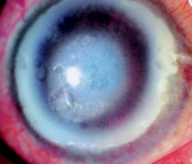
Figure 1 .
On slit lamp examination, right eye shows arcus lipoides and diffuse, central corneal opacity with subepithelial crystals.
Figure 2 .
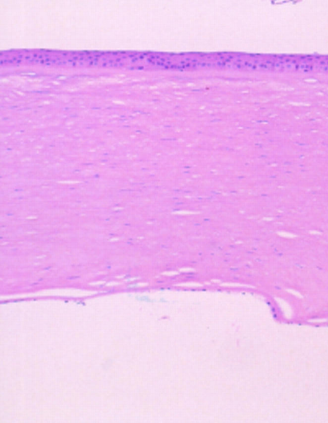
Photomicrograph of right corneal button. Loosened spaces representing dissolved lipids were present in the stroma (haematoxylin and eosin; original magnification, × 40).
Figure 3 .
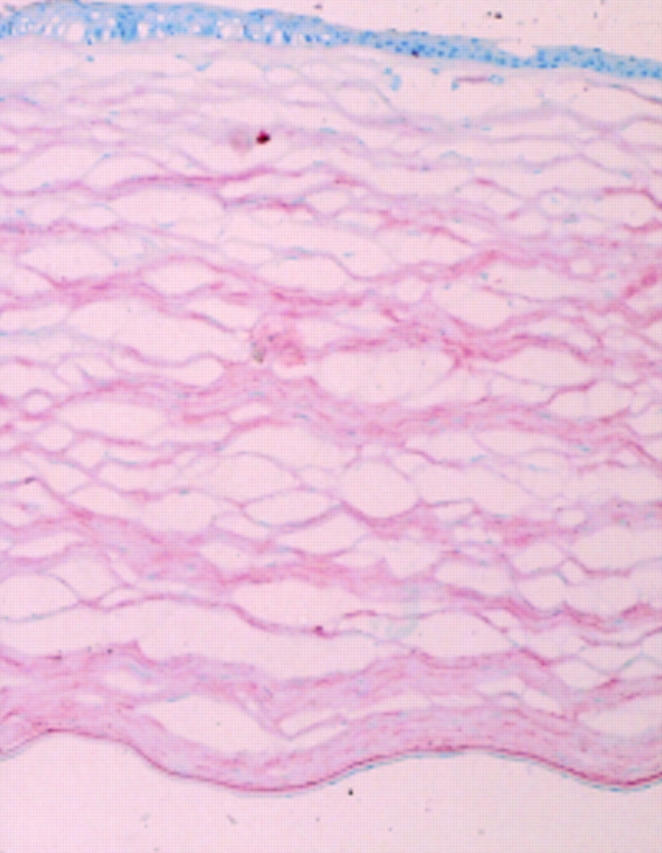
Photomicrograph of frozen section of the same corneal button stained with oil red O shows panstromal reactivity (oil red O; original magnification, × 40).
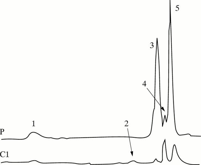
Figure 4 .
Chromatograms of neutral lipid analysis. (P) patient; (C1) control 1. 1 = esterified cholesterol; 2 = free fatty acids; 3 = unesterified cholesterol; 4 = monoglycerides + diglycerides; and 5 = phospholipids.
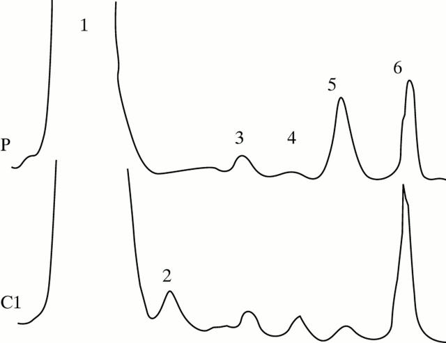
Figure 5 .
Chromatograms of phospholipids analysis. (P) patient; (C1) control 1. 1 = neutral lipids; 2 = phosphatidylethanolamine; 3 = phosphatidylserine + phosphatidylinositol; 4 = phosphatidylcholine; 5 = sphingomyelin; and 6 = origin.
Selected References
These references are in PubMed. This may not be the complete list of references from this article.
- ADAMS C. W. A perchloric acid-naphthoquinone method for the histochemical localization of cholesterol. Nature. 1961 Oct 28;192:331–332. doi: 10.1038/192331b0. [DOI] [PubMed] [Google Scholar]
- Barchiesi B. J., Eckel R. H., Ellis P. P. The cornea and disorders of lipid metabolism. Surv Ophthalmol. 1991 Jul-Aug;36(1):1–22. doi: 10.1016/0039-6257(91)90205-t. [DOI] [PubMed] [Google Scholar]
- Bron A. J. Corneal changes in the dislipoproteinaemias. Cornea. 1989;8(2):135–140. [PubMed] [Google Scholar]
- Bron A. J., Williams H. P., Carruthers M. E. Hereditary crystalline stromal dystrophy of Schnyder. I. Clinical features of a family with hyperlipoproteinaemia. Br J Ophthalmol. 1972 May;56(5):383–399. doi: 10.1136/bjo.56.5.383. [DOI] [PMC free article] [PubMed] [Google Scholar]
- Delleman J. W., Winkelman J. E. Degeneratio corneae cristallinea hereditaria. A clinical, genetical and histological study. Ophthalmologica. 1968;155(5):409–426. doi: 10.1159/000305324. [DOI] [PubMed] [Google Scholar]
- FOLCH J., LEES M., SLOANE STANLEY G. H. A simple method for the isolation and purification of total lipides from animal tissues. J Biol Chem. 1957 May;226(1):497–509. [PubMed] [Google Scholar]
- Freddo T. F., Polack F. M., Leibowitz H. M. Ultrastructural changes in the posterior layers of the cornea in Schnyder's crystalline dystrophy. Cornea. 1989 Sep;8(3):170–177. [PubMed] [Google Scholar]
- Garner A., Tripathi R. C. Hereditary crystalline stromal dystrophy of Schnyder. II. Histopathology and ultrastructure. Br J Ophthalmol. 1972 May;56(5):400–408. doi: 10.1136/bjo.56.5.400. [DOI] [PMC free article] [PubMed] [Google Scholar]
- Gaynor P. M., Zhang W. Y., Weiss J. S., Skarlatos S. I., Rodrigues M. M., Kruth H. S. Accumulation of HDL apolipoproteins accompanies abnormal cholesterol accumulation in Schnyder's corneal dystrophy. Arterioscler Thromb Vasc Biol. 1996 Aug;16(8):992–999. doi: 10.1161/01.atv.16.8.992. [DOI] [PubMed] [Google Scholar]
- McCarthy M., Innis S., Dubord P., White V. Panstromal Schnyder corneal dystrophy. A clinical pathologic report with quantitative analysis of corneal lipid composition. Ophthalmology. 1994 May;101(5):895–901. [PubMed] [Google Scholar]
- Pentchev P. G., Comly M. E., Kruth H. S., Vanier M. T., Wenger D. A., Patel S., Brady R. O. A defect in cholesterol esterification in Niemann-Pick disease (type C) patients. Proc Natl Acad Sci U S A. 1985 Dec;82(23):8247–8251. doi: 10.1073/pnas.82.23.8247. [DOI] [PMC free article] [PubMed] [Google Scholar]
- Rodrigues M. M., Kruth H. S., Krachmer J. H., Vrabec M. P., Blanchette-Mackie J. Cholesterol localization in ultrathin frozen sections in Schnyder's corneal crystalline dystrophy. Am J Ophthalmol. 1990 Nov 15;110(5):513–517. doi: 10.1016/s0002-9394(14)77874-2. [DOI] [PubMed] [Google Scholar]
- Rodrigues M. M., Kruth H. S., Krachmer J. H., Willis R. Unesterified cholesterol in Schnyder's corneal crystalline dystrophy. Am J Ophthalmol. 1987 Aug 15;104(2):157–163. doi: 10.1016/0002-9394(87)90008-0. [DOI] [PubMed] [Google Scholar]
- Shantha N. C. Thin-layer chromatography-flame ionization detection Iatroscan system. J Chromatogr. 1992 Oct 30;624(1-2):21–35. doi: 10.1016/0021-9673(92)85672-g. [DOI] [PubMed] [Google Scholar]
- Shearman A. M., Hudson T. J., Andresen J. M., Wu X., Sohn R. L., Haluska F., Housman D. E., Weiss J. S. The gene for schnyder's crystalline corneal dystrophy maps to human chromosome 1p34.1-p36. Hum Mol Genet. 1996 Oct;5(10):1667–1672. doi: 10.1093/hmg/5.10.1667. [DOI] [PubMed] [Google Scholar]
- Silva-Araújo A., Tavares M. A., Lemos M. M., Soares M. I., Castro-Correia J., Salgado-Borges J. Primary lipid keratopathy: a morphological and biochemical assessment. Br J Ophthalmol. 1993 Apr;77(4):248–250. doi: 10.1136/bjo.77.4.248. [DOI] [PMC free article] [PubMed] [Google Scholar]
- Weiss J. S., Rodrigues M. M., Kruth H. S., Rajagopalan S., Rader D. J., Kachadoorian H. Panstromal Schnyder's corneal dystrophy. Ultrastructural and histochemical studies. Ophthalmology. 1992 Jul;99(7):1072–1081. doi: 10.1016/s0161-6420(92)31848-2. [DOI] [PubMed] [Google Scholar]
- Weiss J. S. Schnyder crystalline dystrophy sine crystals. Recommendation for a revision of nomenclature. Ophthalmology. 1996 Mar;103(3):465–473. doi: 10.1016/s0161-6420(96)30670-2. [DOI] [PubMed] [Google Scholar]
- Weiss J. S. Schnyder's dystrophy of the cornea. A Swede-Finn connection. Cornea. 1992 Mar;11(2):93–101. doi: 10.1097/00003226-199203000-00001. [DOI] [PubMed] [Google Scholar]
- Weller R. O., Rodger F. C. Crystalline stromal dystrophy: histochemistry and ultrastructure of the cornea. Br J Ophthalmol. 1980 Jan;64(1):46–52. doi: 10.1136/bjo.64.1.46. [DOI] [PMC free article] [PubMed] [Google Scholar]
- Winder A. F., Garner A., Sheraidah G. A., Barry P. Familial lecithin:cholesterol acyltransferase deficiency. Biochemistry of the cornea. J Lipid Res. 1985 Mar;26(3):283–287. [PubMed] [Google Scholar]


