

Abstract
AIMS—To examine the course taken by individual retinal ganglion cell axons through the human lamina cribrosa. METHODS—Retinal ganglion cell axons were labelled using the retrograde tracer horseradish peroxidase applied directly to the optic nerve in two normal human eyes removed during the course of treatment for extraocular disease. RESULTS—A majority of axons took a direct course through the lamina cribrosa but a significant minority, in the range 8-12%, deviated to pass between the cribrosal plates in both central and peripheral parts of the optic disc. CONCLUSIONS—It is postulated that these axons would be selectively vulnerable to compression of the lamina cribrosa in diseases such as glaucoma in which the intraocular pressure is increased. Keywords: retina; optic nerve; glaucoma; lamina cribrosa
Full Text
The Full Text of this article is available as a PDF (108.8 KB).
Figure 1 .

Camera lucida drawing showing the path taken by individual HRP labelled axons through the optic nerve head. Transverse section. Optic nerve showing the location of sample areas a, b, and c Scale bar 1 mm. A, B, and C are areas in the region of the lamina cribrosa drawn at higher magnification. Stippled areas show regions corresponding to the location of the cribrosal plates. BV= blood vessel (filled areas in all figures). Scale bar 50 µm.
Figure 2 .
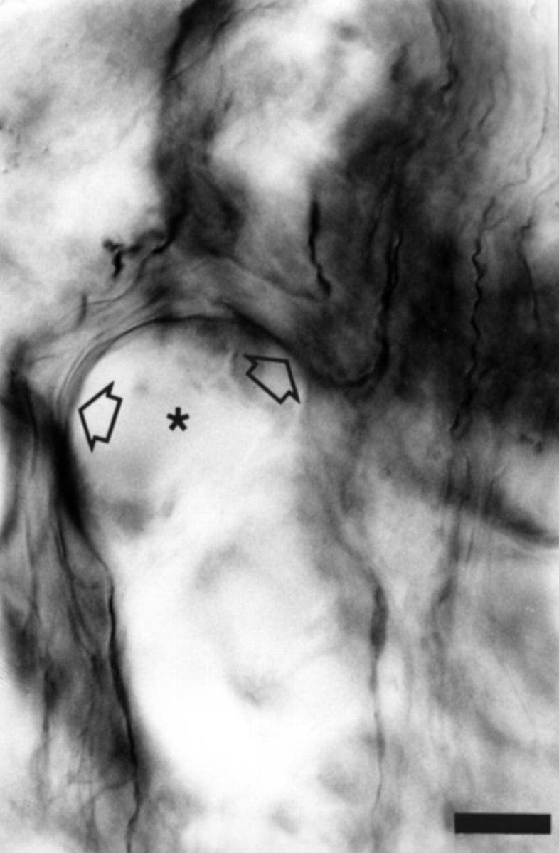
Photomicrograph from the optic nerve head showing deviation of HRP labelled axons in the vicinity of a cribrosal plate (asterisk). Arrows highlight the axon of interest. The top of the image represents more anterior optic nerve. Scale bar 2 µm.
Figure 3 .

Photomicrograph of single deviating axon in the vicinity of a cribrosal plate taken from the optic nerve in Figure 1. The top of the image represents more anterior optic nerve. Arrows highlight deflections in the axon of interest. Scale bar 2 µm.
Selected References
These references are in PubMed. This may not be the complete list of references from this article.
- Dichtl A., Jonas J. B., Naumann G. O. Course of the optic nerve fibers through the lamina cibrosa in human eyes. Graefes Arch Clin Exp Ophthalmol. 1996 Sep;234(9):581–585. doi: 10.1007/BF00448803. [DOI] [PubMed] [Google Scholar]
- Horton J. C., Greenwood M. M., Hubel D. H. Non-retinotopic arrangement of fibres in cat optic nerve. Nature. 1979 Dec 13;282(5740):720–722. doi: 10.1038/282720a0. [DOI] [PubMed] [Google Scholar]
- Mesulam M. M., Rosene D. L. Sensitivity in horseradish peroxidase neurohistochemistry: a comparative and quantitative study of nine methods. J Histochem Cytochem. 1979 Mar;27(3):763–773. doi: 10.1177/27.3.113450. [DOI] [PubMed] [Google Scholar]
- Ogden T. E., Duggan J., Danley K., Wilcox M., Minckler D. S. Morphometry of nerve fiber bundle pores in the optic nerve head of the human. Exp Eye Res. 1988 Apr;46(4):559–568. doi: 10.1016/s0014-4835(88)80012-5. [DOI] [PubMed] [Google Scholar]
- Ogden T. E. Nerve fiber layer of the macaque retina: retinotopic organization. Invest Ophthalmol Vis Sci. 1983 Jan;24(1):85–98. [PubMed] [Google Scholar]
- Ogden T. E. Nerve fiber layer of the primate retina: morphometric analysis. Invest Ophthalmol Vis Sci. 1984 Jan;25(1):19–29. [PubMed] [Google Scholar]
- Quigley H. A., Addicks E. M. Chronic experimental glaucoma in primates. II. Effect of extended intraocular pressure elevation on optic nerve head and axonal transport. Invest Ophthalmol Vis Sci. 1980 Feb;19(2):137–152. [PubMed] [Google Scholar]
- Quigley H. A., Addicks E. M., Green W. R., Maumenee A. E. Optic nerve damage in human glaucoma. II. The site of injury and susceptibility to damage. Arch Ophthalmol. 1981 Apr;99(4):635–649. doi: 10.1001/archopht.1981.03930010635009. [DOI] [PubMed] [Google Scholar]
- Quigley H. A., Dunkelberger G. R., Green W. R. Chronic human glaucoma causing selectively greater loss of large optic nerve fibers. Ophthalmology. 1988 Mar;95(3):357–363. doi: 10.1016/s0161-6420(88)33176-3. [DOI] [PubMed] [Google Scholar]
- Quigley H. A., Nickells R. W., Kerrigan L. A., Pease M. E., Thibault D. J., Zack D. J. Retinal ganglion cell death in experimental glaucoma and after axotomy occurs by apoptosis. Invest Ophthalmol Vis Sci. 1995 Apr;36(5):774–786. [PubMed] [Google Scholar]
- Quigley H. A., Sanchez R. M., Dunkelberger G. R., L'Hernault N. L., Baginski T. A. Chronic glaucoma selectively damages large optic nerve fibers. Invest Ophthalmol Vis Sci. 1987 Jun;28(6):913–920. [PubMed] [Google Scholar]
- Triviño A., Ramírez J. M., Salazar J. J., Ramírez A. I., García-Sánchez J. Immunohistochemical study of human optic nerve head astroglia. Vision Res. 1996 Jul;36(14):2015–2028. doi: 10.1016/0042-6989(95)00317-7. [DOI] [PubMed] [Google Scholar]
- Yan D. B., Coloma F. M., Metheetrairut A., Trope G. E., Heathcote J. G., Ethier C. R. Deformation of the lamina cribrosa by elevated intraocular pressure. Br J Ophthalmol. 1994 Aug;78(8):643–648. doi: 10.1136/bjo.78.8.643. [DOI] [PMC free article] [PubMed] [Google Scholar]