

Abstract
AIM—To explore the pathogenic role of delayed tear clearance. METHODS—By comparing 10 patients with punctal obstruction and 20 asymptomatic normals, delayed tear clearance was diagnosed in 70 patients without apparent punctal obstruction using fluorescein clearance test. RESULTS—The majority were older (71.4 (SD 1.2) years) and women (66%). Frequent complaints included redness, itching, mucus discharge, and crusting, which tended to be worse upon awakening. Common associated problems were medicamentosa (13%), drug induced pseudo pemphigoid, ocular hypertension (27%), and glaucoma (7%). Topical non-preserved 1% methylprednisolone resulted in subjective (83%) and objective (80%) improvement and resolution of delayed tear clearance (87%). CONCLUSION—These results indicate strong association of delayed tear clearance with intrinsically and extrinsically generated ocular surface inflammation. The presence of delayed tear clearance may set up a vicious cycle to aggravate the existing inflammation. Future prospective studies are needed to delineate the pathogenic role of delayed tear clearance in various ocular surface disorders. Keywords: medicamentosa; ocular irritation; tear clearance; tear turnover
Full Text
The Full Text of this article is available as a PDF (262.7 KB).
Figure 1 .
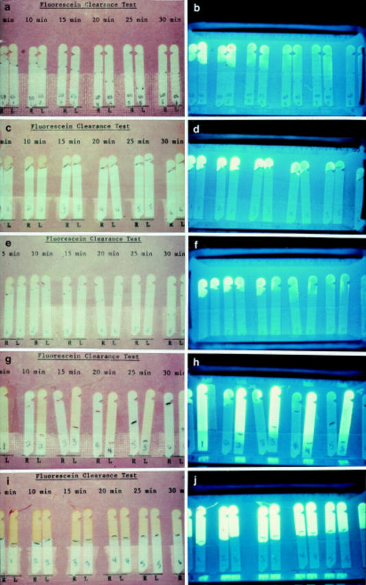
Representative fluorescein clearance test (FCT) results of normal tear secretion and clearance (a, b), severe aqueous tear deficiency (c, d), mild aqueous tear deficiency (e, f), unilateral (left eye) delayed tear clearance (g, h), and bilateral delayed tear clearance (i, j). For each case, six sets of Schirmer paper strips were used in a period of 30 minutes, with one set per 5 minute interval. For each set, the first and second strip was obtained from right and left eye, respectively. The detail of how FCT was performed is given in the Methods section. The entire six sets of strips were taped onto the record card with the wetting length marked by pencil (left panel), and the fluorescein intensity can be better appreciated when photographed under blue light (right panel).
Figure 2 .
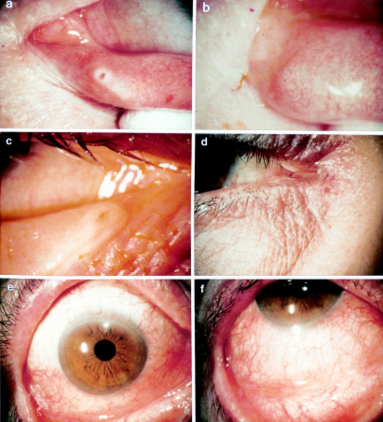
External photographs of a representative case with delayed tear clearance showing no apparent obstruction at the punctum (a). The corresponding fluorescein clearance test (FCT) is shown in Figure 1(g) and (h). For comparison, another representative case with delayed tear clearance showing punctal obstruction (b), for which the corresponding FCT is shown in Figure 1(i) and (j). The remaining photographs illustrate the typical appearance of a swollen punctum yielding a slit-like appearance (c), the subcutaneous oedema of the adjacent skin (c, d), bulbar conjunctival injection preferentially in the inferior aspect (e), and tarsal conjunctiva with a mild papillary response (f).
Figure 3 .

External appearance of case example showing diffuse conjunctival injection of both eyes (a) which was also preferentially distributed in the inferior tarsal conjunctiva with a mixed papillary and follicular response (b) and the entire bulbar conjunctiva (c) before the use of methylprednisolone. After treatment for 2 weeks, the entire bulbar conjunctiva became white and quiet (d). Fluorescein clearance test revealed marked delayed clearance of fluorescein (e), which was markedly improved after treatment (f).
Figure 4 .

Hypothetical scheme of the pathophysiology of delayed tear clearance. Delayed tear clearance can be induced by ineffective or decreased blinking (functional block) or mucosal inflammation and oedema in the nasolacrimal drainage system (partial anatomical block). The former is contributed by such risk factors as old age, female sex, decreased corneal sensitivity, and lid laxity including floppy lids. The latter can intrinsically be generated by allergy, atopy, rosacea, and meibomian gland dysfunction, or extrinsically caused by medication toxicity. A vicious cycle can form between delayed tear clearance and mucosal inflammation and oedema leading to ocular irritation, medicamentosa, drug induced pseudopemphigoid, and steroid induced ocular hypertension.
Selected References
These references are in PubMed. This may not be the complete list of references from this article.
- BOBERG-ANS J. On the corneal sensitivity. Acta Ophthalmol (Copenh) 1956;34(3):149–162. doi: 10.1111/j.1755-3768.1956.tb03346.x. [DOI] [PubMed] [Google Scholar]
- Barton K., Monroy D. C., Nava A., Pflugfelder S. C. Inflammatory cytokines in the tears of patients with ocular rosacea. Ophthalmology. 1997 Nov;104(11):1868–1874. doi: 10.1016/s0161-6420(97)30014-1. [DOI] [PubMed] [Google Scholar]
- Burstein N. L. Corneal cytotoxicity of topically applied drugs, vehicles and preservatives. Surv Ophthalmol. 1980 Jul-Aug;25(1):15–30. doi: 10.1016/0039-6257(80)90072-7. [DOI] [PubMed] [Google Scholar]
- Cho P., Yap M. Schirmer test. I. A review. Optom Vis Sci. 1993 Feb;70(2):152–156. doi: 10.1097/00006324-199302000-00011. [DOI] [PubMed] [Google Scholar]
- Chrai S. S., Patton T. F., Mehta A., Robinson J. R. Lacrimal and instilled fluid dynamics in rabbit eyes. J Pharm Sci. 1973 Jul;62(7):1112–1121. doi: 10.1002/jps.2600620712. [DOI] [PubMed] [Google Scholar]
- Collins M., Seeto R., Campbell L., Ross M. Blinking and corneal sensitivity. Acta Ophthalmol (Copenh) 1989 Oct;67(5):525–531. doi: 10.1111/j.1755-3768.1989.tb04103.x. [DOI] [PubMed] [Google Scholar]
- Doane M. G. Blinking and the mechanics of the lacrimal drainage system. Ophthalmology. 1981 Aug;88(8):844–851. doi: 10.1016/s0161-6420(81)34940-9. [DOI] [PubMed] [Google Scholar]
- Fraunfelder F. T. Extraocular fluid dynamics: how best to apply topical ocular medication. Trans Am Ophthalmol Soc. 1976;74:457–487. [PMC free article] [PubMed] [Google Scholar]
- Holly F. J., Lamberts D. W., Esquivel E. D. Kinetics of capillary tear flow in the Schirmer strip. Curr Eye Res. 1982;2(1):57–70. doi: 10.3109/02713688208998380. [DOI] [PubMed] [Google Scholar]
- Holly F. J., Lemp M. A. Tear physiology and dry eyes. Surv Ophthalmol. 1977 Sep-Oct;22(2):69–87. doi: 10.1016/0039-6257(77)90087-x. [DOI] [PubMed] [Google Scholar]
- Hoshina Y., Sakuma Y. Changes in photically evoked blink reflex during sleep and wakefulness. Jpn J Ophthalmol. 1991;35(2):182–187. [PubMed] [Google Scholar]
- Jones L. T. The lacrimal secretory system and its treatment. Am J Ophthalmol. 1966 Jul;62(1):47–60. doi: 10.1016/0002-9394(66)91676-x. [DOI] [PubMed] [Google Scholar]
- Jordan A., Baum J. Basic tear flow. Does it exist? Ophthalmology. 1980 Sep;87(9):920–930. doi: 10.1016/s0161-6420(80)35143-9. [DOI] [PubMed] [Google Scholar]
- Kuppens E. V., Stolwijk T. R., de Keizer R. J., van Best J. A. Basal tear turnover and topical timolol in glaucoma patients and healthy controls by fluorophotometry. Invest Ophthalmol Vis Sci. 1992 Nov;33(12):3442–3448. [PubMed] [Google Scholar]
- Kuppens E. V., van Best J. A., Sterk C. C., de Keizer R. J. Decreased basal tear turnover in patients with untreated primary open-angle glaucoma. Am J Ophthalmol. 1995 Jul;120(1):41–46. doi: 10.1016/s0002-9394(14)73757-2. [DOI] [PubMed] [Google Scholar]
- Lemp M. A., Hamill J. R., Jr Factors affecting tear film breakup in normal eyes. Arch Ophthalmol. 1973 Feb;89(2):103–105. doi: 10.1001/archopht.1973.01000040105007. [DOI] [PubMed] [Google Scholar]
- Lemp M. A., Weiler H. H. How do tears exit? Invest Ophthalmol Vis Sci. 1983 May;24(5):619–622. [PubMed] [Google Scholar]
- Lindén C., Alm A. The effect of reduced tear drainage on corneal and aqueous concentrations of topically applied fluorescein. Acta Ophthalmol (Copenh) 1990 Dec;68(6):633–638. doi: 10.1111/j.1755-3768.1990.tb01686.x. [DOI] [PubMed] [Google Scholar]
- Meyer D. R., Antonello A., Linberg J. V. Assessment of tear drainage after canalicular obstruction using fluorescein dye disappearance. Ophthalmology. 1990 Oct;97(10):1370–1374. doi: 10.1016/s0161-6420(90)32408-9. [DOI] [PubMed] [Google Scholar]
- Millodot M. The influence of age onthe sensitivity of the cornea,. Invest Ophthalmol Vis Sci. 1977 Mar;16(3):240–242. [PubMed] [Google Scholar]
- Mishima S., Gasset A., Klyce S. D., Jr, Baum J. L. Determination of tear volume and tear flow. Invest Ophthalmol. 1966 Jun;5(3):264–276. [PubMed] [Google Scholar]
- NOVER A., JAEGER W. Kolorimetrische Methode zur Messung der Tränensekretion (Fluoreszein-Verdünnungstest). Klin Monbl Augenheilkd Augenarztl Fortbild. 1952;121(4):419–425. [PubMed] [Google Scholar]
- Norn M. S. Desiccation of the precorneal film. I. Corneal wetting-time. Acta Ophthalmol (Copenh) 1969;47(4):865–880. doi: 10.1111/j.1755-3768.1969.tb03711.x. [DOI] [PubMed] [Google Scholar]
- Norn M. S. Lacrimal apparatus tests. A new method (lacrimal streak dilution test) compared with previous methods. Acta Ophthalmol (Copenh) 1965;43(4):557–566. [PubMed] [Google Scholar]
- Norn M. S. Tear secretion in normal eyes. Estimated by a new method: the lacrimal streak dilution test. Acta Ophthalmol (Copenh) 1965;43(4):567–573. doi: 10.1111/j.1755-3768.1965.tb03693.x. [DOI] [PubMed] [Google Scholar]
- Occhipinti J. R., Mosier M. A., LaMotte J., Monji G. T. Fluorophotometric measurement of human tear turnover rate. Curr Eye Res. 1988 Oct;7(10):995–1000. doi: 10.3109/02713688809015145. [DOI] [PubMed] [Google Scholar]
- Patton T. F., Robinson J. R. Influence of topical anesthesia on tear dynamics and ocular drug bioavailability in albino rabbits. J Pharm Sci. 1975 Feb;64(2):267–271. doi: 10.1002/jps.2600640215. [DOI] [PubMed] [Google Scholar]
- Pflugfelder S. C., Tseng S. C., Pepose J. S., Fletcher M. A., Klimas N., Feuer W. Epstein-Barr virus infection and immunologic dysfunction in patients with aqueous tear deficiency. Ophthalmology. 1990 Mar;97(3):313–323. doi: 10.1016/s0161-6420(90)32595-2. [DOI] [PubMed] [Google Scholar]
- Puffer M. J., Neault R. W., Brubaker R. F. Basal precorneal tear turnover in the human eye. Am J Ophthalmol. 1980 Mar;89(3):369–376. doi: 10.1016/0002-9394(80)90006-9. [DOI] [PubMed] [Google Scholar]
- Reed S., Lissner G. Clinical study on the effectiveness of tear drainage with a single canalicular system under environmental stress. Ophthal Plast Reconstr Surg. 1993;9(1):27–31. doi: 10.1097/00002341-199303000-00003. [DOI] [PubMed] [Google Scholar]
- Sack R. A., Tan K. O., Tan A. Diurnal tear cycle: evidence for a nocturnal inflammatory constitutive tear fluid. Invest Ophthalmol Vis Sci. 1992 Mar;33(3):626–640. [PubMed] [Google Scholar]
- Tomlinson A., Trees G. R., Occhipinti J. R. Tear production and evaporation in the normal eye. Ophthalmic Physiol Opt. 1991 Jan;11(1):44–47. [PubMed] [Google Scholar]
- Tucker N. A., Codère F. The effect of fluorescein volume on lacrimal outflow transit time. Ophthal Plast Reconstr Surg. 1994 Dec;10(4):256–259. doi: 10.1097/00002341-199412000-00006. [DOI] [PubMed] [Google Scholar]
- Webber W. R., Jones D. P. Continuous fluorophotometric method of measuring tear turnover rate in humans and analysis of factors affecting accuracy. Med Biol Eng Comput. 1986 Jul;24(4):386–392. doi: 10.1007/BF02442693. [DOI] [PubMed] [Google Scholar]
- Webber W. R., Jones D. P., Wright P. Fluorophotometric measurements of tear turnover rate in normal healthy persons: evidence for a circadian rhythm. Eye (Lond) 1987;1(Pt 5):615–620. doi: 10.1038/eye.1987.95. [DOI] [PubMed] [Google Scholar]
- White W. L., Glover A. T., Buckner A. B. Effect of blinking on tear elimination as evaluated by dacryoscintigraphy. Ophthalmology. 1991 Mar;98(3):367–369. doi: 10.1016/s0161-6420(91)32287-5. [DOI] [PubMed] [Google Scholar]
- Wojno T. H. Allergic lacrimal obstruction. Am J Ophthalmol. 1988 Jul 15;106(1):48–52. doi: 10.1016/s0002-9394(14)76387-1. [DOI] [PubMed] [Google Scholar]
- Xu K. P., Tsubota K. Correlation of tear clearance rate and fluorophotometric assessment of tear turnover. Br J Ophthalmol. 1995 Nov;79(11):1042–1045. doi: 10.1136/bjo.79.11.1042. [DOI] [PMC free article] [PubMed] [Google Scholar]
- Xu K. P., Yagi Y., Toda I., Tsubota K. Tear function index. A new measure of dry eye. Arch Ophthalmol. 1995 Jan;113(1):84–88. doi: 10.1001/archopht.1995.01100010086025. [DOI] [PubMed] [Google Scholar]
- Zappia R. J., Milder B. Lacrimal drainage function. 2. The fluorescein dye disappearance test. Am J Ophthalmol. 1972 Jul;74(1):160–162. [PubMed] [Google Scholar]
- van Best J. A., Benitez del Castillo J. M., Coulangeon L. M. Measurement of basal tear turnover using a standardized protocol. European concerted action on ocular fluorometry. Graefes Arch Clin Exp Ophthalmol. 1995 Jan;233(1):1–7. doi: 10.1007/BF00177778. [DOI] [PubMed] [Google Scholar]
- van Bijsterveld O. P. Diagnostic tests in the Sicca syndrome. Arch Ophthalmol. 1969 Jul;82(1):10–14. doi: 10.1001/archopht.1969.00990020012003. [DOI] [PubMed] [Google Scholar]