Abstract
AIMS—Ophthalmodynamometry has been used extensively since the last century; however, controversy surrounds what it actually measures. This study was set up to determine the relation between ophthalmodynamometric (ODP) and systemic blood pressures. METHODS—Aortic pressure was continuously monitored and altered by phlebotomy in six anaesthetised dogs, while ophthalmodynamometry was performed, by directly altering intraocular pressure. Maxillary artery pressure was monitored in two animals. All pressure transducers were zeroed at eye level. RESULTS—Mean ODP was 96.6% (1.6%) (95% confidence interval, n = 49) of aortic pressure. Mean maxillary artery pressure was 95.7% (5.5%) (95% CI, n = 16) of aortic pressure. ODP was 1.9 (0.6) mm Hg (95% CI, n = 33) higher than maxillary artery pressures. CONCLUSION—ODP was only slightly below aortic pressure and not significantly different from maxillary artery pressure, the analogue of the internal carotid artery in humans. These results also suggest a retinal artery collapse pressure of at least 1.9 mm Hg. Keywords: ophthalmodynamometry; dog; arterial pressure
Full Text
The Full Text of this article is available as a PDF (97.1 KB).
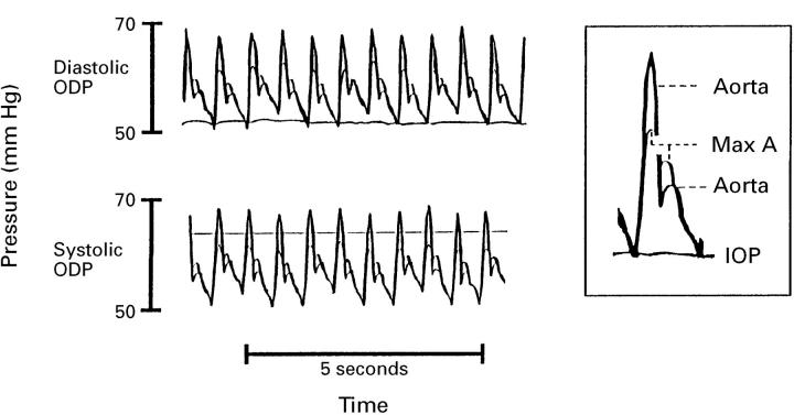
Figure 1 .
Typical traces of aortic and maxillary artery (max A) pressures. First CRA pulsation (diastolic ODP) was seen when IOP was elevated to a level equal to aortic and maxillary diastolic pressure. Complete CRA closure (systolic ODP) was seen when IOP was elevated to a level above maxillary artery but below aortic systolic pressure.
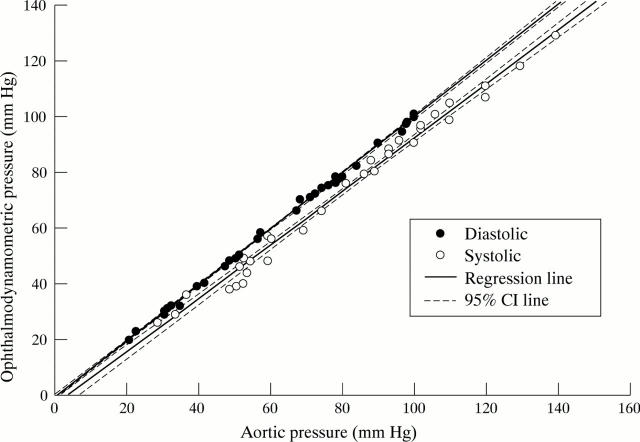
Figure 2 .
Ophthalmodynamometric (ODP) diastolic versus aortic diastolic pressure and ODP systolic versus aortic systolic pressure, with regression and 95% confidence interval lines. It can be seen that diastolic ODP is almost equivalent to aortic diastolic pressure, and that the systolic ODP is slightly less than aortic systolic pressure.
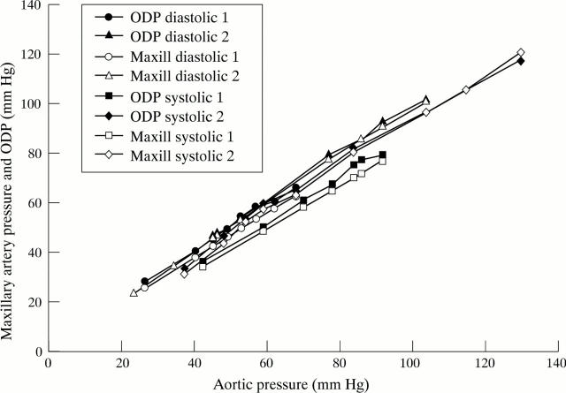
Figure 3 .
Ophthalmodynamometric (ODP) and maxillary artery (maxill) pressures versus aortic pressure in two animals. The aortic pressure was varied by phlebotomy. Systolic and diastolic pressures are shown. Both systolic and diastolic maxillary artery pressures are seen to be slightly less than ODP.
Selected References
These references are in PubMed. This may not be the complete list of references from this article.
- AbuRahma A. F., Robinson P. A., Short Y., Lucente F. C., Boland J. P. Cross-filling of circle of Willis and carotid stenosis by angiography, duplex ultrasound, and oculopneumoplethysmography. Am J Surg. 1995 Mar;169(3):308–312. doi: 10.1016/S0002-9610(99)80164-0. [DOI] [PubMed] [Google Scholar]
- Best M., Blumenthal M., Futterman H. A., Galin M. A. Critical closure of intraocular blood vessels. Arch Ophthalmol. 1969 Sep;82(3):385–392. doi: 10.1001/archopht.1969.00990020387018. [DOI] [PubMed] [Google Scholar]
- Cennamo G., Rosa N., De Palma L., Pastena B. Echographic and ophthalmodynamometric study in the empty sella syndrome. Ophthalmologica. 1993;206(1):29–32. doi: 10.1159/000310358. [DOI] [PubMed] [Google Scholar]
- DeLano F. A., Schmid-Schönbein G. W., Skalak T. C., Zweifach B. W. Penetration of the systemic blood pressure into the microvasculature of rat skeletal muscle. Microvasc Res. 1991 Jan;41(1):92–110. doi: 10.1016/0026-2862(91)90011-y. [DOI] [PubMed] [Google Scholar]
- Drzewiecki G., Hood R., Apple H. Theory of the oscillometric maximum and the systolic and diastolic detection ratios. Ann Biomed Eng. 1994 Jan-Feb;22(1):88–96. doi: 10.1007/BF02368225. [DOI] [PubMed] [Google Scholar]
- Duke-Elder W. S. THE OCULAR CIRCULATION: ITS NORMAL PRESSURE RELATIONSHIPS AND THEIR PHYSIOLOGICAL SIGNIFICANCE. Br J Ophthalmol. 1926 Oct;10(10):513–572. doi: 10.1136/bjo.10.10.513. [DOI] [PMC free article] [PubMed] [Google Scholar]
- Gore R. W. Pressures in cat mesenteric arterioles and capillaries during changes in systemic arterial blood pressure. Circ Res. 1974 Apr;34(4):581–591. doi: 10.1161/01.res.34.4.581. [DOI] [PubMed] [Google Scholar]
- Grunwald J. E., Furubayashi C. Effect of topical timolol maleate on the ophthalmic artery blood pressure. Invest Ophthalmol Vis Sci. 1989 Jun;30(6):1095–1100. [PubMed] [Google Scholar]
- Hedges T. R., Weinstein J. D., Kassell N. F., Langfitt T. W. Correlation of ophthalmodynamometry with ophthalmic artery pressure in the rhesus monkey. Am J Ophthalmol. 1965 Dec;60(6):1098–1101. doi: 10.1016/0002-9394(65)92819-9. [DOI] [PubMed] [Google Scholar]
- Holladay J. T., Arnoult J. B., Ruiz R. S. Comparative evaluation of current ophthalmodynamometers. Am J Ophthalmol. 1979 May;87(5):665–674. doi: 10.1016/0002-9394(79)90301-5. [DOI] [PubMed] [Google Scholar]
- Hu H. H., Sheng W. Y., Yen M. Y., Lai S. T., Teng M. M. Color Doppler imaging of orbital arteries for detection of carotid occlusive disease. Stroke. 1993 Aug;24(8):1196–1203. doi: 10.1161/01.str.24.8.1196. [DOI] [PubMed] [Google Scholar]
- Lovasik J. V., Kothe A. C., Kergoat H. Comparison of noninvasive methods to derive the mean central retinal artery pressure in man. Optom Vis Sci. 1993 Dec;70(12):1005–1011. doi: 10.1097/00006324-199312000-00003. [DOI] [PubMed] [Google Scholar]
- Morgan W. H., Yu D. Y., Cooper R. L., Alder V. A., Cringle S. J., Constable I. J. Retinal artery and vein pressures in the dog and their relationship to aortic, intraocular, and cerebrospinal fluid pressures. Microvasc Res. 1997 May;53(3):211–221. doi: 10.1006/mvre.1997.2010. [DOI] [PubMed] [Google Scholar]
- Morgan W. H., Yu D. Y., Cooper R. L., Alder V. A., Cringle S. J., Constable I. J. The influence of cerebrospinal fluid pressure on the lamina cribrosa tissue pressure gradient. Invest Ophthalmol Vis Sci. 1995 May;36(6):1163–1172. [PubMed] [Google Scholar]
- Mullie M. A., Kirkham T. H. Ophthalmodynamometry revisited. Can J Ophthalmol. 1983 Jun;18(4):165–168. [PubMed] [Google Scholar]
- Robinson F., Riva C. E., Grunwald J. E., Petrig B. L., Sinclair S. H. Retinal blood flow autoregulation in response to an acute increase in blood pressure. Invest Ophthalmol Vis Sci. 1986 May;27(5):722–726. [PubMed] [Google Scholar]
- Vander Werff T. J. The pressure measured in ophthalmodynamometry. Arch Ophthalmol. 1972 Mar;87(3):290–292. doi: 10.1001/archopht.1972.01000020292011. [DOI] [PubMed] [Google Scholar]
- Weinberger J. Clinical applications of noninvasive carotid artery testing. J Am Coll Cardiol. 1985 Jan;5(1):137–148. doi: 10.1016/s0735-1097(85)80097-8. [DOI] [PubMed] [Google Scholar]
- Wiebers D. O., Folger W. N., Forbes G. S., Younge B. R., O'Fallon W. M. Ophthalmodynamometry and ocular pneumoplethysmography for detection of carotid occlusive disease. Arch Neurol. 1982 Nov;39(11):690–691. doi: 10.1001/archneur.1982.00510230016005. [DOI] [PubMed] [Google Scholar]
- Williamson T. H., Baxter G. M., Pyott A., Wykes W., Dutton G. N. A comparison of colour Doppler imaging of orbital vessels and other methods of blood flow assessment. Graefes Arch Clin Exp Ophthalmol. 1995 Feb;233(2):80–84. doi: 10.1007/BF00241476. [DOI] [PubMed] [Google Scholar]


