Abstract
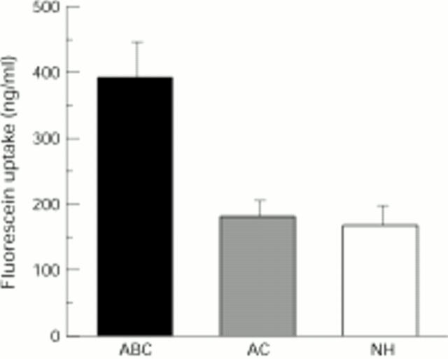
AIMS—To assess the integrity of the ocular surface epithelium in patients with atopic dermatitis from the viewpoint of its barrier function. METHODS—49 patients with atopic dermatitis with blepharoconjunctivitis (ABC group), 27 age matched patients with seasonal or perennial allergic conjunctivitis (AC group), and 20 volunteers with normal healthy eyes (NH group) were assigned to this study. Ocular surface epithelium barrier function was evaluated by the fluorophotometric method using a slit lamp fluorophotometer. 3 µl of 0.5% sodium fluorescein was instilled into the conjunctival sac of the right eye and fluorescein uptake (ng/ml) 30 minutes later (20 minutes after eye washing) was measured in the central cornea and the temporal bulbar conjunctiva. Fluorophoto metric measurements performed were analysed in each group and compared between the groups. RESULTS—The ABC group showed significantly higher fluorescein uptake (mean 28.2 (SEM 3.3) ng/ml) in the cornea than the AC (11.4 (2.2), p=0.001) and NH groups (9.3 (2.1), p=0.001). There was no significant difference between the AC and NH groups (p=0.930). The ABC group also showed significantly higher fluorescein uptake in the bulbar conjunctiva (393.4 (54.0)) than the AC (182.9 (24.6), p=0.011) and NH groups (169.3 (29.1), p=0.012). There was also no significant difference in fluorescein uptake between the AC group and the NH group (p=0.987). CONCLUSION—This study suggested that ocular surface epithelium barrier function is impaired in patients with atopic dermatitis with blepharoconjunctivitis. Keywords: atopic blepharoconjunctivitis; ocular surface epithelium; barrier function; fluorophotometry
Full Text
The Full Text of this article is available as a PDF (86.4 KB).
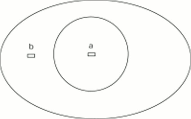
Figure 1 .
A schema indicating the site of fluorophotometric measurement. Fluorescein uptake was measured in the central cornea (a) and temporal bulbar conjunctiva 3 mm away from the limbus (b). The measurement area was 0.15 mm × 0.3 mm (vertical × horizontal section)
Figure 2 .
Comparison of corneal fluorescein uptake between atopic dermatitis patients with atopic blepharoconjunctivitis (ABC), seasonal or perennial allergic conjunctivitis patients (AC), and volunteers with normal healthy eyes (NH). A significant difference was noted in fluorescein uptake between the three groups (p<0.0001), with markedly higher fluorescein uptake in the ABC than in the AC or NH groups (p=0.001 and p=0.001, respectively). There was no significant difference between the AC and NH groups (p=0.930).
Figure 3 .
Comparison of conjunctival fluorescein uptake between atopic dermatitis patients with atopic blepharoconjunctivitis (ABC), seasonal or perennial allergic conjunctivitis patients (AC), and volunteers with normal healthy eyes (NH). A significant difference was noted in the fluorescein uptake between the three groups (p=0.0003), with markedly higher fluorescein uptake in the ABC than in the AC or NH group (p=0.011 and p=0.012, respectively). There was no significant difference between the AC and NH group (p=0.987).
Selected References
These references are in PubMed. This may not be the complete list of references from this article.
- Abelson M. B., Schaefer K. Conjunctivitis of allergic origin: immunologic mechanisms and current approaches to therapy. Surv Ophthalmol. 1993 Jul-Aug;38 (Suppl):115–132. doi: 10.1016/0039-6257(93)90036-7. [DOI] [PubMed] [Google Scholar]
- Foster C. S., Calonge M. Atopic keratoconjunctivitis. Ophthalmology. 1990 Aug;97(8):992–1000. doi: 10.1016/s0161-6420(90)32477-6. [DOI] [PubMed] [Google Scholar]
- Friedlaender M. H. Conjunctivitis of allergic origin: clinical presentation and differential diagnosis. Surv Ophthalmol. 1993 Jul-Aug;38 (Suppl):105–114. doi: 10.1016/0039-6257(93)90035-6. [DOI] [PubMed] [Google Scholar]
- HOGAN M. J. Atopic keratoconjunctivitis. Am J Ophthalmol. 1953 Jul;36(7 1):937–947. doi: 10.1016/0002-9394(53)92176-x. [DOI] [PubMed] [Google Scholar]
- HOGAN M. J. Atopic keratoconjunctivitis. Trans Am Ophthalmol Soc. 1952;50:265–281. [PMC free article] [PubMed] [Google Scholar]
- Montan P. G., van Hage-Hamsten M. Eosinophil cationic protein in tears in allergic conjunctivitis. Br J Ophthalmol. 1996 Jun;80(6):556–560. doi: 10.1136/bjo.80.6.556. [DOI] [PMC free article] [PubMed] [Google Scholar]
- Roat M. I., Ohji M., Hunt L. E., Thoft R. A. Conjunctival epithelial cell hypermitosis and goblet cell hyperplasia in atopic keratoconjunctivitis. Am J Ophthalmol. 1993 Oct 15;116(4):456–463. doi: 10.1016/s0002-9394(14)71404-7. [DOI] [PubMed] [Google Scholar]
- Tanihara H., Yokoi N., Komuro A., Honda Y., Kinoshita S. Prolonged impairment of peripheral corneal epithelium barrier function after successful trabeculectomy. Am J Ophthalmol. 1997 Apr;123(4):487–493. doi: 10.1016/s0002-9394(14)70174-6. [DOI] [PubMed] [Google Scholar]
- Trocme S. D., Kephart G. M., Allansmith M. R., Bourne W. M., Gleich G. J. Conjunctival deposition of eosinophil granule major basic protein in vernal keratoconjunctivitis and contact lens-associated giant papillary conjunctivitis. Am J Ophthalmol. 1989 Jul 15;108(1):57–63. doi: 10.1016/s0002-9394(14)73261-1. [DOI] [PubMed] [Google Scholar]
- Trocmé S. D., Gleich G. J., Kephart G. M., Zieske J. D. Eosinophil granule major basic protein inhibition of corneal epithelial wound healing. Invest Ophthalmol Vis Sci. 1994 Jun;35(7):3051–3056. [PubMed] [Google Scholar]
- Trocmé S. D., Hallberg C. K., Gill K. S., Gleich G. J., Tyring S. K., Brysk M. M. Effects of eosinophil granule proteins on human corneal epithelial cell viability and morphology. Invest Ophthalmol Vis Sci. 1997 Mar;38(3):593–599. [PubMed] [Google Scholar]
- Trocmé S. D., Kephart G. M., Bourne W. M., Buckley R. J., Gleich G. J. Eosinophil granule major basic protein deposition in corneal ulcers associated with vernal keratoconjunctivitis. Am J Ophthalmol. 1993 May 15;115(5):640–643. doi: 10.1016/s0002-9394(14)71463-1. [DOI] [PubMed] [Google Scholar]
- Tuft S. J., Kemeny D. M., Dart J. K., Buckley R. J. Clinical features of atopic keratoconjunctivitis. Ophthalmology. 1991 Feb;98(2):150–158. doi: 10.1016/s0161-6420(91)32322-4. [DOI] [PubMed] [Google Scholar]
- Udell I. J., Gleich G. J., Allansmith M. R., Ackerman S. J., Abelson M. B. Eosinophil granule major basic protein and Charcot-Leyden crystal protein in human tears. Am J Ophthalmol. 1981 Dec;92(6):824–828. doi: 10.1016/s0002-9394(14)75637-5. [DOI] [PubMed] [Google Scholar]
- Yokoi N., Kinoshita S., Akiyama K. [A new slit-lamp fluorophotometer for the clinical evaluation of corneal epithelial barrier function]. Nippon Ganka Gakkai Zasshi. 1994 Jul;98(7):641–647. [PubMed] [Google Scholar]
- Yokoi N., Kinoshita S. Clinical evaluation of corneal epithelial barrier function with the slit-lamp fluorophotometer. Cornea. 1995 Sep;14(5):485–489. [PubMed] [Google Scholar]


