Abstract
AIM—To study the refractive status and corneal topography in Down's syndrome. METHOD—A matched cohort subgroup of 50 individuals with Down's syndrome in the Manchester area aged 15-22 years was studied by refraction, corneal topography, A-scan biometry, slit lamp examination, and orthoptic examination. RESULTS—(1) A linear relation was found between axial length and spherical equivalent refraction. There was no statistical relation between keratometry and the axial length. (2) 80% of the group had a hyperopic refraction (mean +2.46 D, range +0.5 to +7.5 D); 18% were myopic (mean −2.75 D, range −0.5 to −8.0 D); and 2% were emmetropic (within plus or minus 0.5 D of zero). The overall mean spherical equivalent refraction was +1.43 (SD 2.86) D. 63% of eyes could see 6/12 or better and 66% of the individuals had a binocular vision of 6/12 or better. (3) Corneal topography was generally of a regular "bow tie" pattern, but there was a high incidence of oblique cylinders. Mean cylinder strength was 1.14 (1.15) D. (4) The prevalence of overt keratoconus was 2%. 6% had corneal topography with inferior steepening which may be a preclinical keratoconic process. CONCLUSIONS—In this cohort of late teenagers with Down's syndrome, emmetropisation has failed to occur in most individuals. In a similar aged group of non-disabled individuals one would expect about 83% emmetropic (plus or minus 0.25 D), 13% myopic, and 4% hyperopic. The wide spread of oblique cylinders and the small proportion of with the rule astigmatism is probably related to this failure of emmetropisation. The prevalence of 2% keratoconus in Down's syndrome compares with that found by other authors of between 5.5 and 15%. The 6% with inferior steepening on topography will be followed up over the next few years to see if there is any development of clinical keratoconus. Hence we will see if corneal topography is useful as a screening tool for preclinical keratoconus in this high risk group. Keywords: Down's syndrome; emmetropisation; refraction; keratoconus
Full Text
The Full Text of this article is available as a PDF (120.1 KB).
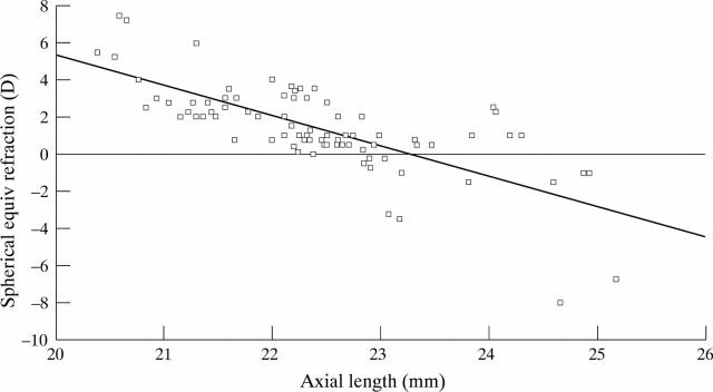
Figure 1 .
Plot of spherical equivalent refraction v axial length as measured by A-scan biometry.
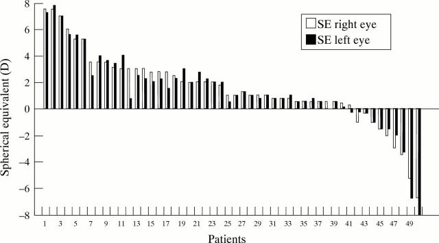
Figure 2 .
Bar chart of spherical equivalent refraction for each eye.
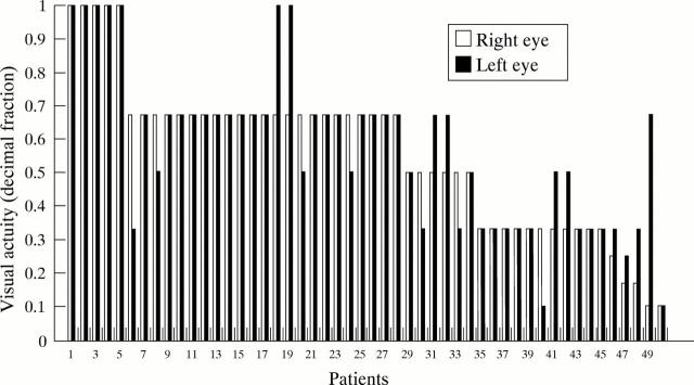
Figure 3 .
Bar chart of visual acuity for each eye.
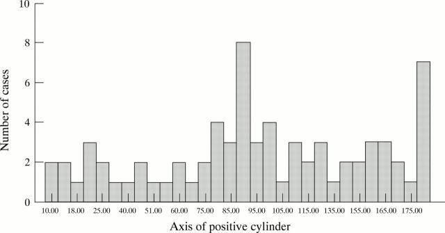
Figure 4 .
Bar chart of axes of the refractive cylinders.
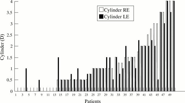
Figure 5 .
Bar chart of magnitude of the refractive cylinders.
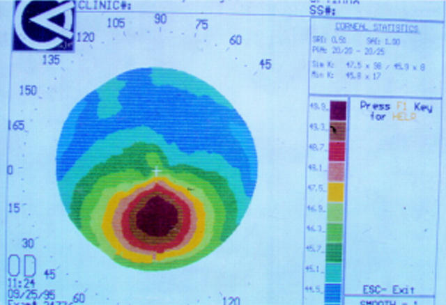
Figure 6 .
Corneal topography of subject with clinical keratoconus.
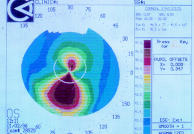
Figure 7 .
Corneal topography of subject with inferior steepening
Selected References
These references are in PubMed. This may not be the complete list of references from this article.
- CULLEN J. F., BUTLER H. G. MONGOLISM (DOWN'S SYNDROME) AND KERATOCONUS. Br J Ophthalmol. 1963 Jun;47:321–330. doi: 10.1136/bjo.47.6.321. [DOI] [PMC free article] [PubMed] [Google Scholar]
- Catalano R. A. Down syndrome. Surv Ophthalmol. 1990 Mar-Apr;34(5):385–398. doi: 10.1016/0039-6257(90)90116-d. [DOI] [PubMed] [Google Scholar]
- EISSLER R., LONGENECKER L. P. The common eye findings in mongolism. Am J Ophthalmol. 1962 Sep;54:398–406. doi: 10.1016/0002-9394(62)93757-1. [DOI] [PubMed] [Google Scholar]
- Hayashi K., Hayashi H., Hayashi F. Topographic analysis of the changes in corneal shape due to aging. Cornea. 1995 Sep;14(5):527–532. [PubMed] [Google Scholar]
- Hosaka A. The growth of the eye and its components. Japanese studies. Acta Ophthalmol Suppl. 1988;185:65–68. doi: 10.1111/j.1755-3768.1988.tb02667.x. [DOI] [PubMed] [Google Scholar]
- Kennedy R. H., Bourne W. M., Dyer J. A. A 48-year clinical and epidemiologic study of keratoconus. Am J Ophthalmol. 1986 Mar 15;101(3):267–273. doi: 10.1016/0002-9394(86)90817-2. [DOI] [PubMed] [Google Scholar]
- Pierse D., Eustace P. Acute keratoconus in mongols. Br J Ophthalmol. 1971 Jan;55(1):50–54. doi: 10.1136/bjo.55.1.50. [DOI] [PMC free article] [PubMed] [Google Scholar]
- Rabinowitz Y. S., Maumenee I. H., Lundergan M. K., Puffenberger E., Zhu D., Antonarakis S., Francomano C. A. Molecular genetic analysis in autosomal dominant keratoconus. Cornea. 1992 Jul;11(4):302–308. doi: 10.1097/00003226-199207000-00005. [DOI] [PubMed] [Google Scholar]
- Shapiro M. B., France T. D. The ocular features of Down's syndrome. Am J Ophthalmol. 1985 Jun 15;99(6):659–663. doi: 10.1016/s0002-9394(14)76031-3. [DOI] [PubMed] [Google Scholar]
- Teasdale T. W., Goldschmidt E. Myopia and its relationship to education, intelligence and height. Preliminary results from an on-going study of Danish draftees. Acta Ophthalmol Suppl. 1988;185:41–43. doi: 10.1111/j.1755-3768.1988.tb02660.x. [DOI] [PubMed] [Google Scholar]
- Turner S., Sloper P., Cunningham C., Knussen C. Health problems in children with Down's syndrome. Child Care Health Dev. 1990 Mar-Apr;16(2):83–97. doi: 10.1111/j.1365-2214.1990.tb00641.x. [DOI] [PubMed] [Google Scholar]
- Walsh S. Z. Keratoconus and blindness in 469 institutionalised subjects with Down syndrome and other causes of mental retardation. J Ment Defic Res. 1981 Dec;25(Pt 4):243–251. doi: 10.1111/j.1365-2788.1981.tb00114.x. [DOI] [PubMed] [Google Scholar]


